Chapter 4 Knee
Structured oral examination question 1#
TK Ain valgus knee
This is a radiograph of a 72-year-old lady complaining of pain and gradual deformity of her left knee ( Figure 4.1a and 4.1b). She has been referred to your clinic to be considered for total knee arthroplasty. What can you see?
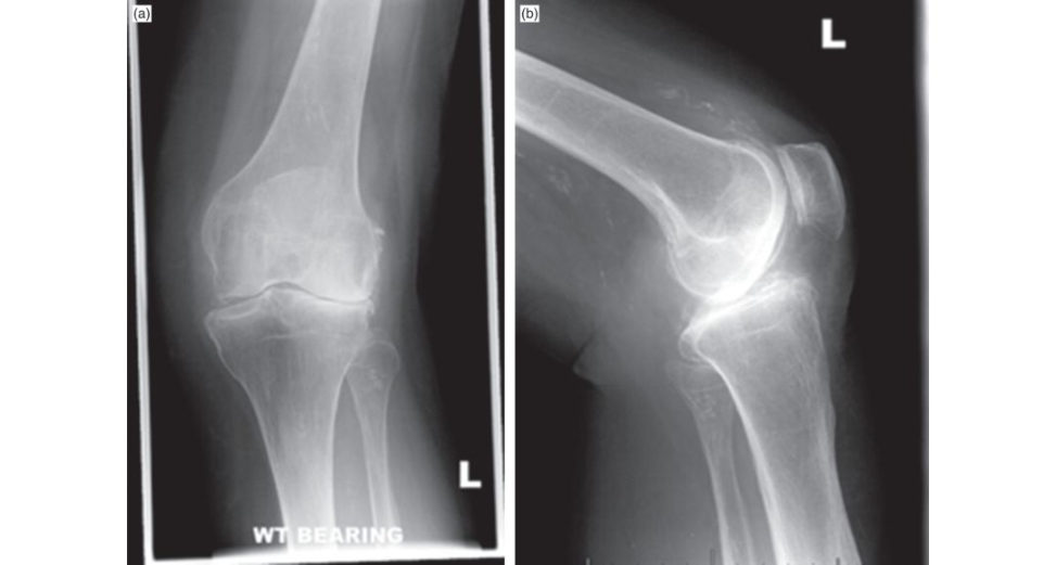
This is a weight-bearing AP radiograph of a 72-year-old female demonstrating severe osteoarthrosis of the left knee with moderate valgus deformity. The cardinal features of osteoarthritis are demonstrated here, which include loss of joint space, subchondral sclerosis, osteophyte formation and subchondral cysts.
How will you manage this patient?
Firstly, I would establish what are the symptoms the patient is suffering from. I would focus on pain, loss of function and severity of symptoms. I would like to know the exact location of the pain, alleviating and relieving factors, and where the pain is radiating to. How the pain is affecting activities of daily living such ascu ting toenails, puting shoes and socks on, how easy it is to go up and downstairs are all questions I would ask. Treatment to date is also important; has the patient had any physiotherapy/rehabilitation, trialled an y analgesics? Previous surgical procedures need to be established. Assessment of the effect of osteoarthritis on the patient’s function, quality of life, occupation, mood relationships and leisure activities is also important. Clinical examination findings such as assessment of the soft tissue envelope is also important. Severity of the deformity in the coronal plane will need to be established. A fixed flexion deformity should also be noted. The competency of the knee collateral ligaments and degree of deformity correction should be assessed in order to plan the type of implants.
OK, after the history and examination, what are you actually going to do?
The management should commence with non-surgical options and generally should be exhausted before surgical options are explored. As per the NICE guidelines, strengthening, low- impact aerobic exercise and neuromuscular education should all be recommended. Acetaminophen and NSAIDS and Tramadol should be the first analgesics offered. Interventions to lose weight, as there is moderate evidence that this will influence osteoarthritis of the knee. Obese patients generally have an increased risk of infection and t end to have an earlier onset of symptoms [1]. Other non-pharmacological treatments include education, social support physical therapy, exercise and orthotics devices. Acupuncture and herbal remedies have limited evidence; however, they are still popular methods of treatment.
What conditions are associated with this pattern of joint disease?
The valgus deformity of the knee with arthritis is commonly seen in women and in inflammatory joint conditions such as rheumatoid arthritis. It can also occur in primary osteoarthritis, overcorrection of high tibial osteotomy (HTO), post-traumatic arthritis following lateral meniscectomy and osteonecrosis of the lateral femoral condyle.
What are your technical goals of treatment?
My aims are the following: Restoring neutral mechanical axis of 0° (±3°). Balancing the flexion/extension gap. Ensuring the joint line perpendicular to the mechanical axis. Preserving the joint line height. Balancing ligaments (2–3 mm symmetrical opening). Restoring normal joint alignment and Q angle.
How would you restore the mechanical alignment?
The bony cuts of the femur and tibia should both be made perpendicular to the mechanical axis of the limb. The mechanical axis is a line bisecting the centre of the hip, knee and ankle. This is to ensure equal loading and even contact pressure of both medial and lateral compartments. The natural tibia has 3° of natural varus to the mechanical axis; however, we do cut perpendicular to the anatomical/mechanical axis. Deformity within the tibia needs to betaken into consideration and the cut should be made perpendicular to the mechanical axis and not the anatomical axis. Longleg mechanical axis views can be helpful for preoperative planning. The mechanical and anatomical axes of the femur are different, and it is this difference which creates the distal femoral valgus angle. This is usually 5–7°. The femoral component must be externally rotated by 3° with respect to the femoral neutral axis to create a rectangular flexion gap with the tibia. As the tibia was cut perpendicular to the mechanical axis, hence in 3° of valgus relative to the native plateau, the femoral component rotation will create a rectangular flexion gap.
Tell me more about the intraoperative considerations.
In valgus knees the lateral femoral condyle is often deficient. This is important to remember because if you perform posterior femoral condylar referencing for femoral component rotation the resultant position of the femoral component will be internally rotated with reference to the transepicondylar axis. In this situation, the AP axis (Whiteside line) can be used to prevent malrotation in the form of internal rotation. The medial structures are stretched while lateral and posterior structures are contracted. The vastus lateralis acts as a subluxing or dislocating force to patella. In severe valgus deformity (7–10°) a distal femoral cut of 5 or 6° can improve patella tracking and avoid the need for lateral retinacular release. Patients with severe valgus deformity may require a lateral retinacular release to achieve normal patella tracking. Excessive PCL release usually requires cruciate-sacrificing implants in order to balance the knee. With correction of significant valgus deformity, one has to be cautious of the common peroneal nerve palsy in the postoperative period. It may be wise to identify the nerve to ensure no increased tension or damage occurs.
OK, so what will be your choice of implant?
I would use a cemented implant. From the latest report of our National Joint Registry, over 84% of the primary TKA procedures performed are cemented, with less than 5% being uncemented and the remaining being a hybrid fixation. The advantages are that the cemented knee isless prone to aseptic loosening. The reis, however, growing evidence for the use of uncemented knees [2]. There is an association with higher failure rates of cemented TK Rin younger, heavier men. There are theoretical advantages of mobile bearing devices over fixed bearing devices, such as reduction in shear stresses and subsequent wear as the tibial insert will be ablet o rotate on a smooth tibial plafoorm. However, I would use a fixed bearing insert; the Knee Arthroplasty Trial Group, after alarge multic entre RCT, concluded that there is no advantage with the mobile bearing designs [3–5]. There are advantages and disadvantages of both cruciate-retaining and posterior stabilized knee replacements; however, I use a posterior stabilized knee, i.e. a cruciate-sacrificing knee replacement. There are many advantages, such as this implant design will facilitate deformity correction and provide anterior posterior stability with the CAM-post mechanism. I find it is technically easier to balance a PCL-sacrificing TK Rand the results are more consistent. It can also be used inpatients with previous patellectomy.
How would you manage gap imbalance?
Ligament balancing is essential for proper knee stability and range of movement. When both the flexion and extension gaps are tight options would be to reduce the polyethylene thickness or resect proximal tibia as this will equally alter both the flexion and extension gaps. If the extension and flexion gap are both equally loose, a larger polyethylene insert can be used. A tight flexion gap with a normal extension gap would require downsizing of the femoral component or shitiing of the femoral component anteriorly [6,7]. With normal knee flexion, a flexion contracture would indicate a tight extension gap and may be an indication of an overstuffed extension spacer. Options of management would be to remove posterior osteophytes initially and release some posterior capsule. If the knee is still tight inextension the next step would be to resect some distal femur. Again, a normal flexion gap but a loose extension gap would indicate excessive distal femoral resection and distal femoral augmentation would be required. Flexion instability results when the flexion gap is larger than the extension gap and is often a result of undersized implants, incompetent PC Lor excessive tibial slope. When the extension gap is normal the femoral component can be shifted posteriorly or one can augment the posterior condyles. It is imperative to ensure there is no anterior femoral cortex notching as a result of this shift.
Figure 4.1a,b Anteroposterior (AP) and lateral radiographs of left knee.
References#
1. Perry KI, MacDonald SJ. The obese pa tienta problem of larger consequence. Bone Joint J. 2016;98(1
Supple A):3–5.
2. Arnold JB, Walters JL, Solomon LB, Thewlis D. Does the method of component fixation influence clinical outcomes after total knee replacement? A systematic literature review. J Arthroplasty. 2013;28(5):740–746.
3. Campbell MK, Fiddian N, Fitzpatrick R, et al. The Knee Arthroplasty Trial (KAT): design features, baseline characteristics and tw o-year functional outcomes after alternative approaches to knee replacement. J
Bone Joint Surg Am. 2009;91(1):134–141.
4. Fransen BL, van Duijvenbode DC, Hoozemans MJ, Burger BJ. No differences between fixed-and mobile-bearing total knee arthroplasty. Knee Surg Sports Traumatol Arthrosc. 2017;25(6):1757–1777.
5. Van der Voort P, Pijls BG, Nou taKA, Val starER Jacobs WC, Nelissen RG. A systematic review and meta-regression of mobile-bearing versus fixed-bearing total knee replacement in 41 studies. Bone Joint J. 2013
Sep 1;95(9):1209–1216.
6. Bercik MJ, Joshi A, Parvizi J. Posterior cruciate-retaining versus posterior-stabilized total knee arthroplasty: a meta-analysis. J Arthroplasty. 2013;28(3):439–444.
7. Huang T, Long Y, George D, Wang W. Meta-analysis of gap balancing versus measured resection techniques in total knee arthroplasty. Bone Joint J. 2017;99(2):151–158.
Structured oral examination question 2#
Basic science: anatomy
This young gentleman sustained a varus type injury to the knee and there is a suspected posterolateral (PLC) injury. Can you describe the lateral structures of the knee by layers (Figure 4.2)?
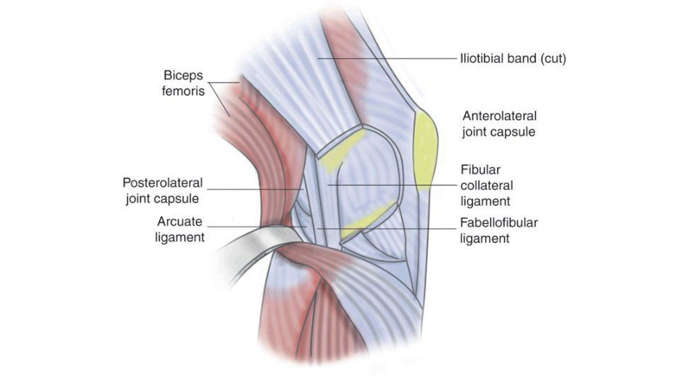
The lateral side of the knee comprises three main layers. Layer one, i.e. the most superficial layer, consists of the iliotibial tr act and biceps femoris. The patella retinaculum is the primary constituent of the second layer. The common peroneal nerve lies between layers one and two. Layer three is split into superficial and deep layers. The lateral collateral ligament (LCL), fabellofibular ligament and anterolateral ligament (ALL) lie superficially. Within the deep layer are the arcuate ligament, coronary ligament, popliteus tendon and popliteofibular ligament. The lateral genicular artery lies between the deep and superficial layers.
How about the medial side of the knee?
The medial side of the knee, similarly to the lateral side of the knee, is split into three layers. Layer one, most superficial, contains the sartorius and patella retinaculum. La yer two contains the semimembranosus, superficial MC Land the MPFL. The deepest layer consists of the deep MCL, capsule and the coronary ligament. Gracilis, semitendonosis and the saphenous nerve run between layers one and two.
So, what is the function of the PL C and how commonly is it injured?
The role of the PL Cis to resist external tibial rotation, varus and posterior tibial translation. The integrity of varus and external rotation stability ultimately depends on the integrity of the fibular collateral ligament (FCL), popliteus, the popliteofibular ligament and the lateral capsule. The PL Cis rarely injured in isolation and is often associated with multi ligament knee injuries; the PC Lis injured less often than the ACL.
Are you aware of any clinical tests to detect a PLC injury?
Hyperextension of the limb on passive extension testing can indicate a PLC injury. Initial g ait examination may reveal a varus thrust. Lateral compartment gapping is assessed and compared with the contralateral side. If there is more gapping of the lateral compartment inflexion, then injury to the secondary stabilizers such as popliteus should be suspected. The dial test essentially measures external rotation of the tibia relative to the femur. I place the patient prone, and flex both knees to 30°. If there is more than 10° external rotation compared with the normal side, a PLC injury is diagnosed. I then flex both knees to 90° and repeat this manoeuvre. An increase in external rotation would suggesta combined PC Land PLC injury. I also use the reverse pivot shift testI place the patient supine with the knee flexed to 90°. A valgus load is applied through the knee joint as well as an external rotation moment around the tibia. The knee is then brought out into extension. If the posteriorly subluxed tibia reduces at 40° of extension, this is interpreted as a positive result. The tibia reduces as a result of the ITB changing from a knee flexor to a knee extender.
What investigations would you perform?
A routine radiograph work up should be performed including anteroposterior (AP), lateral and axial views. Mechanical axis longleg views should also be performed to assess the lower limb alignment. If there is any malalignment, an osteotomy should be performed prior to or at the time of reconstruction. These should be donet o exclude fractures. Varus and PCL stress radiographs are reliable to assist in PC Land PLC injuries respectiv ely.1 Lateral compartment varus gapping of 2.7– 4 mm is consistent with isolated FCL injuries, whereas more than 4 mm indicates a severe-grade PLC injury. Kneeling stress radiographs are best to assess PCL injuries and an opening of 4–12 mm would indicate an isolated PCL injury. Arthroscopy and MRI also have a role to assess for associated chondral, ligamentous and meniscal pathology. Double varus injuries can lead to a varus thrust as the LC Lis injured. A triple varus can also lead to hyperextension and recurvatum, indicating a PL C injury.
What are the options of management?
PLC injuries are usually part of a mulft-lig ament injury spectrum. It is rare to sustain an isolated PLC injury; however, if this were expected, the majority can be managed non-operativ ely.2 If part of a mulft-lig ament injury, the PLC should be addressed. Patients with ACL ruptures should be assessed for PLC injuries and, depending on the grading of injury, should have a reconstruction to assess this deficiency. Grade 3 injuries are a different entity and the reis good evidence to suggest poor functional outcomes and degenerative changes if non-operativ e measures are pursued. Options of operative approaches for grade 3 PLC injuries include acute repairs, which generally are reserved for bone or soft tissue avulsion injuries such as fibular head avulsions. This is normally done within 2–3 weeks and offers the advantage of fixing avulsed bony fragments and other structures anatomically. The AC Land PCL will need to be reconstructed acutely. Delayed reconstruction involves reconstruction of all structures including the PC LAC Land PLC. Earlier evidence by Levy and Stannard et al. have both demonstrated higher failure rates with repair compared to delayed reconstruction. 3,4 However, more recent evidence by Wester mannet al. has suggested good outcomes can be achieved with both repair and reconstruction of PL C injuries treated concurrently with ACL reconstruction a t 6- year follow-up.5 Anatomical reconstruction using tendon allograft and all three static stabilizers has been shown biomechanically to restore native knee biomechanics with improved outcomes. The common peroneal nerve needs to be identified and protected. I would use a modified Larson’s technique for a grade I associated PLC injury, an Arciero for grade II and a LaPrade PLC reconstruction for a grade III injury.
Figure 4.2 Diagram representing lateral side of the knee.
References#
1. LaPrade RF, Heikes C, Bakker AJ, Jakobsen RB. The reproducibility and repeatability of varus stress radiographs in the assessment of isolated fibular collateral ligament and grade-III posterolateral knee injuries: an in vitro biomechanical study. J Bone Joint Surg Am. 2008;90(10):2069–2076.
2. Krukhaug Y, Mølster A, Rodt A, Strand T. Lateral ligament injuries of the knee. Knee Surg Sports
Traumatol Arthrosc. 1998;6(1):21–25.
3. Levy BA, Dajani KA, Morgan JA, Shah JP, Dahm DL, Stuart MJ. Repair versus reconstruction of the fibular collateral ligament and posterolateral corner in the multilig ament-injured knee. Am J Sports Med.
2010;38(4):804–809.
4. Geeslin AG, LaPrade RF. Outcomes of treatment of acute grade-III isolated and combined posterolateral knee injuries: a prospective case series and surgical technique. J Bone Joint Surg Am. 2011;93(18):1672–1683.
5. Westermann RW, Spindler KP, Huston LJ, Wolf BR. Posterolateral corner repair versus reconstruction: 6-year outcomes from a prospective multic enter cohort. Orthop J Sports Med. 2017;5(7_suppl6).
Structured oral examination question 3#
Meniscus
Tell me about the anatomy and function of the meniscus.
The menisci are crescentic cartilaginous structures interposed between the tibia and femoral condyles. They are triangular in cross-section. The peripheral borders are attached to the joint capsule. The medial meniscus is nearly semicircular with a wider posterior than anterior horn. This is attached anterior to the ACL insertion while the mid aspect is firmly attached to the deep MCL. The lateral meniscus is circular with a larger surface than the medial meniscus. The posterior horns of both menisci attach to the posterior intercondylar eminence. The attachment of the lateral meniscus to the capsule is interrupted by the popliteus tendon. Due to the loose attachment to the capsule, the lateral meniscus has twice the excursion to that of the medial meniscus. The anterior horns of both menisci are connected by intermeniscal ligaments. Histologically, the menisci have an extracellular matrix composed mainly of water (70%) and primarily type 1 collagen fibres (60%), proteoglycans, elastin and glycoproteins. The main cellular component is the fibrochondrocytes that synthesize and maintain extracellular matrix. The blood supply to the meniscus comes from the lateral, middle and medial geniculate vessels with 20–30% of the peripheral portion being vascular. The main functions of the menisci are load transmission with estimated 50% inextension and 85% inflexion, joint conformity and articular congruity, distribution of synovial fluid aiding nutrition and joint lubrication. The menisci also have proprioceptive function, act as shock absorbers and prevent soft tissue impingement during joint motion.
What are the vascular zones of meniscus?
The menisci are relatively avascular structures with peripheral blood supply from the premeniscal capillary plexus formed by branches from lateral and medial geniculate vessels. Studies have shown that the degree of peripheral vascular penetration is 10 –30% of medial meniscal width and 10–25% of lateral meniscal width. This gives rise to the three zones of meniscal vasculature from peripheral to central, namely red–red, red–white and white–white. Therefore, peripheral tears are suitable for repair while central tears may not be suitable due to lack of healing capacity.
These are the images which belong to a young professional footballer (Figures 4.3b and 4.3c). What can you see?
The first is asa gift al T1 MRI image and the secondis a T2-weighted image. The coronal image does not show any medial meniscal tissue indicating a peripheral detachment. There is a ‘double PCL signon the sagift al image suggestive of bucket handle tear of the medial meniscus.
How would you manage this patient?
I would start by taking a detailed history and clinical examination ... [ EXAMINER interrupts].
How would you treat this patient? [ EXAMINER geting impatient]
I would offer this patient EUA, arthroscopy, repair or excision of bucket handle tear.
Good. What factors affect the prognosis following meniscal repair?
Firstly, the patient age dictates what I would do for the bucket handle tear. As he is a young professional footballer I would do my best to preserve the meniscus. Older patients haveless cellularity and decreased healing response. Re-tears are common inpatients above the age of 30. Location of the t ear and the tear pattern are also important factors. The chronicity of the tear will also dictate what I do. I am also much more inclined to preserve the lateral meniscus than the medial meniscus. Other factors include location and pattern, and associated pathology, i.e. ligamentous malalignment and pre-existing arthritis.
Are you aware of any meniscal repair techniques?
The four main meniscal repair methods are open repair, inside out, outside in and all inside. The ‘outside in’ method is versatile and safe but is reserved for anterior horn tears. [ EXAMINER interrupts again]
Let’s move on ...
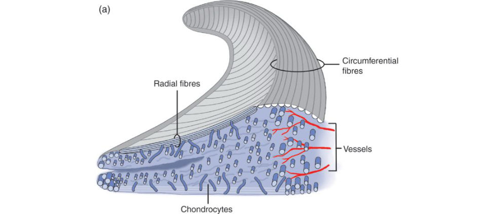
Figure 4.3a Cross-sectional anatomy of the meniscus.
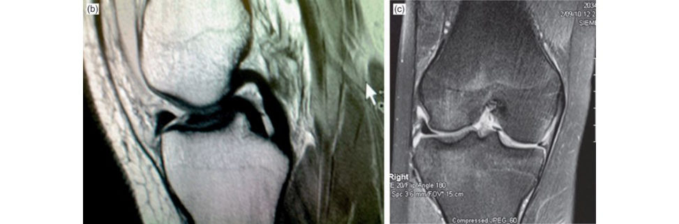
Figure 4.3b and 4.3c T2-weighted sagift al and coronal MRI scan images of the knee.
References#
Arnoczky SP, Warren RF. Microvasculature of the human meniscus. Am J Sports Med. 1982;10:90–95.
Structured oral examination question 4#
Infected total knee arthroplasty (TKA)
This gentleman had aTKA performed 3 months ago. Ten days after surgery he developed a large blister that was drained on the ward. Two weeks later he developed a draining abscess at the proximal aspect of the wound. He was taken to theatre and the abscess was washed out. The skin was debrided and closed primarily. Have you got any concerns?
This patient has a periprosthetic joint infection (P JI) until proven otherwise. He clearly developed an acute infection following the surgery. An acute infection is defined by the American Academy of Orthopaedic Surgeons and the International Consensus on P JI as infection within 3 weeks of the procedure, or in the case of late haematogenous infection, within 3 weeks of development of symptoms. Any infection developing after this time is considered as late. This is irrespective of bone stock and stability of the components.
These are the most recent radiographs and there are no other postoperative radiographs available (Figures 4.4a and 4.4b). What would you like to do for this patient?
I would start by taking a detailed history of the peri operative events, general health as well as current problem. I would like to know the date of index operation, if there was prolonged discharge from the wound, redness or persistent swelling in the immediate postoperative period. A pain-free interval after the operation followed by sudden deterioration may be suggestive of haematogenous spread precipitated by bacteraemia from UTI, URTI or dental procedure.
Are you aware of any staging systems with the use of periprosthetic joint infection?
McPherson advocated the concept of staging, which consists of timing of infection, i.e. early , acute haematogenous or late. The systemic medical and immune status of the patient is used. Systemic compromising factors include excessive alcohol, diabetes, smoking, liver, renal and lung failure. Immune status assessment includes CD4 count and neutrophil counts. Local compromising factors are also recorded and include whether active infection is present, synovial cutaneous fistula presence and a subcutaneous abscess of more than 8 cm2. Each category has different grades according to what they score.1
So how would you diagnose a periprosthetic joint infection?
I would always start with the history and clinical examination of the patient. I would focus on the events around the procedures this gentleman has had. Even without signs of infection, a painful TKR can manifestas a low-grade infection. I am a ware of the AAOS clinical guideline practice summary for diagnosis of periprosthetic joint infection of the knee. The working group strongly recommend: Testing E SR and CRP. Joint aspiration. The use of intraoperative frozen sections.
Obtaining multiple intraoperative cultures, against initiating antibiotic treatment until after cultures and against the use of intraoperative Gram stain.
Nuclear imaging was weakly recommended as an option in patients in whom diagnosis of periprosthetic joint infection has not been established and who are not scheduled for reoperation. 2
I refer to the diagnostic criteria setout by the workgroup of the MSK infection society. The major criteria include either a sinus tract communicating with the prosthesis or if a pathogen is isolated by culture from at least two separate tissue or fluid samples obtained from the affected prosthetic joint, or four of the following six minor criteria: (a) elevated CR Pand ESR, (b) elevated synovial leucocyte count, (c) elevated synovial neutrophil percentaged) presence of purulence in the affected joint, (e)
isolation of a microorganism in one culture of periprosthetic joint fluid or tissue and (f ) > 5 neutrophils per high power field observed at ×400 magnification. 3 The isolation of the infecting organism and the corresponding antibiotic profile are essential.
Anything else?
Analysis of the joint fluid which should include a leucocyte esterase assessment ...
Anything more recent in the literature?
The alpha-defensin test, which is an immunoassay, has recently been shown to have a sensitivity and specificity of 100% in the diagnosis of P JI. Advantages include the specimen could also be assessed when blood was in the synovial fluid.4
So how are you going to manage this case?
It is unfortunate, but I think this gentleman cannot be treated as an acute infection. He has missed the window of opportunity of debridement, antibiotic treatment and implant retention. He has already undergone a soft -tissue debridement and I am worried about the soft tissue envelope . It may require a plastic surgeon to review the wound. I would offer this pa tienta revision knee arthroplasty.
And how would you do this?
It can be done over a one-stage or two-stage procedure. One stage has many advantages including low treatment costs and shorter hospital stays. The infecting organism and its sensitivity should be established preoperatively. There are, however, contraindications to this, including infection with a highly virulent organism, sepsis with substantial systemic manifestations and cultur e- negative PJI where appropriate antibiotic treatment cannot be determined.
So ... ?
I would plan for a two-stage exchange procedure where I would remove all the material, including the cement, and perform an aggressive debridement of the soft tissues and bone. I would insert an antibiotic-loaded dynamic cement spacer. This should preserve the joint space, reduce soft -tissue contractures, and provide better ROM and knee function scores while eluting high doses of IV antibiotics. 5 Debridement is the most essential part of the procedure with meticulous handling of soft tissues to remove all septic membranes.
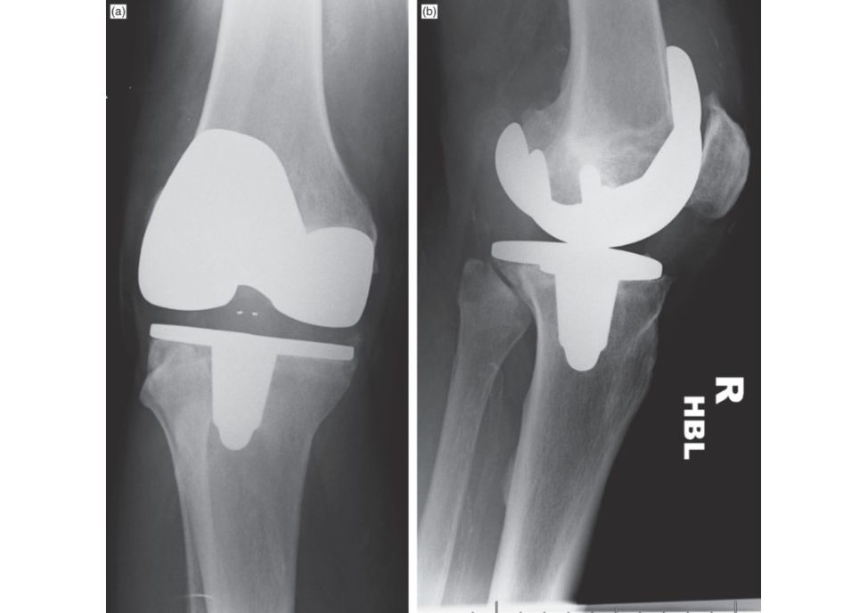
Figure 4.4a and 4.4b Anteroposterior (AP) and lateral radiographs of left TKA .
References#
1. McPherson EJ, Woodson CHol tom P, Roid isN, Shufelt C, Patzakis M. Periprosthetic total hip infection:
outcomes using a staging system. Clin Orthop Relat Res. 2002;403:8–15.
2. Della Valle C, Parvizi J, Bauer TW, et al. Diagnosis of periprosthetic joint infections of the hip and knee. J
Am Acad Orthop Surg. 2010;18(12):760–770.
3. Parvizi J, Zmistowski B, Berbari EF, et al. New definition for periprosthetic joint infection: from the
Workgroup of the Musculoskeletal Infection Society. Clin Orthop Relat Res. 2011;469(11):2992.
4. Deirmengian C, Kardos K, Kilmartin P, et al. The alpha-defensin test for periprosthetic joint infection outperforms the leukocyte esterase test strip. Clin Orthop Relat Res. 2015;473(1):198–203.
5. Ding H, Yao J, Chang W, Liu F. Comparison of the efficiency of static versus articular spacers in two-stage revision surgery for the treatment of infection following total knee arthroplasty: a meta-analysis. J Orthop
Surg Res. 2017;12(1):151.
Structured oral examination question 5#
Unicondylar knee arthroplasty (UKA) vs high tibial osteotomy (HTO)
This is a radiograph of a 42-year-old man who works as a bricklayer (Figure 4.5). He complains of pain over the medial aspect of the knee and has failed non-surgical management. He has come to your clinic for a consultation. What can you see?
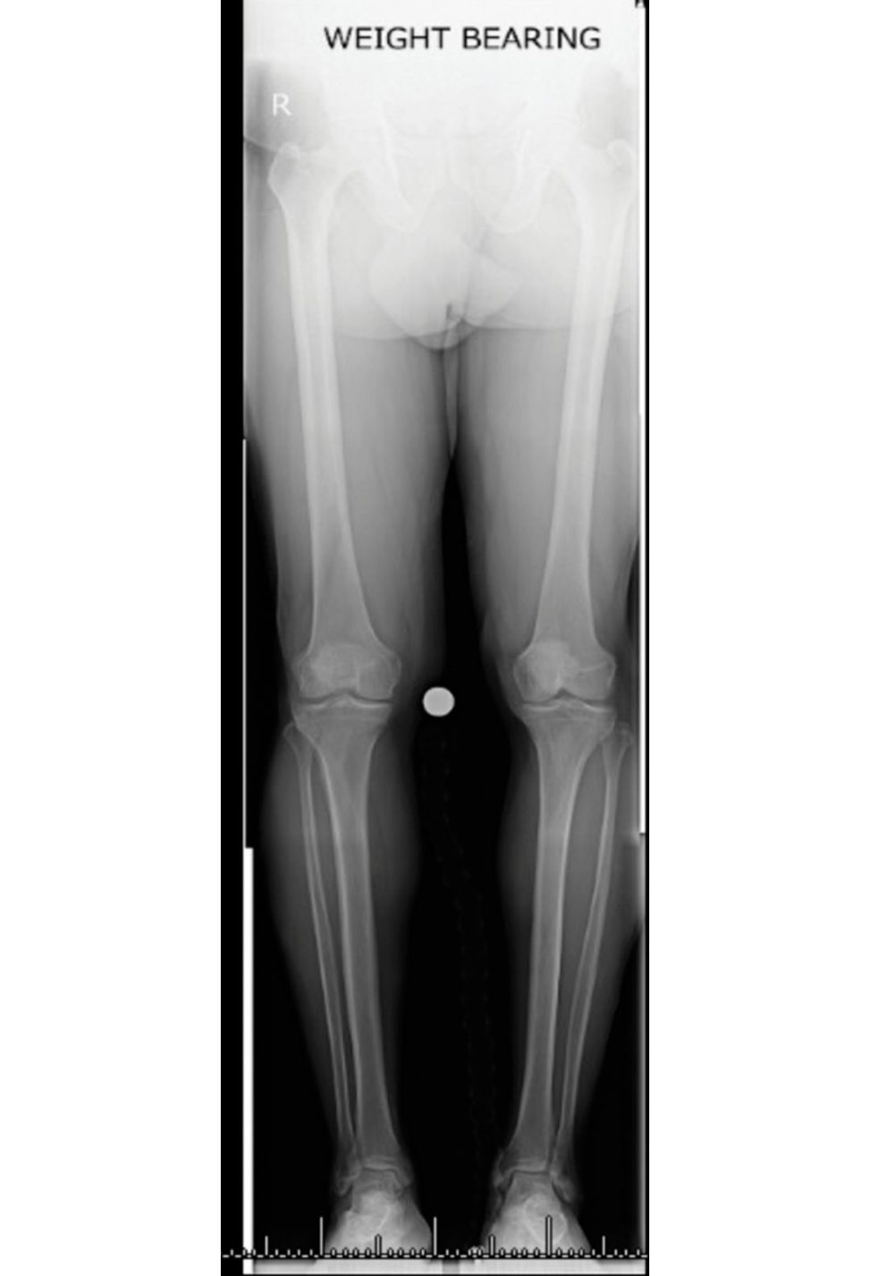
These are longleg x-rays demonstrating moderate medial tibiofemoral compartment osteoarthritis. The lateral compartment appears normal. Crude assessment of the tibiofemoral angle appears to show a varus deformity of less than 10°. I cannot comment on the patellofemoral joint, hence I would like to see a lateral and a skyline view. There is no joint subluxation, long bone deformity or adjacent joint disease. The soft tissues appear to have normal body habitus. I would like to take a targeted history and examine the patient The examination should be focused on eliciting localized tenderness in the medial compartment, range of motion, if the varus deformity is correctable and the stability of the knee.
The patient is fit and well, states that the pain is affecting his job and he would like to consider surgical option 5. What would you offer him?
The options of surgical management include a valgizing osteotomy such as a high tibial osteotomy (HTO), medial unicondylar knee arthroplasty or total knee replacement. As this patient has a high-demand physical job and is young, I would offer him an osteotomy.
What are the prerequisites of an HTO? And what are your indications?
The ideal candidate for HTO is apa tien t with physiological age of < 65 years, fixed varus deformity < 15° or valgus deformity < 12°, fixed flexion deformity of < 15° and a flexion arc of more than 90°. Patients with pain primarily on the medial aspect of the knee and less than 4 mm of space on the medial compartment on weight-bearing views with mechanical overload and varus deformity are prime candidates for a valgizing osteotomy. Young patients undergoing medial meniscal transplantation and articular cartilage restoration procedures may require an unloading osteotomy. Preoperative varus must exist and even patients with as liti leas 4° of varus can benefit from an HTO.
Are you aware of any contraindication for HTO?
I would avoid an HTO in apa tien t with marked decreased range of movement, i.e. flexion of less than 90° if possible. The main contraindications are inflammatory arthropathy such as rheumatoid arthritis and psoriatic arthropathy incompetent medial collateral ligament or ACL, large varus thrust with coronal subluxation of > 1 cm, although there are times one can adjust the tibial slope to compensate for ligament deficiency. Obesity is also a relative contraindication because valgus knee is poorly tolerated due to medial thigh contact. However, ensuring stable fixation can improve the success in obese patients. A small lateral plate to fix the hinge in addition to the medial plate in a medial opening wedge HTO has been described. Any patient who smokes really should stopas this can cause a non-union. Consideration of a closing osteotomy or cancellous gratiing are options.
The patient tells you that he has heard about closing wedge osteotomy arthroscopies and partial knee replacements and is keen to consider the alternative options. How do you proceed?
I would explain that UKA is an option however, I would not recommend UKA for this particular patient because of his young age and the high physical demand of his job could result in accelerated wear of a UKA. In the absence of an acute fixed flexion deformity and locked knee I would not offer him an arthroscopy. There have been numerous randomized controlled trials failing to show benefit when comparing arthroscopic debridement and partial meniscal resections against other controlled interventions. Thorlund et al. published a systematic review and meta-analysis of the benefits and harms of arthroscopy to the degenerative knee [1]. The evidence does not support this option in this situation. The reis also a recent randomized controlled trial, by Barton et al., demonstrating that an arthroscopy in the year prior to aTKA were linked with significantly reduced Oxford Knee scores; also this cohort had a higher revision rate over a four-year period [2].
So which patients would you offer UKA?
The indications and prerequisites for HTO an dUKA are more orless the same between the ages of 55 and 65 years of age. However, women prefer the UKA because they do not tolerate the angular deformity created by an HTO very well. In addition, patients who have low physical demands may benefit from a UKA. A frank discussion regarding the pros and cons of each procedure needs to occur between the patient and surgeon. A meta-analysis by Fu et al. demonstrated with careful patient selection that both HTO an dUKA demonstrate effective and reliable results [3].
Are there any contraindications to performing a UKA?
Accepted contraindications include inflammatory arthritis, large fixed flexion deformities, ligamentous laxity and prior meniscectomy in the contralateral compartment. Previously old age, high activity le vel [4], obesity and pattern of OA were seen as contraindications However, recent evidence has shown this is not the case [3]. There is good evidence to offer octogenarians a UKA with good outcome data [5]. Increasing BMI has also been found not to be associated with an increased failure rate [6].
What are the advantages of a UKA?
By retaining the cruciate ligaments and remaining healthy joint surfaces, UKA restores the normal ligament driven kinematics of the native knee [7]. The procedure generally requires a smaller incision, avoiding the need to evert the patella. There is a shorter hospital stay and lower rates of infection, morbidity and mortality. Patients undergoing UKA are halfas likely to have a major complication such as a myocardial infarction ors troke within the first 30 days after surgery. There is also evidence to suggest better PROMs and increased range of movement inpatients undergoing UKA. It is technically not too challenging to revise a UKA to aTKA.
So how about the outcomes? Are they comparable? And what does the NJR state?
There is a lot of evidence published from the designer group with regards to the outcomes of UKR. Lisowski et al. published a 10–15-year outcome prospective study. These are results from a non-designer group. Only media lUKAs were included. PROMs including the OKS, VAS and WOMAC scores were recorded. There was a mean follow-up of 11.7 years and results demonstrated excellent long-term functional and radiological outcomes with a 90% 15-year survival rate [8]. The 10-year revision rate published in the 13th annual report of the National Joint Registry was 12.38%. It has been suggested by Baker et al. that high-volume centres and surgeons specializing in UKA have superior results compared to their low-volume counterparts. The low-volume surgeon and low- volume centres will have skewed the NJR revision rate [9].
Right let’s get back to our patient how about the results of TKA following HTO? Are they worse?
There is evidence to suggest the outcomes of TKA following HTO, especially an opening wedge osteotomy, are as good as TKA without a HTO [10]. However, there is added difficulty with performing the procedure such as additional removal of hardware if a one-staged procedure is decided upon. [11]. There is no evidence demonstrating an y differences in the outcomes between opening and closing wedge osteotomies [4].
Let’s say this patient has decided to go ahead with a HTO. How would you go about this? What would be your preference?
I always obtain full-length weight-bearing mechanical axis hip–knee–ankle radiographs. The mechanical and anatomical axes should be drawn. Native tibiofemoral varus alignment, medial joint space degeneration and lateral capsuloligamentous laxity need to be assessed. This can be compared with the contralateral side. The Coventry group suggest overcorrection to 8° of anatomical valgus. The difference between the preoperative anatomic axis and the planned anatomic axis should be calculated. The angle of correction can also be calculated using the Miniaci method. Firstly, the true mechanical axis is drawn from the centre of the femoral head to the centre of the tibiot alar joint, Line A. A second line is dropped from the centre of the femoral head topass through the knee at the intended Mikulicz point (in a varus knee, aiming for 55% – width from medial tibial plateau), Line B. Now we need to create a hinge point, Point H. Two lines creating an angle are drawn connecting the caudal points of Line A and B.
You mentioned difficulty with conversion of HT Oto TKA. Tell me more about this.
Problems which can be encountered include patella baja which can make eversion of the patella and access very difficult, also tracking of the patella will be more challenging. There will be more distorted landmarks for proper tibial component orientation. The incision may well not be ideal for my joint replacement. I may need to consider staging the removal of metalwork and taking some deep samples to ensure infection is not present. Bone stock may be deficient laterally following a closing wedge osteotomy. The anterior tibial slope is also of importance and should be addressed. The tibial shaft offset can be medially deviated, which may require offset stems. Soft tissue balancing can be more challenging. More recent studies show that closing wedge osteotomy increases patellar height, whereas opening wedge osteotomy lowers patellar height, and this can have implications following TKA. Van Raaij et al. performed a systematic review and reported prolonged surgical time, extra operative procedures and less postoperative knee range of motion (R OM), but no increase in revision surgeries for patients receiving TKA after prior HTO compared topa tien ts receiving primary
TKA.
Figure 4.5 Standing anteroposterior (AP) lower limb alignment views.
References
1. Thorlund JB, Juhl CB Roos EM, Lohmander LS. Arthroscopic surgery for degenerative knee: systematic review and meta-analysis of benefits and harms. Br J Sports Med. 2015;49(19):1229–1235.
2. Barton SB, McLauchlan GJ, Canty SJ. The incidence and impact of arthroscopy in the year prior to total knee arthroplasty. Knee. 2017;24(2):396–401.
3. FuD, LiG, Chen K, Zhao Y, Hua Y, Cai Z. Comparison of high tibial osteotomy and unicompartmental knee arthroplasty in the treatment of unicompartmental osteoarthritis: a me ta-analysis. J Arthroplasty.
2013;28(5):759–765.
4. Prest onS, Howard J, Naudie D, Somerville L, McAuley J. Total knee arthroplasty after high tibial osteotomy: no differences between medial and lateral osteotomy approaches. Clin Orthop Relat Res. 2014;472(1):105–110.
5. Tadros BJ, Dabis J, Twyman R. Short-term outcome of unicompartmental knee arthroplasty in the octogenarian population . Knee Surg Sports Traumatol Arthrosc. 2018;26(5):1571–1576.
6. Ali AM, Pandit H, Liddle AD, et al. Does activity affect the outcome of the Oxford unicompartmental knee replacement? Knee. 2016;23(2):327–330.
7. Pandit H, Jenkins C, Gill HS, et al. Unnecessary contraindications for mobile-bearing unicompartmental knee replacement. J Bone Joint Surg Br. 2011;93(5):622–628.
8. Lisowski LA, Meijer LI, Bekerom MP, Pilot P, Lisowski AE. Ten- to 15-year results of the Oxford Phase III mobile unicompartmental knee arthroplasty: a prospective study from a non-designer group. Bone Joint J.
2016; 98B(10 Supple B):41–47.
9. Baker P, JamesonS, Critchley R, Reed M, Gregg P, Dee hanD. Center and surgeon volume influence the revision rate following unicondylar knee replacement: an analysis of 23,400 medial cemented unicondylar knee replacements. J Bone Joint Surg Am. 2013;95(8):702–709.
10. Meding JB, Wing JT, Rifter MA. Does high tibial osteotomy affect the successor survival of a total knee replacement? Clin Orthop Relat Res. 2011;469(7):1991–1994.
11. Niinimäki T, Eskelinen A, Ohtonen P, Puhto AP, Mann BS, Leppilahti J. Total knee arthroplasty after high tibial osteotomy: a registry-based case–control study of 1,036 knees. Arch Orthop Trauma Surg.
2014;134(1):73–77.
Structured oral examination question 6#
Dislocate dUKA PE spacer
Have a look at these radiographs (Figure 4.6a and 4.6b). What can you see?
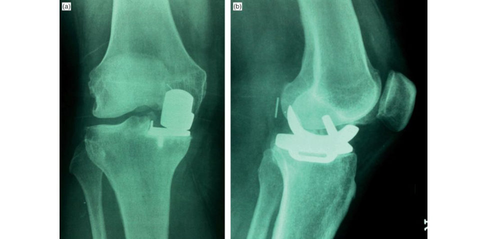
Weight-bearing AP and lateral radiographs of 54-year-old man showing a left media lUKA in situ. The components look well fixed and aligned. There are no obvious periprosthetic fractures. The lateral compartment and PFJ look relatively normal.
What else can you see?
[A bit hesitant and moving closer to the computer screen. This is followed by a period of silence before the Examiner prompts]
The patient tells you that he fell while coming down the stairs, sustaining injury to the left knee. He complains of global pain and swelling of the left knee and inability to flex it. What’s going through your mind?
There is a faint radio-opaque line behind the femoral component and on the AP radiograph, aver tical lucency can beseen proximal to the medial tibial eminence. I would like to compare this with previous radiographs. The history and radiographs are suggestive of dislocation of the mobile- bearing spacer.
How common is meniscal dislocation?
The meniscal dislocation rate for media lUKA is 1 in 200 and 10% in latera lUKA. Dislocation in media lUKA is usually anterior and rare in other directions. There may be a history of trauma and it usually presents with severe onset of knee pain with difficulty weight-bearing. Mobile-bearing knee implants are less tolerant of soft tissue imbalance. The reis increased risk of bearing instability and dislocation with poor soft tissue balancing or unequal flexion and extension gaps.
Any other possible causes for dislocation?
Other causes could include intraoperative MCL damage, progressive stretching of the medial collateral ligament or bearing impingement on unresected osteophytes.
Why is there an increased risk of dislocation in latera lUKA?
The increased dislocation rate in latera lUKA is mainly due to the fact that the medial collateral ligament is tight and lateral collateral ligament lax inflexion. Therefore, the medial compartment gets distracted about 2 mm on average in comparison with 7 mm on the lateral side.
How are we going to manage this case?
The spacer could be exchanged for a larger one, but it may dislocate again with continued flexion/extension imbalance.
Definitive management would be revision toPS knee replacement.
Figure 4.6a and 4.6b Anteroposterior (AP) and lateral radiographs UKA.
Structured oral examination question 7#
Posterior cruciate ligament and anterior cruciate ligament reconstruction
These are images of a 26-year-old rugby player who has given a history of falling awkwardly. What can you see (Figures 4.7a–4.7d)?
These are plain radiographs and MR Iof the right knee which show a displaced avulsion fracture of the posterior intercondylar tibial spine. This is where the PCL inserts, with the origin of the PCL being the lateral wall of the medial femoral condyle.
How would you treat this patient?
I would offer this patient reattachment of the PCL avulsion through open procedure.
What approach would you use?
The posterior approach.
Tell me about the posterior approach to the knee.
The indications include removal of popliteal cysts and neoplasms, posterior synovectomy, open reduction and internal fixation of posterior tibial plateau shear fractures, fixation of bone avulsions associated with a posterior cruciate ligament (PCL) injury, repair of posterior vascular injuries, and more recently, posterior inlay PCL reconstructions. The patient is usually positioned prone with tourniquet high up in the thigh. The S-shaped incision is centred over the popliteal fossa with the oblique section overlying the joint. The incision can be extended proximally along the tendon of semitendinosus and continued distally over the head of the lateral head of gastrocnemius. The medial sural cutaneous nerve is identified beneath the fascia. Just lateral to the nerve is the short saphenous vein. The deep fascia is incised in the midline. The medial sural cutaneous nerve is traced proximally where it pierces deep fascia from the tibial nerve trunk. At the apex of the fossa, the common peroneal nerve separates from the tibial nerve. The tibial nerve lies posterior to the popliteal vein, which inturn is superficial to the popliteal artery. Popliteal vessels are displaced laterally and this usually requires ligation of the middle geniculate and superior medial geniculate vessels. The medial head of the gastrocnemius is identified, traced proximally and can be detached from its origin then retracted towards the midline to expose the medial joint capsule. Similarly, the lateral head of the gastrocnemius can be detached to expose the posterolateral corner of the joint. The main structures at risk are the popliteal vessels, small saphenous vein and common peroneal nerve and tibial nerve.
Can you tell me about the anatomy of the ACL?
The AC Lis an intra-articular structure but extra-synovial with a blood supply from the middle genicular artery. It arises from the medial border of the lateral femoral condyle and inserts proximally into the tibial spines. It has an anteromedial (AM) and a posterolateral (PL) bundle. The AM bundle tighten sin flexion and the PL bundle tighten sin extension. This, however, is an oversimplification. It has been recently described as a ribbon-like structure. It primarily functions to prevent excessive anterior translation relative to the femur. It also primarily resists internal rotation of the tibia relative to the femur. It is a secondary stabilizer to varus and valgus stress. The main consistency is type 1 collagen; however, there is some type III collagen.
When considering an ACL reconstruction, which gr aft would you use and why?
The two main options are autograft and allograft The two most popular autogratis are hamstring and bone–patella–bone tendon gratis (BPBG) [ 1]. The advantages of using autogratis are that they are biologically friendly and readily available, there are no additional costs and no risk of disease transmission. There are advantages of a BPBG over the hamstring graft, such as direct bone- to-bone healing within the femoral and tibial tunnels and a voiding loss of hamstring proprioception and strength, which can be detrimental to a high-level athlete. There is said to be less anterior knee pain with hamstring reconstructions. Smaller incisions and a higher maximum load to failure being almost twice that of the BPBG are other potential advantages. However, hamstring gratis have slow healing properties because of tendon to bone incorporation, which can take 8–12 weeks. There are several studies comparing outcome of BPTB versus hamstring graft. Most studies show arthroscopic reconstruction with either gr aft results in similar functional outcome but increased morbidity in BPT Bin the form of early OA and increased knee laxity [2–4].
How about in revision ACL reconstruction? Does the type of gr aft affect the outcome?
Grassie tal. [5] published a meta-analysis looking into just this. Autogratis had better outcomes than allogratis in revision ACL reconstruction. There were lower rates of postoperative laxity and lower rates of re-operations and complications. Interestingly , if irradiated allogratis are excluded, outcomes are similar between auto- and allogratis.
Now tell me about the optimum tunnel placement of your bony tunnels in an ACL reconstruction.
The principles of ACL reconstruction are placement of tunnels anatomically and isometrically, using biologically active gratis which are adequately tensioned to allow early rehabilitation. My aim ist o stabilize the knee, restore normal kinematics and prevent early-onset degenerative arthrosis. The clockface method is a method for assessing femoral tunnel height; however, without proper alignment of the arthroscope it can be imprecise. In order to achieve ‘graft isometry’ the optimal position of the femoral tunnel was thought to be 11 or 1 o’clock depending on the side of surgery. This will place the femoral tunnel high and deep in the lateral femoral condyle. This would be ideal for isometric positioning. As someone whoa dop ts anatomic tunnel placement, I believe isometry is not the most crucial factor. The ACL femoral attachment is oval in appearance and is defined by two bony ridges. These ridges, the lateral intercondylar ridge and the bifurcate ridge, will dictate where I place the femoral tunnel; however, this does require some extensive ACL remnant resection. The preserved remnant can actually enhance biological healing and provides mechanical support to the ACL reconstruction. The most common mistake is to place the femoral tunnel too ‘shallow’ on the so-called ‘resident’s ridge’. This restricts flexion of the knee and may result in elongation of the gr aft causing incompetence and recurrent instability. Similarly, too ‘deep’
a tunnel placement results in excessive tightening of the graft when the knee is extended. It has been shown that an abnormally narrow intercondylar notch correlates directly with increased incidence of
ACL tears. Careful assessment of the notch should be done prior to graft insertion to ensure no impingement on the lateral femoral condyle. The presence of impingement with correct placement of the tunnels necessitates notchplasty of the anterior portion of the lateral femoral condyle.
How would you manage a child with an ACL injury who came to your clinic? Say she was 12 years old?
I would counsel the patient with regards to rehabilitation and activity modification . However, compliance with bracing and limitation of activity can be very difficult. My aim is to limit damage to the menisci and chondral surfaces. One should always consider the physis when planning reconstruction, as damage to the distal femoral physis can cause deformity. Reconstructions can be either extraphyseal or intraphyseal or even extra- or intra-articular . Extra-articular la teral-based procedures like a Macintosh oraLe maire tenodesis are an option. In tra-articular reconstructions can be physeal-sparing. If transphyseal reconstruction is considered, then soft tissue gratis need to be used and bone plugs need to be avoided.
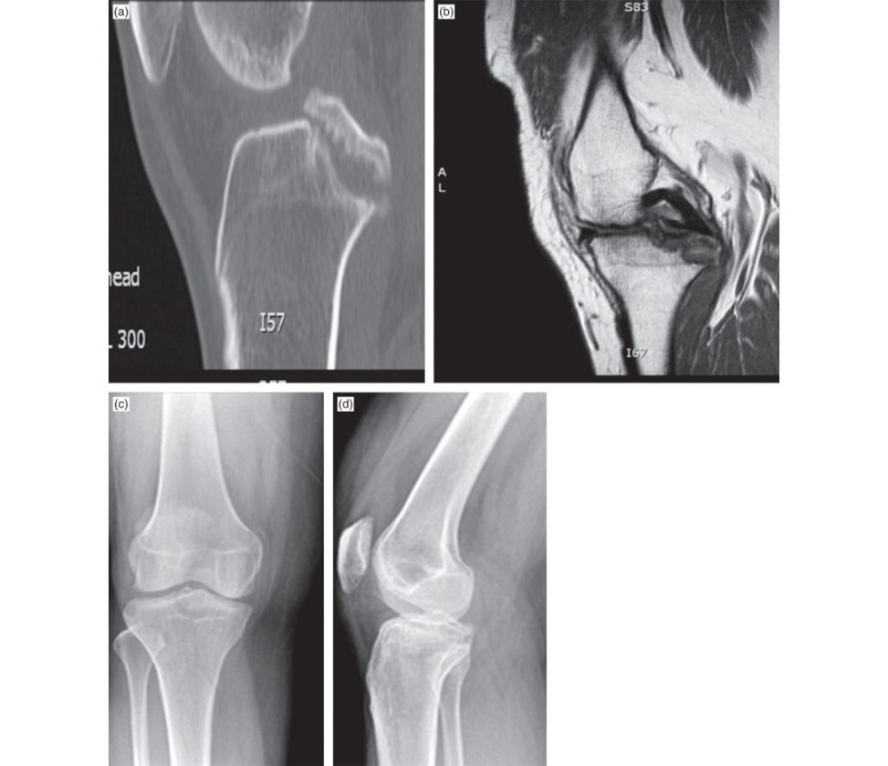
Figure 4.7a, 4.7b, 4.7c and 4.7d CT, MRI and plain radiographs of right knee.
References#
1. Xie X, Liu X, Chen Z, Yu Y, PengS LiQ. A meta-analysis of bone–patellar tendon–bone autograft versus four-strand hamstring tendon autograft for anterior cruciate ligament reconstruction . Knee.
2015;22(2):100–110.
2. Xie XX iao Z, LiQ, et al. Increased incidence of osteoarthritis of knee joint after ACL reconstruction with bone–patellar tendon–bone autogratis than hamstring autogratis: a me ta-analysis of 1,443 patient sat a minimum of 5 years. Eur J Orthop Surg Traumatol. 2015;25(1):149–159.
3. Howell SM, Taylor MA. Failure of reconstruction of the anterior cruciate ligament due to impingement by the intercondylar roof. J Bone Joint Surg Am. 1993;75(7):1044–1055.
4. Webster KE, Feller JA, Hartnett N, Leigh WB, Richmond AK. Comparison of Patellar tendon and hamstring tendon anterior cruciate ligament reconstruction: a 15- year follow-up of a randomized controlled trial. Am J Sports Med. 2016;44(1):83–90.
5. Gras siA, Nit riM, Moulton SG, et al. Does the type of graft affect the outcome of revision anterior cruciate ligament reconstruction? A me ta-analysis of 32 studies. Bone Joint J. 2017;99B(6):714–723.
Structured oral examination question 8#
Revision knee replacement
Have a look at these images and tell me what you can see (Figure 4.8a and 4.8b).
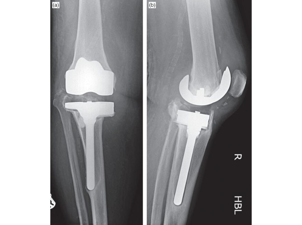
These are AP and lateral radiographs of a failed left total knee replacement. The implants appear to be loose with widespread osteolysis and bone loss on both the femoral and tibial sides. The tibial component is stemmed and has a medial augment, suggesting that this in itself is a revision implant. Bone stock on the tibial side is certainly an issue which needs to be addressed. There is calcification of soft tissues including the popliteal vessels. I would like to see immediate postoperative and most recent radiographs for comparison. The radiographs are suggestive of infection un til proven otherwise.
OK. After investigation you conclude this is aspetic loosening. The patient is keen to consider single-stage revision surgery. What are your concerns with regards to these radiographs?
The collateral ligaments are likely to be dysfunctional, especially the MC Las the tibial component has subsided significantly and may be compromising the insertion of the MCL. Therefore, a constrained knee replacement may be required. The soft tissue envelope shadow on the radiograph appears contracted and calcified which may lead to wound complications. I would always take deep samples in all revisions to exclude infection asP JI can often be misdiagnosed.1 The extensive bone loss will require a stemmed implant to promote load-sharing of the diaphysis. A fracture of the tibia may occur; however, the use of the stem will bypass this area. A metaphyseal sleeve will be needed to help with significant bone loss. I would beworried about the state of the extensor mechanism and if the patella tendon would remain attached after removal of the implants. The joint line needs to be restored and this may be a challenge.
And what are your goals of surgery?
I would want to restore the joint line by achieving well-fixed implants and achieve a stable knee joint in a balanced fashion. I would also like to restore bone deficiencies and extract all the components with minimal bone loss while preserving the extensor mechanism.
Are you aware of any classification system for bone loss around knee arthroplasty?
The most commonly used classification system is that of the Anderson Orthopaedic Research Institute (AORI), which classifies the femur (F) and tibia (T) separate lyas follows: Type-1 – Intact metaphysical bone with minor defects which will not compromise the stability of a revision component. Type-2 – Damaged metaphysical bone. Loss of cancellous bone in the metaphyseal segment which will need to be filled with cement, augments or a bone graft a t revision in order to restore the joint line. Defects can occur in one femoral condyle or tibial plateau (2A) or in both condyles or plateau (2B).
Type-3 – Deficient metaphysical bone. Bone loss which comprises a major portion of either condyle or plateau. These defects are occasionally associated with detachment of the collateral or patellar ligaments and usually require long-stemmed revision implants with bone gratis or acus tom-made hinged prosthesis.
What are the factors you need to consider in the preoperative planning?
It is important to establish, prior to surgery, what components are present, their sizes, the level of constraint, and the surgical approaches previously used. Wherever tical incisions have been used, I follow the most lateral incision. Transverse incisions can be crossed at right angles. Bone stock and soft tissue integrity should be considered as well as current deformity. The extraction of the implants and fixation methods all need to be planned prior to surgery.
How would you optimize your exposure in a revision knee replacement?
I would utilize a longer skin incision to identify virgin territory. This will also allow the scar tissue to be mobilized from the underlying quadriceps, which we call debulking. A full synovectomy needs to be performed and there is a plane which needs exploiting between the muscle and synovium. A partial lateral release can help release the lateral guft er and aid the mobilization of the patella. If there are concerns about the patella tendon insertion, subluxing the patella may be a better option. I may need to use a rectus snip or quadriceps turndown or even a tibial tubercle osteotomy (TTO). A TTO provides excellent exposure and avoids an extensor lag which may beseen with a V-Y turndown as it avoids any violation of the quadricep s tendon. Patella baja can be corrected with TTO as well. The main problem is a non-union following fixation. The reis a possibility of lengthening or shortening the quadriceps with techniques such as the V-Y turndown. The PCL should be sacrificed to aid balancing and assessment of the tibial plateau.
You mentioned that a constrained implant may be required. What are the levels of constraints?
The constraint ladder within knee implant design includes: PCL-retaining (cruciate-retaining or CR) rotating pla foorm more constrained due to conformity. ↓ PCL-substituting (posterior stabilized or PS). ↓ Unlinked constrained non-hinged condylar implant (varus–valgus constrained) provides anteroposterior and varus–valgus stability (substitute for deficient collaterals), e.g. constrained condylar knee (LCCK, NexGen), TC3 (Figure 4.8c). High central post which will substitute for a MCL deficiency and requires medullary stem support. ↓
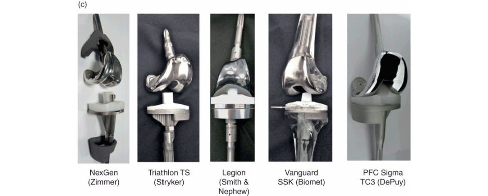
Linked, constrained condylar implant (rotating-hing e knee or RHK). Rarely indicated. Used for global instability (total collateral disruption/r ecurvatum) and severe distal femoral bone loss, osteolysis/fracture.
↓
Fusion – may be required for chronically infected unstable knee with a poor soft tissue envelope .
What is your intraoperative plan for revising a knee replacement with bone loss?
Removal of implants, i.e. extraction, is the first step. Bone preservation is critical and making sure fine or flexible osteotomes and a narrow reciprocating saw are available. My aim is to achieve stable fixation a t the epiphysis, but often one has to use stemmed implants to achieve diaphyseal fixation aswell as metaphysical sleeves to achieve press-fit metaphysical fixation, especially when there is a significant amount of bone loss. Once the tibial pla foorm is established the flexion and extension gaps can be assessed. An intramedullary stem can guide the positioning of the femoral component. Gap balancing should be used to assess flexion and extension gaps. Distal femoral augments can be used to match the flexion gap.
[Going back to the radiographs] What are the principles of management of bone loss in revision knee replacement in this patient?
The options of management of the extensive bone loss are: 1. The use of cement, either alone or combined with screws and mesh. 2. The use of bone gratiing with structural or morsellized graft. 3. The use of modular augmentation of the components with wedges or blocks of metal. Recent studies show modular porous coated press fit metaphyseal sleeves may be used to fill AORI type 2 and 3 defects and provide for stable ingrowth. 4 The utilization of cus tom-made, tumour or hinge implants. The method of reconstruction and the materials for revision surgery are largely dependent on the potential for future further revision and the life expectancy, functional demand and co morbidities of the patient. In this patient who is reasonably young, restoration of bones tock is preferable, because of the likelihood of further revision surgery.
Figure 4.8a and 4.8b Anteroposterior and lateral radiographs revision TKA.
Figure 4.8c Commonly used CCK systems.
References#
1. Koh IJ, ChoWS, Choi NY, Parvizi J, Kim TK; Korea Knee Research Group. How accurate are orthopaedic surgeons in diagnosing periprosthetic joint infection after total knee arthroplasty? A multic enter study.
Knee. 2015;22(3):180–185.
Structured oral examination question 9#
Patellar instability
A 17-year-old lady is referred to your patella clinic by her GP due to recurrent bilateral patella dislocations. How would you assess this patient?
I would start by taking a detailed history followed by clinical examination. In the history, I would enquire about age at first dislocation frequency of dislocations traumatic or a traumatic, an y associated syndromes such as bone or connective tissue dysplasia and generalized joint laxity. I would also enquire about any mechanical symptoms, the presence and localization of pain. I would start with a general hypermobility assessment using the Beighton score. Examination of the patella includes assessment of coronal and rotational alignment and patella height. We should also assess patella apprehension and patellofemoral crepitus. Quadriceps function and hamstring tightness should also be recorded.
What are the risk factors for patella instability?
The risk factors for patellar instability are complex and multifactorial; however, they can be subcategorized in the following sections: 1. Bony factors (static). Trochlear dysplasia. Excessive lateral patella tilt. Patella alta. Femoral or tibial malrotation. 2. Malalignment. Patellar malalignment is an abnormal rotational or translational deviation of the patella along any axis. External tibial t orsion/foot pronation increased femoral anteversion and genu valgum especially in adolescence – miserable malalignment syndrome. Increased Q angle or abnormal tibial tuber osity–trochlea groove (TT–TG) distance. 3. Soft tissue (dynamic). Ligamentous laxity (medial patellofemoral ligament rupture/insufficiency). Muscle and soft tissue imbalance around the knee. Core muscle instability in the hip. 4. Abnormal gait. Walking with a valgus thrust.
5. Genetic factors such as connective tissue disorder syndromes and generalized hypermobility.
Two-thirds of patients are known to have multiple anatomical factors predisposing to recurrent patella dislocations.
Tell me about the important stabilizers of the patella.
Patella stability results from a complex interplay of local, distant, static and dynamic factors. Dynamic muscular restraints, static soft -tissue restraints, bony morphology and lower limb alignment all stabilize the patellofemoral joint. The distant static factors are femoral anteversion, knee rotation and external tibial torsion. The distant dynamic factors include the iliotibial band complex, hip abductors and foot rotation. The primary local static restraint to the lateral patellar displacement is the medial patellofemoral ligament. It provides 50–60% of the total medial restraining force and resists lateral translation in early knee flexion (20–30°). MPFL sectioning can lead to substantial changes in patellar tracking. The femoral attachment of the medial patellofemoral ligament (MPFL) has been the centre of much debate. Amis et al. concluded that the origin was the medial epicondyle [1]. Following on from his work, Schötile et al., in a cadaveric study, identified a radiographic point for the origin, 1 mm anterior to the posterior cortex extension line, 2.5 mm distal to the posterior origin of the medial femoral condyle and proximal to the level of the posterior point of the Blumensaat line on a lateral radiograph with both posterior condyles projected in the same plane, which represented the mean femoral MPFL isometric centre [2]. Intraoperative fluoroscopy can be used to accurately identify this position However, it is now believed to be a point just anterior to the confluence of the Blumensaat line, a curving line of the posterior femoral cortex and posterior to the straight extension line from the posterior cortex in a true lateral radiograph. Hence it is named the confluence point. During acute patellar dislocation the reis a 90–95% incidence of damage to the MPFL. Femoral attachment is commonly affected. In the past 10 years, MPFL reconstruction has become a popular procedure for treatment of recurrent patellar dislocation. Dejour et al. examined CT scans on 134 patients treated for patellar instability. They identified f our common factors of unstable symptomatic knees: (1) trochlear dysplasia (85%), (2) quadriceps dysplasia (83%), (3) patella alta (24%), (4) tibial tuber osity–trochlear groove distance, pathological when greater than or equal to 20 mm (56%) [3].
How would you investigate this patient?
I would perform the following investigations: 1. A true lateral radiograph is the most helpful view for assessment of patella height and trochlear depth.
2. Axial radiographs (Merchant’s view) to assess patellar tilt angle (normal < 10°), congruence, lateral patellofemoral angle (normal 138°) and trochlear dysplasia.
3. Rotational profile CT scans assess femoral anteversion which normally is 5–15°, external tibial torsion, patellar tilt and index, TT–TG (tibial tuber osity–trochlear groove) distance. If the TT–TG is more than 19 mm this is pathological.
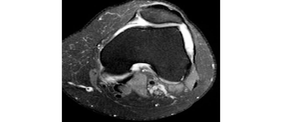
4. MRI for articular surface assessment (OCD), integrity of the MPFL, patella height, TTTG and trochlear dysplasia (Figure 4.9a).
When assessing the patella height, I use the Caton–Deschamps method. This is the ratio between the distance from the inferior border of the patella articular surface to the upper edge of the tibial plateau and the length of the patella articular surface. A ratio of more than 1.2 indicates patella alta.
I use the lateral view with the knee in at least 30° of flexion.
When would you offer a lateral release?
I would only perform a lateral release if there were isolated lateral patellar tilt. I would not perform this procedure independently.
What are the options of surgical treatment?
Generally, surgical procedures for chronic patellofemoral instability are often used in combination and include bony and soft -tissue procedures at the level of the joint, proximal or just distal to it. The realignment procedures are determined by direction of tibial tubercle (TT) transfer: medial transfer to treat malalignment, anteromedial transfer for malalignment and PFJ chondrosis, anterior when there is distal PFJ chondrosis. Operative intervention for acute isolated first-time dislocation is only indicated if there is evidence of a large osteochondral defect. There are several reports of lower redislocation rates following surgical treatment of primary acute patella dislocations however, there is no difference in the long term. Decision-making using patien t-specific instability predictors can identify patients who would benefit from operative intervention. The parameters in the Patellar-Instability Severity Score (PIS- Score) include age, patellar tilt patellar alta, TT–TG distance, trochlear dysplasia and positive anamnesis of contralateral patella dislocation. A sc ore of more than 4 indicates a higher risk of redislocation. MPFL reconstruction is a very successful procedure. An ideal candidate for this should have a history of recurrent dislocation and a physical examination demonstrating excessive lateral patella translation and a normal trochlea [4]. A Caton–Deschamps index up to 1.3 can be acceptable, except where there is a very short trochlea or significant knee hyperextension. The key bony procedures for patella instability are: trochleoplasty, tibial tuberosity osteotomy and femoral osteotomy (derotation or angular) [5].
Figure 4.9a MRI scan, T2 axial view demonstrating lateral patella subluxation, knee effusion and shallow trochlea groove.
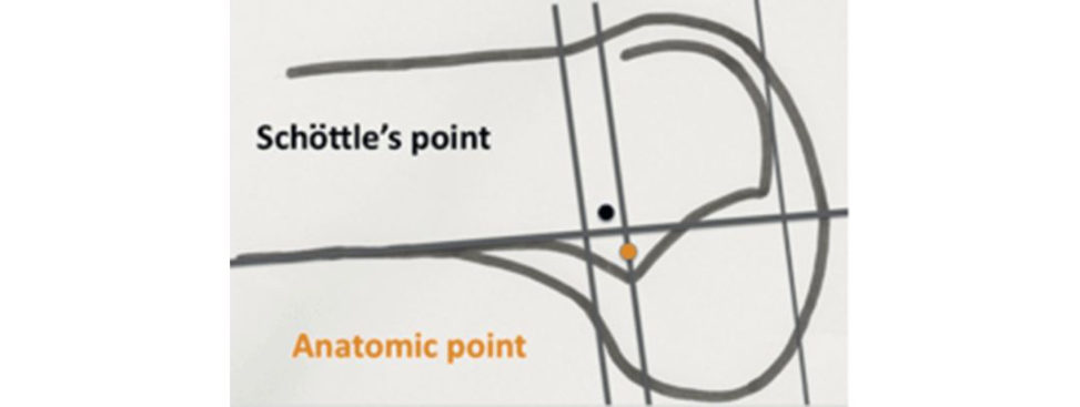
Figure 4.9b Schötile ’s point.
References#
1. Amis AA, Firer P, Mountney J, Senavongse W, Thomas NP. Anatomy and biomechanics of the medial patellofemoral ligament. The Knee. 2003;10(3):215–220.
2. Schötile PB, Schmeling A, Rosenstiel N, Weiler A. Radiographic landmarks for femoral tunnel placement in medial patellofemoral ligament reconstruction . Am J Sports Med. 2007;35(5):801–804.
3. Dejour H, Walch G, Nove-Josserand L, Guier C. Factors of patellar instability: an anatomic radiographic study. Knee Surg Sports Traumatol Arthrosc. 1994;2(1):19–26.
4. Yeung M, Leblanc MC, Ayeni OR, et al. Indications for medial patellofemoral ligament reconstruction: a systematic review. J Knee Surg. 2016;29(7):543–554.
5. Steensen RN, Bentley JC, Trinh TQ, Backes JR, Wilft on gRE. The prevalence and combined prevalences of anatomic factors associated with recurrent patellar dislocation: am agne tic resonance imaging study. Am J
Sports Med. 2015;43:921–927.
Structured oral examination question 10#
Pain after TKA
A 70-year-old gentleman has been referred to your clinic with ongoing pain in his knee following his primary uncomplicated TKA. He states the knee has never been quite right since his surgery three years ago. Please describe these radiographs (Figure 4.10a and 4.10b).
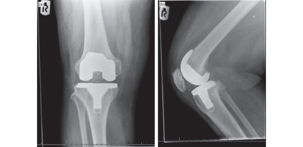
These radiographs demonstrate a cemented cruciate-sacrificing posterior stabilized implant. The patella has been resurfaced with a polyethylene buft on. There is no evidence of gross loosening of the implant neither are there any fractures. There is no evidence of tibial tray overhang or over-/under-sizing of the femoral component. The overall alignment is adequate.
This patient is complaining of generalized knee pain. How would you proceed?
Firstly, I would take a history. I would like to know if there have been wound issues and if antibiotics have been prescribed previously. Were there any issues with wound healing? If infection has been excluded it is important to differentiate between extra- and intra-articular causes of pain. Lack of pain relief immediately after the TKA could suggest that the pain was not actually originating from the knee. It is also important to establish if the patient was initially pain- free, as pain developing after months or years is quite characteristic of component loosening or failure.
OK, so this gentleman states his pain was no different following his TKA. What do you think of that?
Extra-articular causes of pain around the knee need to be excluded. Firstly, the hip should be assessed; pain may be radiating from the hip to the knee via the obturator nerve. Pain can radiate to the anterior or posterior aspects of the knee. Pathologies such as osteoarthritiss tress fractures, dysplasia and implant loosening after total hip replacement can all cause radiating paint o the knee. Posterior tibial tendon dysfunction causing planovalgus will lead to a valgus hindfoot alignment. This will subsequently cause changes in the ground reaction forces and increased lateral loading of the knee [1].
So, you have suggested causes of pain above and below the knee. What else can cause the pain?
Vascular insufficiency with peripheral artery disease can cause leg pain in the elderly. Thrombosis is very common following TK Rand should be excluded. An ankle–brachial pressure index is non-invasive and not costly. The history will also identify lumbar spinal stenosis which may require further imaging for assessment. Other neurological disorders such as chronic regional pain syndrome and cutaneous neuromas contribute to the incidence of knee pain following TKR. Posterolateral knee pain secondary to a posterolateral tibial tray overhang, a retained posterolateral osteophyte or cementophyte can cause biceps tendonitis and/ or popliteus tendon impingement. Posteromedial pain can be caused by semimembranosus tendonitis. There are multiple periarticular bursae such as the infrapatellar, pes anserine and semimembranosus bursae which can cause pain and inflammation Lateral pain with movement between 20 and 80° classically is a result of iliotibial band dysfunction.
OK, but these causes are all outside the knee. What are the intrinsic pathologies which you should exclude?
Aseptic loosening is common and is frequently described as ‘start-up pain’. A higher constrained implant, higher BMI, varus alignment and malrotation can contribute to implant loosening. Malalignment and wear are frequently responsible for late loosening.
I agree. Aseptic/ septic loosening needs to be excluded. What else?
Pain can be caused by instability as the soft tissues are overloaded. Early failure related to instability may be associated with trauma or more importantly surgical technique. Surgical considerations such as suboptimal align mentor positioning , improper balancing of flexion–extension gaps and medial collateral and posterior cruciate ligament rupture can cause persistent pain.
Tell me how you would assess for malalignment of components?
I would organize a standard radiological series including a skyline view and a CT scan.
What are the consequences of rotational malalignment?
Rotational alignment of the tibial and femoral component plays an important role in TKA. Once correct frontal alignment and proper soft tissue balancing has been achieved , the rotational placement of the components represents the ‘third dimension’ in knee TKA. Femoral component malposition has been implicate din patellofemoral maltracking following TKA, which is associated with anterior knee pain, subluxation, fracture, wear and aseptic loosening [ 1,2]. Internal rotation of the femoral component by resection of excessive amounts of posterior lateral femoral condyle or insufficient resection of the posterior medial femoral condyle moves the anterior femoral patellar groove portion of the femoral component medially, making it more difficult for a relatively laterally placed patella to be captured by the patellofemoral groove. In addition, internal rotation of the femoral component results in a tight flexion gap on the medial side of the knee.
Do you know anything about how you determine rotational alignment of aTKA on CT?
Rotational component malalignment is difficult to access on standard AP and lateral radiographs. Patients with painful early TKA should be assessed for evaluation of mal rotated components.
Anything else?
[Silence] Bergeret al. have described methodology to quantitatively measure component rotational alignment using CT scanning [3].
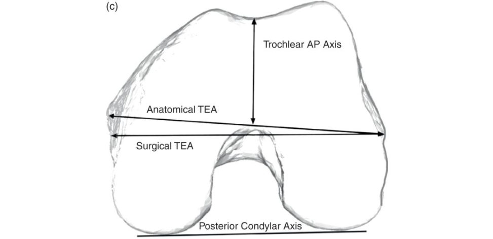
Based on the published values, essentially the surgical transepicondylar axis (TEA) is on average 3° externally rotated in relation to the posterior condylar axis while the perpendicular axis to the trochlear anteroposterior axis is 4° externally rotated to the posterior condylar axis and finally the anatomical TEA is on average 5° externally rotated to the posterior condylar axis (Figure 4.10c). None of the above are entirely reliable, especially the TEA. Surgeons may have to use more than one reference point when placing the femoral and tibial components.
The rotation of the tibial component (tibial posterior component axis) is measured in relation to the posterior tibial plateau axis, medial/middle third of the tibial tubercle, patellar tendon, PCL attachment, transverse axis of the tibia, midsulcus of the tibial spine and anteromedial margin of the tibial plateau.
Tibial component axis normal value = 18°.
Figure 4.10a and 4.10b Anteroposterior and lateral radiographs right TKA.
Figure 4.10c Femoral component rotational alignment.
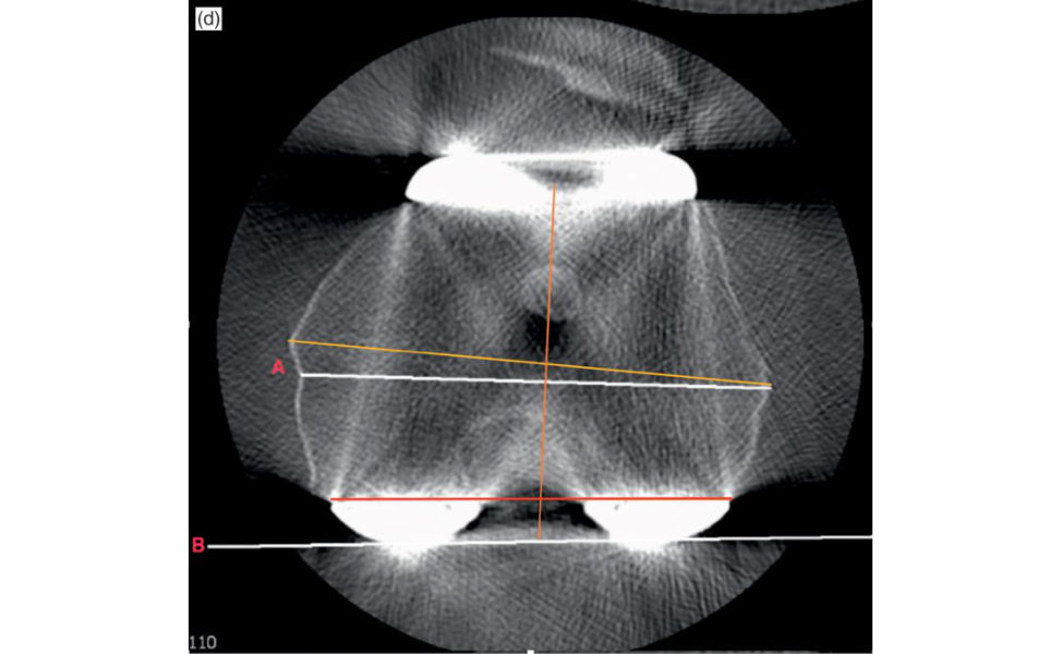
Figure 4.10d CT measurement of femoral implant rotation. The posterior condylar bone cut (redline) is 1–2° internally rotated in relation to the surgical TEA (whiteline A). However, the whiteline B represents the line of the posterior condyle of the femoral component. The yellow line represents the anatomical TEA and the orange line represents Whiteside’s line.
References#
1. Mandalia V, Eyres K, Schranz P, Toms AD. Evaluation of patients with a painful total knee replacement. J
Bone Joint Surg Br. 2008;90(3):265–271.
2. Nicoll D, Rowley DI. Internal rotational err or of the tibial component is a major cause of pain after total knee replacement. J Bone Joint Surg Br. 2010;92B:1238–1244.
3. Berger RA, Crossett LS Jacobs JJ, Rubash HE. Malrotation causing patellofemoral complications after total knee arthroplasty. Clin Orthop Rel Res. 1998;356:144–153.
Structured oral examination question 11#
Osteotomies around the knee
A 28-year-old gentleman who works as a postman attends your clinic, complaining of knee pain. He has had it for many years but is now unable to work. What do you think (Figure 4.11a)?
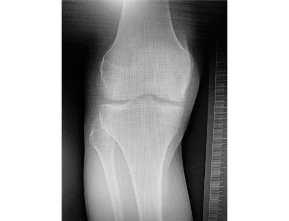
The weight-bearing AP radiograph demonstrates narrowing of the medial tibiofemoral joint space. There is a defect in the lateral aspect of the medial femoral condyle and it seems to be involving the subchondral bone.
What are the pertinent features in the history and examination?
I would like to find out if this is traumatic or a traumatic, take a detailed pain history including site, severity and if he has mechanical symptoms. Clinically I would like to assess his overall alignment and see if he is tender over the medial femoral condyle (MFC). I would also like to get an MR Iof the knee to elucidate the nature of this defect in the condyle. I would assume this patient has had osteochondritis dissec ans in the past.
OK, let’s assume he has been through all conservative treatment and is keen to undergo surgery. Tell me about your management plan.
Firstly, I would like to assess his lower limb alignment. If I were to offer him a chondral procedure, I would certainly ensure his overall alignment is not in varus.
What would you like to see?
I would like to see some longleg alignment views including the hip and ankle to perform some deformity analysis. I would also like to see a lateral and some further imaging of the joint surface would be helpful to plan further surgery.
OK, so what information would you like?
Firstly, I would like to assess the deformity in the frontal plane, and according toPa ley, should include the mechanical axis of the lower limb, overall malalignment, lateral distal femoral angle (LDFA), medial proximal tibial angle (MP TA) and the joint line convergence angle (JLCA). The tibial width is gi vena percentage of 100% starting medially (0%) and ending laterally (100%). Where the mechanical axis crosses the tibial plateau, this is named the Mikulicz point (and is a percentage value). I would also assess the sagift al alignment for the anatomical posterior proximal tibia angle (aPPTA), which reflects the tibial slope, and the anatomical posterior distal femur angle (aPDFA), which represents the flexion/extension positioning of the distal femur.
Tell me, what are the key biomechanical parameters and normal values for frontal alignment?
The LDFA is normally 87° (85–90°) and theM PTA is normally 87° (85–90°). The JLCA is 0–2° [1] (Table 4.1).
So, what do you think of this patient’s full-leg standing radiographs (Figure 4.11b)?
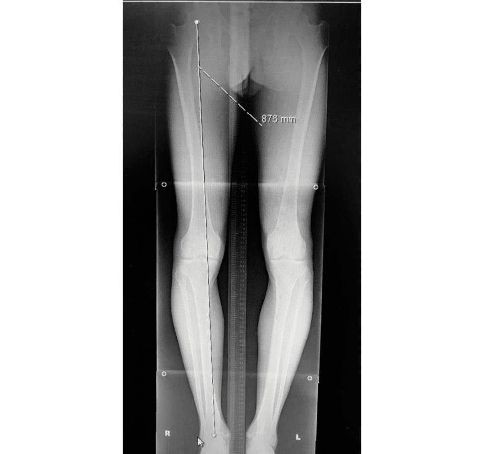
He looks to be in varus. The weight-bearing line is passing well into the medial compartment.
So, the MRI demonstrates the OC Din the MFC, the meniscus is preserved and there is some chondral thinning of the medial tibial plateau. What would you offer him?
Firstly, I would offer him a valgizing osteotomy. Once his alignment is corrected, I will be able to safely perform some form of chondral surgery, if needed.
OK ... Please explain the rationale.
Well, as this patient is in varus, I would like to offload them and perform a valgizing osteotomy in the form of a high tibial osteotomy or distal femoral osteotomy. It depends where the deformity lies.
Explain?
I perform an osteotomy in the bone where the deformity lies. As we can see from the limb alignment analysis (Table 4.1), theM PTA is 80°, which isabnormal. If we performed a femoral osteotomy, we would be introducing joint line obliquity to the knee and ultimately uneven joint loading. As theM PTA is 80°, normal being 87°, the deformity lies in the proximal tibia. The distal femur is normal and the proximal tibia is inv arus.
What kind of osteotomy would you offer him?
We can perform opening and closing wedge osteomies to both the medial and lateral sides of the femur and tibia. Given the proximal tibia is inv arus, and this is where the deformity lies, we will need to increase theM PTA to the normal 87°. The two options are a medial opening wedge HTO or a lateral closing wedge HTO. The most efficient way to do this would be a medial opening wedge HTO.
Why?
The opening wedge HTO has gained popularity because it does not require a fibular osteotomy, common peroneal nerve dissection disruption of proximal tibiofibular joint, and bone stock loss (Bonasia et al., 2014). If we were to perform a lateral closing wedge, this would create two bone surfaces which would be indirect contact and promote healing. Opening a wedge and potentially leaving a gap could reduce the timet o union; this, however, has not been proven in the literature [3], therefore I may consider filling the gap, with either synthetic or allograft.
So, if you’ve decided on a medial opening wedge HTO, how would you go about it and tell me the considerations intra operatively?
I would prepare the patient supine on a radiolucent table under tourniquet with intravenous antibiotics a t induction. Ani mage intensifier would be available to me approaching from the lateral side. I would approach the proximal tibia via a direct anteromedial oblique approach. I would identify the pes and release the hamstrings. I would release the MCL distal fibres subperiosteally in one layer with diathermy until I can see the posterior border of the tibia. After careful dissection posteriorly, I would use a curved brown-handled osteotome to clear the posterior structures off the tibia and place a radiolucent Homann in position to protect the neurovascular bundle. I would mark out my biplane osteotomy to increase the stability of the construct. A precision saw is safe and should be used with the radiolucent retractor. To ensure the trajectory of the saw cut is in the plane of the joint, I place 2 K-wires parallel with the joint under fluoroscopy. The callipers are then used to measure the exact distance for the opening wedge as per the preoperative plan to 0.1 mm (see
Figure 4.11c).
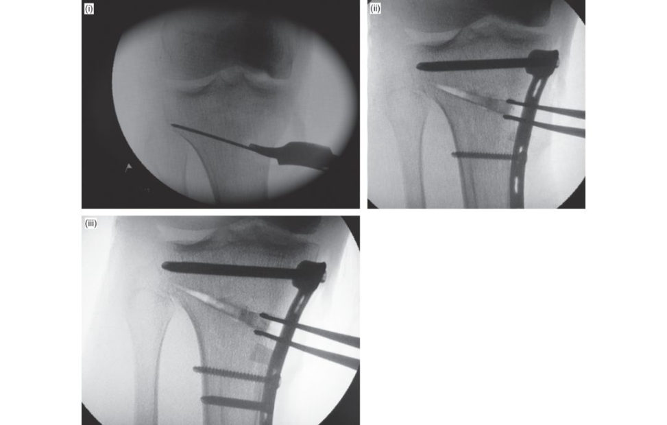
What would be your postoperative rehabilitation programme?
Rest, ice, a cryotherapy cuff would be advantageous, routine observations including neurovascular monitoring. Using a plate like the Tomofix system allows full weight bearing immediately. I would like to see an X-ray at the six weeks mark to ensure there has been no less of correction.
Figure 4.11a Weight-bearing anteroposterior (AP) radiograph of the right knee.
Figure 4.11b Standing anteroposterior (AP) longleg radiographs.
Table 4.1 Limb alignment analysis.
Angle
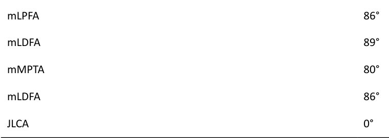
(i) Positioning of the first guide wire, note the position of the hinge point.
(ii) Application of the T omofix plate following opening of the wedge.
(iii) Image demonstrating maintenance of the gap opening following gap filling and application of the plate.
Figure 4.11c Intraoperative images of opening wedge HTO.
References#
1. Paley D, Herzenberg JE, Tetsworth K, McKie J, Bhave A. Deformity planning for frontal and sagift al plane corrective osteotomies. Orthop Clin North Am. 1994;25(3):425–465.
2. Bonasia DE, Dettoni FS ito G, et al. Medial opening wedge high tibial osteotomy for medial compartment overload/arthritis in the varus knee: prosthetic factors. Am J Sports Med. 2014;42(3):690–698.
3. Staubli AE, Jacob HAC. Evolution of open-w edge high-tibial osteotomy: experience with a special angular stable device for internal fixation without interposition material. IntOrt hop. 2010;34(2):167–172.