Chapter 3 Hip
Introduction#
There has been a change in emphasis in the oral questions in the last 2 years to higher-order thinking and judgement. Exam revision should be less book reading and more being practical and adept at managing complex clinical conditions. Examiners would argue if you have been well trained in the basics it isn’t too difficult to apply these basic principles to various clinical situations that you may be tested on in the oral exam. If you haven’t managed periprosthetic joint infection (PJI) then it’s going to be doubly difficult to answer the real-life practical questions that are related to managing apa tien t with this condition.
We have aimed the candidates’ answers for a 7–8 score, so they are significantly more detailed than what would be required for a bare pass. Aiming for the minimum topass will generally be unsuccessful and is not recommended.
Structured oral examination question 1#
These are the radiographs of a 65-year-old gentleman who had a primary left THA 14 years ago (Figure 3.1a and b). Over the last 2 weeks, he has had increasing pain in the left hip and he has contacted his GP who has referred him back to you on an urgent basis Postoperatively, he was progressing well, and he has no history of any trauma. He is now unable to weight bear fully on the left leg and has night pain.
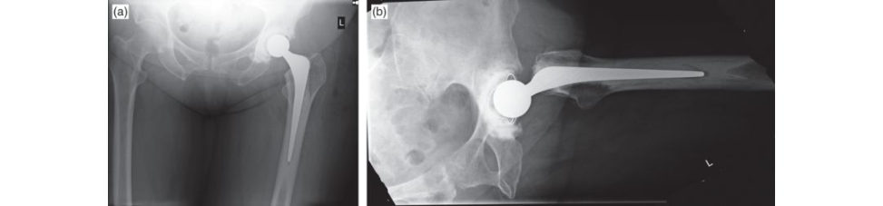
This is an AP pelvic radiograph showing a left cemented Exeter THA. I am concerned that there is an area of radiolucency around the tip of the greater trochanter. The acetabular component appears well fixed. My worry here is that this gentleman may have a periprosthetic joint infection. Every painful prosthetic joint is potentially infected.
So how are you going to proceed with this patient?
I would want to take the patient to theatre and perform an aspiration of the hip to rule out infection.
You are sure? Are you not jumping in abit fastIs there anything else you might want to find out beforehand?
I would want to take a full history from the pa tientA number of patients who develop infection have early wound problems such as prolonged redness, indurations welling or discharge. There may be a history of repeated courses of antibiotics. Thew ound may have become frankly infected requiring washout in theatre. Onset of hip pain following a problem-free interval and an episode of sepsis is suggestive of haematogenous seeding of infective organisms from elsewhere. I would enquire if there was a history of bacteraemia from a UTI, chest infection or dental extraction. Pain from an infected prosthesis is typically non-mechanical and unrelated to physical activity and not relieved by restI would also like to explore more fully host risk factors for infection such as diabetes, rheumatoid arthritis depression, obesity, hypothyroidism, immunosuppression (steroids, DMARDs), smoking and poor dention. Lifestyle factors such as heavy alcohol intake and smoking. Having taken a full history, I would perform a comprehensive clinical examination, looking a t the scar for any evidence of infection such as erythema, warmth, a sinus, etc. What is the hip ROM and is there pain associated with ROM? I would also like to examine the abdomen, knee and lumbar spine to exclude other potential sources of haematogenous infection/ cause for the hip pain. I would then request an FBC, CR Pand ESR to look for evidence of raised inflammatory markers.
How helpful are these?
They have relatively low sensitivity and low specificity as markers of prosthetic joint infection. Ber barie tal. (level 2) published a systematic review in the JBJS American edition in 2010 on the use of inflammatory markers for diagnosis of prosthetic joint infection. 1 They concluded that IL-6 is a much more sensitive test for infection. Unfortunately, IL-6 assays are not readily available in most NHS hospitals and certainly not in my trust at present.
What about diabetes as a risk factor for PJI?
The association between diabetes and PJI may be mediated by impaired leukocyte function and microvascular complications which may impair wound healing. In addition, factors associated with diabetes, such as hyperglycaemia, hyperlipidaemia, hypertension and increased oxidative stress, upregulate cellular and inflammatory reactions and play a part in atherothrombosis that may cause impaired wound healing. Hyperglycaemia has also been shown to increase biofilm formation.
The paper actually reported that IL-6 was more accurate than CR Por ESR rather than sensitive The wound was oozy postoperatively but setiled down. A large part of picking up periprosthetic infection is obtaining a good history and examination along with a high index of clinical suspicion.2 How useful is a hip aspiration in diagnosing infection?
Figure 3.1a and 3.1b Anteroposterior (AP) pelvis and lateral radiographs demonstrating a left Ex eter THA.
Spangehl et al. (level 1) demonstrated a sensitivity of 0.86, a specificity of 0.94, a positive predictive value of 0.67 and a negative predictive value of 0.98 with initial imag e-guided aspiration in 180 patients undergoing revision hip arthroplasties. 3 They reported that aspiration alone is not sufficient for the diagnosis because of the risk of false positive and false negative results. They suggested that aspiration was not necessary in low-probability cases with a normal ES Rand CRP. Aspiration would be indicated if pretest probability for infection was high (acute onset of pain, systemic illness, sinus formation), particularly if the CRP /ESR was normal, or in all cases where the CR Por ESR was high. A normal CR Pand ESR does not always exclude a PJI, however.
How would you perform a hip aspiration?
I would perform this in theatre under II control. I would fully prepare and drape the skin. I would incise the skin with a small nick to minimize the risk of skin contamination and also perform two separate aspirations to reduce the risk of false positives. If both are positive with the same organism this would be suggestive of infection.
What about if one aspirate only is positive?
If only one aspirate is positive then it is a tricky situation. It is a soft positive result and I think it would be reasonable to repeat hip aspiration 2 weeks later.
Joint aspiration did not grow any organisms after 48 h of culture. Is there anything else you may wish to consider in terms of establishing a diagnosis of a periprosthetic joint infection?
48 h for bacterial culture may not belong enough to identify certain bacteria. For example, low-virulence organisms such as Propionibacterium acnes may take up to 2 weeks to culture successfully. I would speak to the microbiology lab and make sure they are performing extended cultures on the material sent.
Isn’t that normal for most labs to do?
My own local hospital policy is to perform extended culture for 5 days on any suspected PJI, but I am not sure if this has been universally agreed on. I would speak to the microbiology lab regardless to make sure I was notified promptly if extended culture grew any organism as this may significantly change my management plans. Synovial fluid culture has a sensitivity of 52% and specificity of 95%. Sensitivity a t 52% is poor. The differential white cell count from synovial fluid analysis is useful in diagnosing a PJI. For hips, a WCC > 4200/μl and/or a granulocyte percentage > 80% has a sensitivity of 85% and a specificity of 90%. I realize there are some difficulties get ing the microbiology department to perform regular counts. Pus swabs are not regarded as useful anymore. The Gram stain itself has a very low sensitivity (< 25%). Use of antibiotics prior to the aspiration of the hip joint can lead to reduced sensitivity of the synovial fluid analysis. Ideally, any antibiotics the patient is still on should be discontinued for up to 2 weeks in order to improve the pick-up rate of any synovial fluid culture. There are modern biomarkers such as α-defensin (Synovasure, Zimmer Biomet), neutrophil elastase 2 (ELA2), bactericidal/permeability increasing protein, neutrophil gelatinase-associa ted lipocalin (NG ALand lactoferrin which have recently shown promise in diagnosing PJI, but there are still no reliable data to prove their sensitivity and specificity to date.4 One potential advantage of such biomarkers is the fact that they are not reliant on the bacteria and, therefore, pre-administered antibiotics should not affect their sensitivity . Synovial tissue biopsy can also betaken which can improve the sensitivity of microbiological culture and samples should also be sent for histology. A minimum of three tissue samples should betaken and ideally more than six samples.
Neutrophil granulocytes are indicators of bacterial infections (in acute infections). Leucocyte esterase has been shown as being as sensitive and specific as Synovasure and much more cost effective.
What is α-defensin?
α-defensin is an antimicrobial peptide secreted by neutrophils to fight infection.
Are there any other tests you might want to perform that could diagnose infection before going ahead with surgery?
The use of nuclear imaging (technetium-99 triple-phase bone sc an, gallium imaging, labelled-leukocyte scans or FDG-PET imaging) for the detection of periprosthetic joint infection is worth considering but controversial. The AAOS clinical practice guidelines summary from 2010 reported a weak recommendation for their use.5,6
How do you classify periprosthetic hip infection?
It may be enough just to mention the uncertainties with nuclear imaging or you may have to quantify your answer more fully. It is a judgement decision, but don’t persist with your answer if the examiners want to move on. Technetium-99 bone sc ans are sensitive but not specific. Some investigators have found that a negative scan rules out infection, others report that a scan can occasionally be negative with infection if the reis inadequate blood supply to the bone. A technetium-99m bone sc an identifies areas of increased bone activity through preferential up take of the diphosphonate by metabolically active bone. Increased uptake occurs with loosening, infection, heterotopic bone formation, P aget’s disease, stress fractures, modulus mismatch of a large uncemented stem, neoplasm, reflex sympathetic dystrophy and other metabolic conditions. In the uncomplicated THA, uptake around the lesser trochanter and shaft is usually insignificant by 6 months, but in 10% of cases, uptake may persist at the greater trochanter, prosthesis tip and acetabulum for more than 2 years. The pattern of uptake has not been found to consistently reflect the presence or absence of infection. Gallium imaging likewise has a poor sensitivity and accuracy. The use of leukocyte scans is generally preferred, having a higher sensitivity (88 –92%) and specificity (73–100%), but their usefulness for the diagnosis of infection continues to be debated. FDG-PET is expensive and limited to a few institutions, and although very sensitive does not allow differentiation between an inflamed aseptically loosened prosthesis and an infected one. Tsukayama et al. proposed a four-stage system consisting of early postoperative, late chronic, and acute hematogenous infections, and positive intraoperative cultures of specimens obtained during revision of a presumed aseptically loose THA.7,8 Early postoperative infection presents less than 1 month after surgery with a febrile patient and a red swollen discharging wound. With late postoperative infection, the patient is well, the wound has healed well, there is a worsening of hip pain and a never pain-free interval. Acute haematogenous infection can occur several years after surgery with a history of bacteraemia (UTI or other source of infection) and severe hip pain in a previously well-functioning hip. P ositiv e intraoperative culture (at least three samples from different locations taken with clean instruments) occurs when a preoperative presumptive diagnosis of aseptic loosening was made.
McPherson et al. have also developed a staging system for periprosthetic hip infections that included three categories: infection type (acute versus chronic), the overall medical and immune health status of the patient and the local extremity (wound) grade.9
How do you diagnose PJI?
I would use the criteria of the musculoskeletal infection society. PJI can be diagnosed when one out of two major and four out of six minor criteria exist (Table 3.1). Major criteria are (1) a sinus tract communicating with the prosthesis and (2) two positive periprosthetic cultures with phenotypically identical organisms. A pathogen is isolated by culture from at least two separate tissue or fluid samples obtained from the affected prosthetic joint. The six minor criteria are (1) elevated ES Rand CRP, (2) elevated synovial leukocyte count, (3) elevated synovial neutrophil percentage (PMN%), (4) presence of purulence in the affected joint, (5) single positive culture, (6) positive histological analysis of periprosthetic tissue. PJI may be present if fewer than four of these criteria are met. TheM SIS criteria were modified in 2013. Essentially three from five minor criteria (Table 3.1).
How will you manage this patient assuming that your diagnosis of an acute PJI is now made?
As the diagnosis of a PJI has been made within 2 weeks and assuming that there are positive microbiological results with a known organism and sensitivities to antimicrobial therapy, there remains the option of undertaking a DAIR-type procedureD AIR stands for Debridement, Antibiotics and Implant Retention, in which the prosthesis is retained with exchange of any mobile components such as a liner and head exchange while keeping the acetabular socket and stem in place. AD AIR should only be undertaken if the PJI is acute and ideally within 3 weeks of the PJI starting , although some units such as Oxford will undertake aD AIR at up to 6 weeks or so. The optimum management of a PJI involves a multidisciplinary team approach with MSK radiologists, bone infection microbiologists histopathologists, plastic surgeons and experienced revision arthroplasty surgeons with a specialist interest in managing PJIs. The success rate of DAIR procedures is around 90–95%.10 The Oxford group have recently published a case control study comparing DAIR versus a two-stage revision and showed a 98% 5-year survivorship rate with DAIR and better outcome with DAIR.11
This patient had a delay in diagnosing the PJI and was only seen 2 months after the onset of their clinical presentation. How would you manage the patient now?
The patient is now outside the window of opportunity to undertake aD AIR. If there was a microbiological result with a known organism and sensitivities, then a single-s tage revision could be undertaken, especially if the patient had multiple co morbidities and the surgical stress of having a two-stage procedure might compromise the patient. In this case, it is critical that all cement is removed as well as all the components and a thorough debridement with complete excision of infected soft tissues is undertaken .
What are the prerequisites for a one-stage procedure?
Prerequisites include a known organism sensitive to antibiotics, no pus present , elderly patients or patients with multiple medical problems. It is also indicated in healthy individuals devoid of re-infection risk who have adequate bone and soft tissue for reconstruction and a lo w-virulence pathogen.
What are the reported success rates for a single-stage revision?
Buchholz et al., who pioneered one-stage revisions at the Endo-Klinic in Hamburg, reported a success rate of 77% in 583 revisions, but only after extensive bone and soft -tissue resection, which compromised long-term function. 12 These results were published in 1981 and can be viewed as somewhat historic now. Raut et al. from Wrighting ton reported a success rate of 86% in 57 cases at average follow-up of 7 years despite many discharging sinuses.13,14 Hanssen and Rand summarized the results of single-stage exchange and found a cumulative success rate of 83% when antibiotic- loaded cement was used, but only 60% when it was not.15 A recent RCT has been started to compare a single- versus two-stage revision.16 In 11 studies with 1225 patients with a hip PJI receiving exclusively one-stage revision, the rate of re-infection a t 2 years was 8.6% (95% CI 4.5–13.9). After two-stage revision exclusively in 28 studies with 1188 patients the rate of re-infection a t 2 years was 10.2% (95% CI 7.7–12.9). The authors conclude that on the basis of a systematic review of published data, there is no difference in the re-infection rate between one-stage and two-stage revision THA for PJI.
What are the advantages to performing a two-stage procedure?
It is particularly important to perform a two-stage revision with more severe infections or virulent organisms, as the success rate of a single-stage procedure is much less in these situations.
That’s not what I asked.
It is more versatile for reconstruction allowing the use of either cemented or cementless components and bone allograft in patients with severe bone loss. It allows clinical assessment of the response to antibiotics prior to re-implantation.
What are the disadvantages of a two-stage procedure?
It can be difficult to nurse patients between stages and the second-stage surgery can be difficult due to soft -tissue scarring, limb shortening, disuse atrophy, loss of bone density and distortion of anatomy.
Have a look at this radiograph below (see Figure 3.1c). What is going on with the right hip?
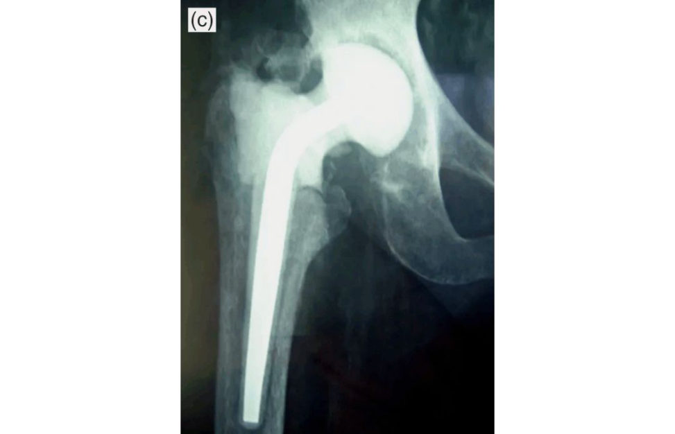
This is a cement spacer in apa tien t who has had a first-stage revision for infection and an articulating cement spacer such as a PROSTALAC spacer has been used. The potential complications of using such a spacer are that it can dislocate or fracture and it costs more money to perform a two- stage procedure. An alternative would be to use a nail or multiple wires with cement coating around the wires to act as a temporary spacer. There is also the option of loosely cement inga THA in place (using a polished tapered stem and cementing a liner for the socket) ((The “Kiwi” Prostalac)) which has the advantage of allowing the patient to mobilize full weight bearing postoperatively and, if necessary, the THA spacer could be left inmore permanently if the patient medical condition precludes further surgery or the patient decided against second-stage revision.
So, you perform the first-stage revision, how long will you keep the patient on antibiotics?
The duration of antibiotic treatment and timing between stages remains controversial. Current practice suggests delaying the second stage for at least 6 weeks pending good clinical progress with antibiotics and wound healing. A number of surgeons re-implant at 3 months, treating the patient with 6 weeks antibiotics and then a further 6 weeks without antibiotics regularly monitoring the CRP/ESR for any signs of elevation and checking clinical progress for any signs of reoccurrence of infection such as sinus discharge or increasing hip pain Antimicrobial therapy will be guided by the microbiology advice given and this is why the management of PJI is best undertaken as part of an MDT review. In general, however, 2 weeks of targeted intravenous antibiotics followed by a further 4 weeks or so of oral antibiotics is the norm.
Five of my last six THAs have become infected. What should I do?
Stop operating and investigate. I would undertake a root-cause analysis to identify the source or cause of these infections.
Go on.
I would want to know if the same organism had been identified in the five cases, particularly if the organism was Staphylococcus aureus as this may suggesta nasal carrier in theatre. Nasal swab cultures would need to betaken of relevant theatre staff and appropriate treatment started. We would want to investigate for a breakdown in theatre sterility. I would involve microbiology and investigate the laminar flow system to see if it was correctly working. There may be issues with the preparation of the instruments set such as packaging integrity and expiry date. A sterilization indicator should be present, and the packaging must be dry. There may be a breakdown in the precautions that must betaken by the scrub practioner during the procedure, such as the sterile field not being constantly observed and too much movement around the sterile field, including the opening and closing of doors and a wide space not being observed between scrubbed staff. Taylor et al. showed that sets opened outside the confines of the laminar hood have significantly higher colony-forming unit (CFU) counts during and after surgery.17 Very few centres follow Sir John
Charnley’s technique of opening the instrument sets under the canopy at each stage of the operation.
Madhavan’s paper from Bristol in the Annals of the Royal College of Surgeons England specifically looked at breakdown in theatre discipline during total joint replacement.18 They noted that slackness had crept into the theatre protocol, such as corridor from changing room to theatre and theatre personnel at ire.
Screening has shown that you, the surgeon, were found to be a Staphylococcus aureus nasal carrier. You have been treated with decolonization and are now clear. Would you mention this to your patients when you are listing them for joint arthroplasty?
Yes.
Are you sure?
Yes, patients should be informed that I was a nasal carrier at the time of listing for surgery.
Are you absolutely sure?
Yes, patients need to be told to make sure they are happy. Wrong answer. It is not expected that a surgeon informs patients when he/she is listing them for surgery that they were previous nasal carriers but have been successfully treated and are now clear.
Do you know any papers that have looked at theatre sterility?
The classic paper on theatre sterility was published by Lidwell et al. in 1982.19 This was an MRC randomized study which showed a decrease in infection rates following joint replacements carried out in ultraclean theatres. The deep infection rate was 3.4% in conventional theatres, 1.7% with ultraclean air and body exhaust and 0.2% when this was combined with prophylactic antibiotics.
That’s fine. Let’s move on.
Reading list
Focus on BJJ 2012:
One stage exchange arthroplasty: the devil is in the detail
D.Kendoff; T. Gehrke
ENDO-Klinik Hamburg
Table 3.1 MSIS Workgroup standard definition for PJI.
Musculoskeletal Infection Society (MSIS) diagnostic criteria
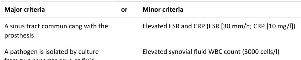
A pathogen is isolated by culture from two separate tissue or fluid samples obtained from the affected prosthetic joint
Elevated synovial fluid neutrophil percentage (65%)
Isolation of a microorganism in one periprosthetic tissue or fluid culture
5 neutrophils per high-powered field in 5 high-power fields observed from histologic analysis of periprosthetic tissue a t ×400 magnification
Figure 3.1c Anteroposterior (AP) radiograph of first-stage PROSTALAC spacer.
Structured oral examination question 2#
This is an AP radiograph of a 52-year-old female who presents to your clinic with non-specific right hip pain. She had a right metal-on-metal hip resurfacing procedure performed 3 years ago (Figure 3.2a).
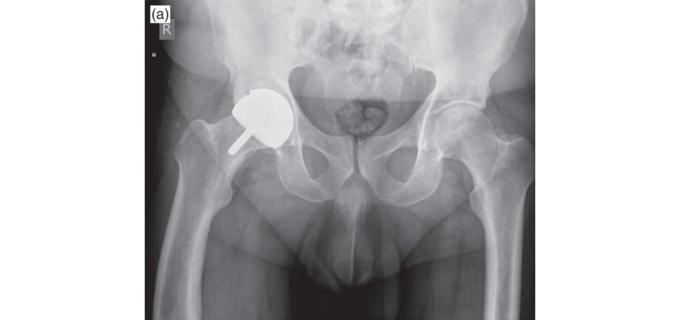
The AP radiograph demonstrates a higher abduction angle lateral opening) than normal. The current recommendations are for an acetabular abduction angle of 40°. Several studies have demonstrated the importance of optimal cup positioning with regard to wear, metal ion levels and the revision rate. High cup angle has been consistently reported to lead to greater wear and higher serum metal ion levels. The head size appears small; the current recommendations are that unless a minimum 46-mm head size can be used the procedure should not be performed because of the risks of ALVAL and pseudotumours. There is no radiolucency about the metaphyseal stem, no obvious narrowing of the neck and no divot sign.
What do you mean by a divot sign?20
A divot sign is a depression in the neck contour just below the junction with the femoral component often associated with a reactive exostosis. It is believed to be caused by repetiv e bone to component abutment due to impingement.
What is a pseudotumour and what is the difference between ALVAL and pseudotumour?
ALVAL (aseptic lymphocy te-dominated vasculitis-associa ted lesion) is a histological diagnosis caused by metal particulate debris. Patients present with localized hip pain and a localized osteolytic reaction. A more severe inflammatory reactionist ermed a pseudotumour. This is diagnosed on an MRI scan. The umbrella term ‘adverse reactions to metal debris’ (ARM Dis now preferred to cover a wide spectrum of destructive involvement including metallosis, pseudotumours and ALVAL. Pseudotumour, aseptic lymphocytic v asculitis-associa ted lesions (ALVAL) and metallosis are terms often used interchangeably to describe the same process. In June 2008, the NJR first introduced the term ARMD for surgeons to select as an indication for revision surgery, given ARM Dis considered the most inclusive term for these abnormal reactions. Several studies have described an association between pseudotumours and increased wear of retrieved components. Influencing factors include implant size and implant design (clearance and cover [arc angle]). In addition, acetabular component positioning and femoral head–neck offset influence the risk of impingement and edge loading usually associated with high wear rates. Despite this, Campbell et al. reported that in 32 THA revised due to pseudotumour several patients demonstrated minimum wear features, suggesting a hypersensitivity c ause.21 Therefore, the origin of pseudotumours is probably multifactorial caused either by excessive wear, metal hypersensitivity , a combination of the two, or an as yet unknown cause. Pseudotumour-like reactions have also been reported in non-metal-on-metal bearings. In these cases, the histological findings showed accumulations of macrophages and giant cells again suggesting an excessive wear origin.
What are the risk factors for pseudotumours?
Significant risk factors for the development of pseudotumour include female sex, ageless than 40 years, small component size, hip dysplasia and specific implant designs (ASR).
How are you going to investigate this patient?
A careful history and examination of the patient is required. It is crucial to determine if the pain is arising from intrinsic (indicating hip pathology) or extrinsic sources (referred pain). Extrinsic sources would include referred pain from the spine or pelvis, peripheral vascular disease, stress fracture, tendinitis or bursitis about the hip. Intrinsic causes include aseptic loosening , avascular necrosis, infection. (Long pause.)
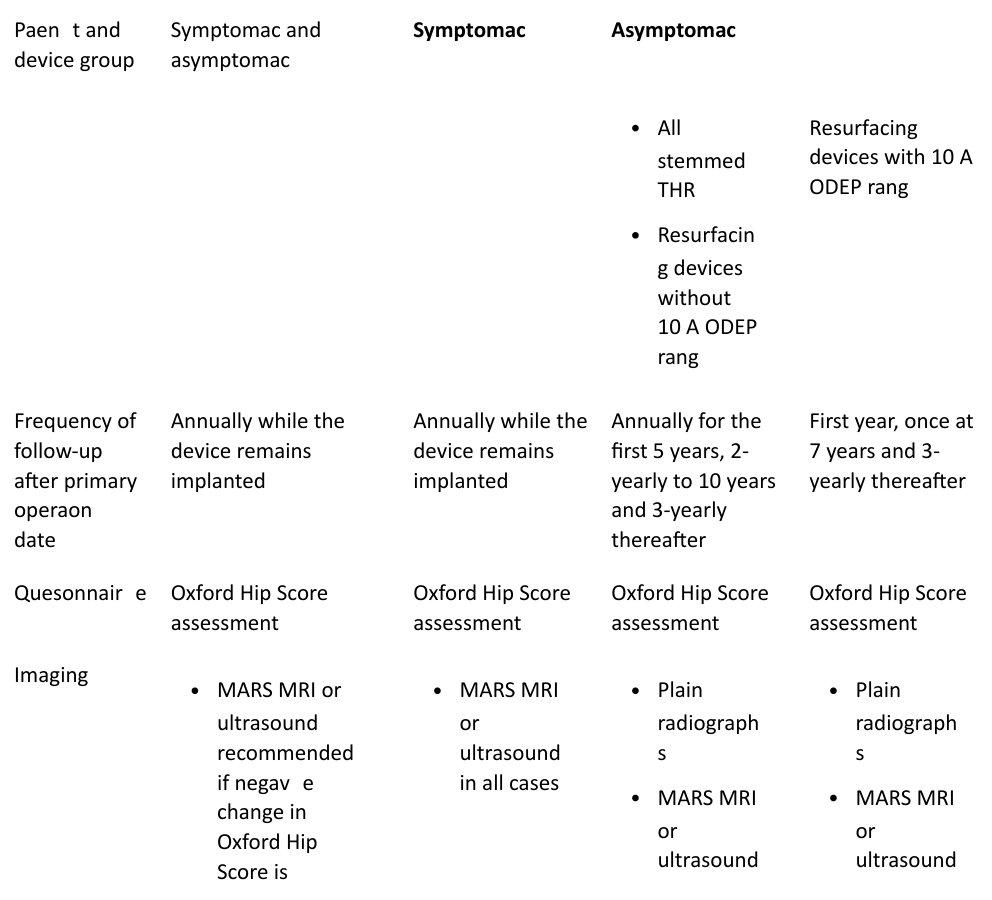
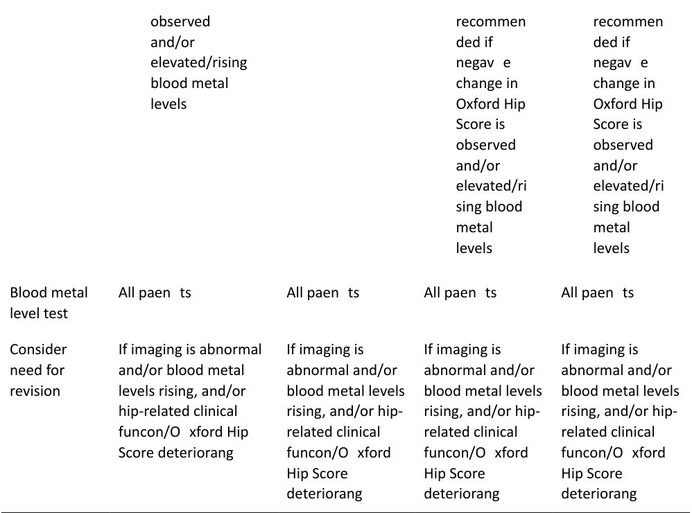
What are the latest Medicines and Healthcare Products Regulatory Agency (MHRA) recommendations (2017) (Prompt)? (Table 3.2.)
In essence, the latest MHRA guidelines with input from the BHS and BOA risk stratifies patients into high-risk and low-risk groups based on patient factors, device implanted and whether they are symptomatic Having an ASR hip is automatically high-risk and therefore patients should have annual surveillance together with cobalt and chromium levels, MARS MRI and OHS. On the other hand, a Birmingham hip resurfacing, which is an ODEP 10 A device, inpatients who are asymptomatic, should bef ollowed-up in the first year, at 7 years and then every 3 years after that. Stemmed large MoM devices are higher risk than resurfacing hips and therefore the follow-up guidance is different, and these patients require closer review.
Anything else?
I am not sure.
ALVAL may occur in both asymptomatic and symptomatic patients and early detection should give a better revision outcome if this is necessary. Essentially all MoM/r esurfacing hips require some sort of clinical follow-up for life and should not be discharged even if well functioning.
Additional clinic slots are required for follow-up of MoM hippa tien ts and this can be a significant burden on resources. Interferes with new/follow-up ratios.
This is the MRI scan obtained. What does it show? (Figure 3.2b and 3.2c.)
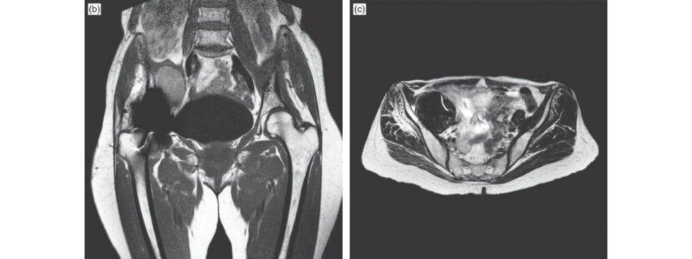
The MRI is a T2-weighted image coronal view, which demonstrates an intra-pelvic mass.
This was a pseudotumour. In fact, the mass could be felt clinically when examining the abdomen. What are you going to do?
This patient requires urgent revision surgery to the hip.
She is very scared of surgery and would prefer to avoid it.
I would stress the importance of early revision surgery as the longer the MoM resurfacing implant is left in place the more extensive the soft -tissue destruction will likely be.
What are the principles of surgery for pseudotumours?
The pseudotumour needs to be managed with aggressive debridement of all involved so tissue. It is important to do a thorough debridement of the abnormal tissue similar to the treatment of infection. The surgery should be performed by an experienced hip surgeon. Although she is still relatively young I would use a metal and PE bearing surface. I would want to use first- or second-generation highly cr oss-linked PE. A ceramic bearing surface has the potential for catastrophic fracture. We are already revising for a rare complication and we don’t want anything to go wrong again. However, I would use an uncemented implant. I would keep the option of using a dual mobility acetabular cup as the soft tissues may be so poorly compromised that the hip is unstable. Another possibility is to use a constrained cup if the hip is unstable with significant abductor muscle destruction, but I would prefer to avoid using this implant if possible as components will loosen early in this situation. It would be sensible to get a second opinion from an experienced hip surgeon as per British Hip Society guidelines to confirmand support the appropriateness of my management plan. In my region, all revision arthroplasty cases are discussed as part of a revision clinical network involving several hospitals within a geographical region. I would certainly ensure that this case is discussed with other colleagues within the revision network meeting.
What is the evidence that highly cross-linked PE improves clinical outcomes?
There was a double-blinded study from New Zealand (level 1 evidence) recently published in the American JBJS edition comparing HXLPE to conventional PE. HXLPE liners had significantly reduced wear present and were associated with a greater implant survival rate at 10 years compared to conventional UHMWPE liner s.22
Why did surgeons bother with MoM hip resurfacing procedures? The old Charnley cemented hip replacement with trochanteric osteotomy worked equally as well with excellent long-term results reported from the surgeons at Wrighting ton.
The perceived advantages of MoM hip resurfacings included better restoration of hip biomechanics, improved proprioceptive feedback, improved wear characteristics with noPE -induced osteolysis, increased levels of postsurgical activity , greater range of movement, reduced risk of dislocation improved femoral bone stock mass because the neck and most of the head are retained and ease of conversion to a THA if the implant should fail.
What are the contraindications for resurfacing?
These include severe osteoporosis, insufficient bone stock in the femoral head, large cysts at the femoral neck or head, a narrow femoral neck, notching of the femoral neck, extensive ON and severe obesity (BMI > 35 kg/m2). Other contraindications include a history of chronic renal disease, metal hypersensitivity , those with anatomical abnormalities in the acetabulum or proximal femur and certainly caution in w omen of childbearing age.
Is resurfacing contraindicated in women of childbearing age?
No, although most surgeons would now avoid a resurfacing procedure in a female regardless of whether they were of childbearing age.
Is there a role for resurfacing at all?
Data from the latest NJR annual report showed that less than 1% of all hip replacements were resurfacings. There may be a role for this implant to be used in young male individuals who are likely to trash their hip.
What do you mean by trashing your hip?
Apa tien t who has to do very heavy manual work as part of his job or who just wants to play football or rugby regardless. Candidates should mention that: Nowadays very few resurfacing hips are performed (< 1% NJR data 14th report 2017). Although they are not contraindicated they would generally only be indicated in a very small, select number of individuals. Surgery should ideally be performed by a specialized hip resurfacing surgeon.23 Cases should ideally be discussed with hip colleagues and a consensus view reached as to whether to proceed with resurfacing. Candidates should be able to discuss: The poorly performing implants (AS Rand subgroups of patient sat risk for ARMD (female sex, age < 40 years, small component size, malpositioning of the acetabular component, hip dysplasia, known nickel allergy). Complications of MoM hips. A follow-up clinic protocol. Large MoM jumbo hip replacements are now contraindicated as a primary procedure due to metal wear and corrosion at the trunnion.
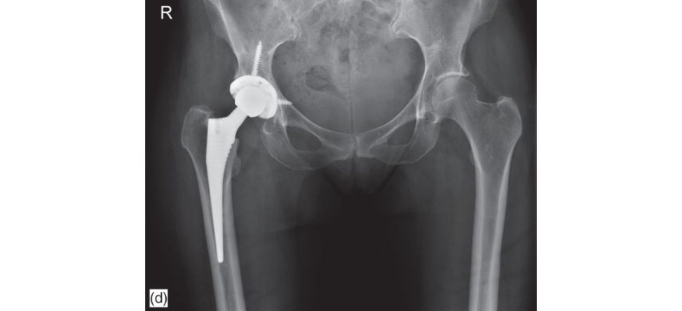
These are her postoperative radiographs (Figure 3.2d). We kept her non-weight bearing for 6 weeks as there was quite an extensive anterior wall defect in the acetabulum, but she has done very well. The hip pain has setiled, and the abdominal mass resolved. We were very lucky as the extensive soft -tissue destruction that sometimes can beseen with this condition was absent24.
What are the outcomes of hip resurfacing compared to conventional THA ?
Several recent studies report identical Harris hip scores but a greater percentage of patients with resurfacing involved in high-demand activities. The reis a higher revision rate in hip resurfacing compared to conventional THA .25
What factors are associated with higher revision rates for hip resurfacing procedures?
These would include ON (relative contraindication), hip dysplasia (technically difficult, more suitable reconstructive options a vailable), female sex, inflammatory arthritis (relative contraindication), increased age (deteriorating bone quality), a small femoral implant and specific implant designs (ASR). Causes of revision primary hip resurfacing procedure include fracture (39%), loosening/lysis 29%, infection 9%, ARMD (6%), pain (5%), dislocation (3%), other (8%). Australian Joint Registry 1999–2008. MoM hips are still a hot topic, but viva themes would be more about how to manage ARMD, when to revise and clinic follow-up protocol. Resurfacing is rarely performed these days (< 1%), so the safest option is not to mention this as a possible arthroplasty option unless specifically brought into the discussion by the examiners.
Figure 3.2a Anteroposterior (AP) radiograph right MoM hip resurfacing implant.
Table 3.2 MHRA management recommendations for patients with metal-on-metal hip replacements.
Device implanted Hip resurfacing (no stem): Hip resurfacing (no stem):
- Female - Male (femoral head diameter > 48 mm) ·
Stemmed total hip replacement (THR):
· - Male (femoral head diameter ≤ 48 mm)
·
- Femoral head diameter < 36 mm ·
- All DePuy ASR hip resurfacing devices
·
Stemmed total hip replacement (THR):
- Femoral head diameter > 3 6 mm
·
Whole blood should be used to test for cobalt and chromium metal levels.
Figure 3.2b and c T1 coronal and transverse MR Iof right MoM hip resurfacing implant demonstrating ALVAL mass.
Figure 3.2d Anteroposterior (AP) radiograph of revised hip demonstrating uncemented THA with screw fixation acetabulum.
Structured oral examination question 3#
This is an AP radiograph of a 78-year-old man presenting with increasing right hip pain. He had a THA performed 17 years ago (Figure 3.3a).
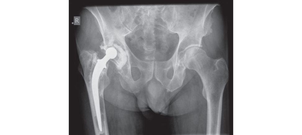
The AP radiograph demonstrates severe osteolysis of both femoral and acetabular components. There are radiolucent lines at the bone cement interface located circumferentially around all three Dee and Charnley zones in the acetabulum. The femoral component has separated from the femoral cement with lucencies in all seven Gruen zones. I am unfamiliar with the type of implant used for the femur. On the radiograph shown i can not determine if a cement plug has been used or not. Score 6 candidates should ideally be able to recognize a Charnley and Exeter stem. Score 7 candidates should recognize a Stanmore prosthesis (banana-shaped). Score 8 candidates should be able to weave in somewhere in the discussion that when revising a Stanmore prosthesis, it is particularly important due to its banana shape to clear the shoulder of the prosthesis removing any cement or bone overhanging the proximal aspect of the greater trochanter. Otherwise stem removal will be obstructed and/or difficult or a greater trochanter fracture will occur.
What are the different generations of cementing techniques?
First-generation cementing techniques involved hand-mixing of cement and finger packing of bone cementin the doughy phase into an unplugged, unwashed femoral canal. Clinical results with first-generation cementing have been variable and in general have produced some disappointing results due to the inability to produce a consistent cement mantle. Second-generation technique involved plugging the medullary canal, cleaning the canal with pulsed lavage and inserting cementin a retrograde manner using a cement gun. This reduced the incidence of gross voids and filling defects in the mantle.
Are you sure pulsed lavage was used?
Yes. Take the hint that your answer may be wrong. Second-generation technique involves essentially using a gun and bone plug (r estrictor) and nothing else. The bone is thoroughly cleaned before cement insertion but not pulsed lavage.
What else?
Third-generation techniques involved porosity reduction via vacuum mixing or centrifugation and cement pressurization. Fourth-generation cementing techniques include stem centralization both proximally and distally to ensure an adequate and symmetrical cement mantle. This is important as uneven and excessively thin cement mantles are associated with early failure and revision.
Distal centralisers are often included as a third generation technique. Some books list only 3 generations of cementing techniques omiting the fourth.
How is cementing technique graded?
The quality of the cement mantle has been described by Harris and Barrack using a scale of A to D.26 Complete filling of the medullary cavity by cementa so-called ‘white-out’ at the cement–bone interface, is graded ‘A’. Slight radiolucency of the cement–bone interface was defined asB’. Radiolucency involving 50–99% of the cement–bone interface or a defective or incomplete cement mantle was graded ‘C’. Grade ‘C2’ was given to a defect where the tip of the s tem abuts the cortex with no intervening cement. Radiolucency at the cement–bone interface of 100% in any projection, or a failure to fill the canal with cement such that the tip of the s tem was not covered, was classified ‘D’.
What are you going to do?
I would want to take a full history from the patient. I would enquire about pain. I would also want to exclude the possibility of infection (septic loosening) and would ask about problems with the hip postoperatively such as wound infection requiring washout or a prolonged course of antibiotics. A history of fever, chills or a sinus tract suggests infection. Night pain, rest pain or constant pain would also suggest infection. With aseptic loosening typically, the pain is aggravated by weight bearing. Pain is significant with the first few steps of walking (start-up pain) which improves slightly with further walking, only to worsen again with further walking. The pain is always improved with rest and rarely constant. With aseptic loosening of a THA examination may reveal shortening of the affected limb, an antalgic gait and a Trendelenberg positive testis usually present. Pain at the extremes of movement suggest loosening. It is important to exclude other causes of local extrinsic hip pain such as trochanteric bursitis, tendinitis or impingement. Extrinsic remote sources of hip pain should also be excluded, particularly the lumbar spine, especially if the pain has neurogenic features such as radiation below the knee, numbness, paraesthesia or dysesthesias. Pulses and skin temperature should be checked to rule out a vascular cause for pain.
Assume there is no infection in the hip and referred causes of pain have been ruled out. What are you going to do?
I would assess the patient Find out how bad the pain is and whether the hip should be revised or whether symptoms are manageable, and the patient can be reviewed regularly at the orthopaedic follow-up clinic. Based on the current radiographic appearances I would have some concerns with the natural progression of the condition and the possibility of catastrophic periprosthetic fracture occurring.
The patient can only walkabout 200 yards before severe pain.
I would offer him revision hip surgery provided comorbidity issues have been optimized and the risks of surgery had been discussed and understood. Both components would need to be revised.
What are the complications that you would need to mention to the patient when consenting for surgery?
I would mention Infection. Dislocation. Usually component malpositioning or laxity of soft tissues around the hip. Fracture/perforation femoral shaft. Nerve palsy (peroneal, sciatic femoral) 2–7%. Vascular injury (femoral, iliac, obturator). Leg-length discrepancy. Heterotopic ossification. Death (cardiac/pulmonary). DVT/PE. In addition, the patient has significant bone loss on both the femoral and acetabular side, so I would plan to use donor femoral head allograft to attempt to restore bone stock. An ETO is helpful if there is a large amount of cement to remove distally but will increase operating time and blood loss. An osteotomy site can also go on to either malunion or non-union. I would prefer to avoid its use in this particular case. I would warn him that he might need a period of partial weight bearing if there were concerns with initial implant stability due to excessive osteolysis and bone loss.
You mentioned about the bone loss. How do you plan for this?
Bone loss can be classified on the femoral side by using either the AAOS (Table 3.3) or the Paprosky classification system (Table 3.4). The Paprosky classification evaluates the femoral diaphysis for its ability to support an uncemented, fully porous coated prosthesis. It isless detailed than the AAOS classification but is more useful indecision making if an uncemented revision is to be performed.
Figure 3.3a Anteroposterior (AP) radiograph of loose cemented right THA.
Table 3.3 AAOS classification system for femoral defects.
I Segmental defect proximal (partial or complete)
intercalary greater trochanter
II Cavitary defect cancellous cortical ectasia (dilatation)
III Combined segmental and cavity defect
IV Malalignment rotational angular
V Femoral stenosis
VI Femoral discontinuity
Table 3.4 Paprosky classification system for femoral defects.
I Minimal metaphyseal cancellous bone loss with normal intact diaphysis
Type I defects are seen after removal of uncemented component without biological ingrowth on surface. Usually seen with Austin Moore type prosthesis or resurfacing procedures. The diaphysis and metaphysis are intact and there is partial loss of the calcar and anteroposterior (AP) bone stock
II Extensive metaphyseal cancellous bone loss with normal intact diaphysis
Often seen after removal of cemented prosthesis. Calcar deficiency and major AP bone loss
IIIA Metaphysis severely damaged with > 4 cm diaphyseal bone for distal fixation
- Grossly loose femoral component
- First-generation cementing techniques
IIIB Metaphysis severely damaged with < 4 cm diaphyseal bone for distal fixation
Type IIIB defects extend slightly further than Type IIIA; however, reliable fixation can be achieved just past the isthmus of the femur
- Cemented with cement restrictor
- Uncemented with substantial distal osteolysis
IV Extensive metaphyseal and diaphyseal bone loss/isthmus non-supportiv e
Extensive defect with severe metaphyseal and diaphyseal bone loss and a widened canal that cannot provide adequate fixation for a long stem
Acetabular bone loss
Acetabular defect classification systems are used to predict the extent of intraoperative bone loss and guide reconstructive options.
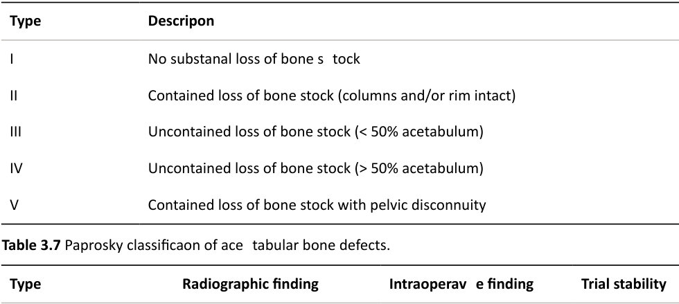
Several classification systems exist; the three most commonly used are the American Academy of
Orthopaedic Surgeons (AAOS) system (Table 3.5), the Gross and associates system (Table 3.6) and the
Paprosky classification system (Table 3.7).
Table 3.5 AAOS classification system for acetabular defects.
Type I Segmental defects
Peripheral – superior/anterior/posterior
Central – medial wall absent
Type II Cavitary defects
Peripheral – superior/anterior/posterior
Central – medial wall intact
Type III Combined segmental and cavitary bone loss
Type IV Pelvic discontinuity
Separation of anterior and posterior columns
Type V Arthrodesis
Table 3.6 Gross and associates classification system.
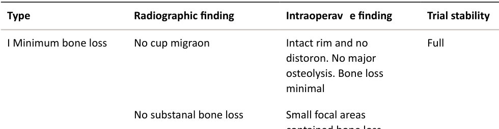
Columns intact
II Anterior and posterior columns intact and supportive
Migration less than 2 cm superomedially or laterally
Minimal ischial lysis
Minimal tardrop lysis
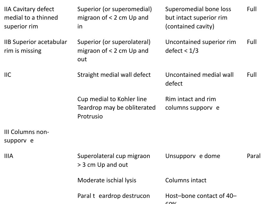
Migration medial toKo hler line
Gross and associates classification system (Table 3.6)
This classification is based on the nature of the bone graft needed for reconstruction determined on standard preoperative AP and lateral radiographs. A bone defect is considered uncontained if morselized bone graft cannot be used to fill the defect.
Paprosky acetabular bone loss classification
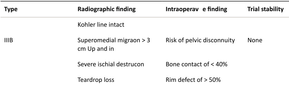
This classification is based on information that can be obtained from AP radiographs. Four radiographic criteria are assessed:
1. Superior migration of the hip centre
Indicates damage to anterior and posterior columns
Supero-medial indicates greater damage to anterior column
Supero-lateral indicates greater damage to posterior column
2. Ischial osteolysis
Bone loss inferior posterior column and posterior column
3. Teardrop osteolysis
Inferior anterior column and medial wall
4. Position of the implant relative toKo hler’s line
Deficiency of anterior column and/or medial wall deficiency
A trial component with full inherent stability does not change position when the surgeon pushes its rim or performs a trial reduction. A trial component with partial inherent stability does not change position with removal of the inserter, but does not withstand the force of pushing on the rim or performance of a trial reduction. A trial component with no inherent stability changes position with the simple act of removing the inserter.
The Paprosky classification (Table 3.7) is often used clinically in preference to the AAOS classification as it not only predicts bone loss encountered intra operatively, but also assists in determining reconstructive options.
How would you plan for surgery?
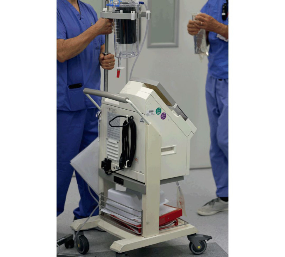
I would counsel the patient regarding the natural history of the condition and recommend revision is undertaken on an urgent basis as the situation is likely to deteriorate and may lead to catastrophic periprostatic fracture. I would get an anaesthetic review to make sure the patient was fit enough for surgery, the risks acceptable and also so they could order any special tests such as echocardiogram or pulmonary function tests if required. I would cross-match for 4 units and make sure the cell saver(Figure 3.3b) was available. I will give tranexamic acid at the time of induction.
What dose of tranexamic acid?
1 g IV and if needed a further dose of 1 g IV at closure. I would order one femoral head frozen allograft and have freeze-dried allograft a vailable if required. I would liaise with my anaesthetic colleagues in case an HDU bed was needed postoperatively. I would obtain the original operative notes to check what surgical approach was used and which implants were inserted. I would make sure the company rep is available at the time of surgery. I would ask the theatre coordinator to make sure there was an appropriate skill mix and experience in theatre on the scrub side to deal with complicated revision hip cases. I would ideally make sure the case wasn’t performed at the weekend when fewer staff are generally available or last case on a Friday with the possibility of a long theatre overrun.27 I would need to make sure the implant removal kit would include curved and straight osteotomies for the cemented cup and femur, ultrasonic tools, high-speed burrs rongeurs, cement sp lifters, reverse hooks, drills and Dall miles cabling system. I would need a flexible light source for visualizing the medullary canal of the femur. I would prefer to use uncemented components if possible if previously cement was used for fixation. The femoral bone surface after cement removal is often sclerotic, hard and resistant to cement interdigitation. I would also hesitate touse cementas long-term results in revision cases can be poorI would plan to use a long-stem uncemented modular tapered fluted revision femoral implant, aiming for a good scratch fit distally. If this could not be achieved, I would use a long-stem uncemented implant with a distal interlocking screw option. I would use a multihole TM revision acetabular shell and a metal on polyethylene bearing surface. I would attempt to use at least a 32-mm head but preferably a 36-mm head, as this will significantly reduce the risk of postoperative dislocation. I would prefer to use a posterior approach, incorporating the old incision in to this if possible. If the risk of dislocation was deemed very high I would consider using an anterolateral approach or more preferably a posterior approach but with the use of a dual-motion acetabular cup. ‘I would’ is what YOU would do. It is probably the most appropriate turn of phrase to use for the exam. I would clear soft tissue and overhanging cement away from the proximal femur to expose the proximal edge of the bone–cement interface. Flexible osteotomes and a small burr can then be used to further disrupt the cement/implant interface.
An ETO would simplify implant and cement removal, but as mentioned, in this particular situation I would prefer to avoid it.
I would use cement sp lifters to remove cement along with ultrasonic tools. Cement is split radially and longitudinally and then removed.
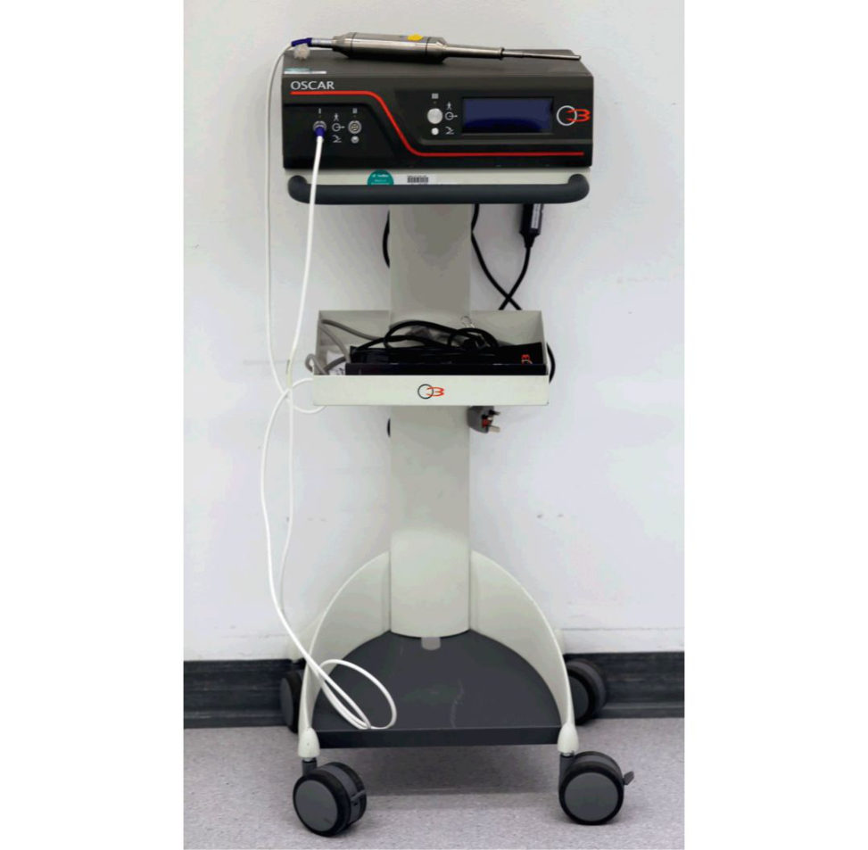
OSCAR (Figure 3.3c) (or a similar ultrasonic cement removal system) is useful forget ing through a distal cement plug. A combination of sharp cement sp lifters, cement osteotomes (straight and curved), a Midas Rex high-speed burr, ultrasonic tools and patience is the key to removing the cement mantle. I would be careful about losing cement down the femoral canal and the use of mastoids down the canal can be helpful. I would avoid levering the cement out as this risks an iatrogenic fracture, especially around the greater trochanteric region.
What about the acetabular component, if it’s cemented?
The safest way is to disrupt the PE cup from the cement using curved gouges. This prevents inadvertent damage to the bone of the acetabulum bed. After removal of the cup the cement is removed piecemeal. Sometimes a threaded acetabular extractor can be used, threading into a drilled hole until its me tal plate is flush with the rim of the acetabular component. The extractor is then toggled to disrupt the fixation interface and allow removal of the component. High-speed burrs are sometimes needed tode bulk cement within acetabular anchoring holes.
And if the socket is uncemented?
The order of removal is component liner removal then screw removal (if there are supplementary screws) and finally metal shell removal. If the liner is polyethylene, then drilling into the centre of the liner followed by inserting a screw into the liner hole will usually liftoff the liner . If the liner is ceramic, then the principle is first to disengage the smooth taper lock by a sharp tap into the liner. This is followed by the use of a suction cup with its attached three scallops that contact the peripheral rim of the metal acetabular to remove the liner. Different implants will have their own extraction devices, so it is critical to know all about the implant one is revising and to ensure that the relevant and appropriate kit is available. That is why it is important to have a good system in place whereby old medical records can be easily retrieved to look over well in advance of surgery. Removing any supplementary screws can be challenging and I would always have the Timex broken screw set available as well as a burr in case the screwheads are gone. Finally, to remove the metal-backed shell, the essential tool required here is the Explant (Figure 3.3d). This has been revolutionary in making the removal of an uncemented acetabular shell easier.
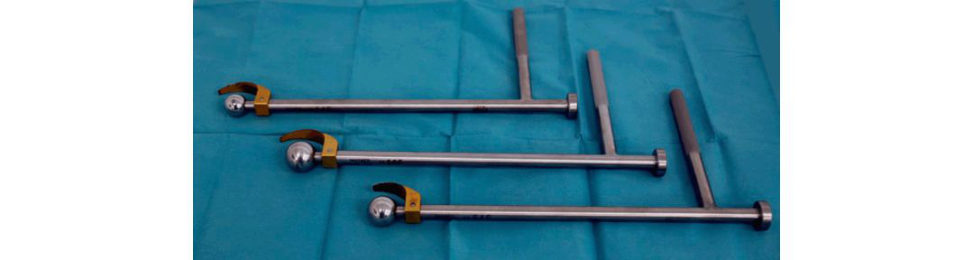
Figure 3.3b Cell saver.
Figure 3.3c OSCAR system.
Figure 3.3d Explant (Innomed) acetabular cup removal system.
Structured oral examination question 4
These are the radiographs of a 78-year-old lady who has been referred to the orthopaedic clinic by her GP because of increasing pain in her right hip. Would you care to comment on the radiographs? (Figure 3.4)
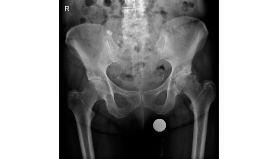
This is an AP radiograph, demonstrating lo wer lumbar vertebrae, both hips and proximal femur. The most obvious features in the right hip are loss of joint space osteophytes, sclerosis and bone cysts. The radiographic features are highly suggestive of osteoarthritis (O A) of the hip.
How is osteoarthritis classified?
OA is classified into primary OA when an obvious cause can be identified, and secondary OA caused by such conditions as osteonecrosis (ON), DDH, postir aumatic, P aget’s disease, slipped capital femoral epiphysis, protrusio acetabuli, and Perthes disease.
What are the percentages of each type of OA?
Various studies have suggested that almost 90% of cases of OA are secondary.
How are you going to manage this patient?
I would take a full history and examination from the patient Specifically, I would want to know the location of pain and exclude referred pain from the spine. Hip pain is classically located in the buft ock or groin radiating to the knee. Pain radiating below the knee to the foot is strongly suggestive of radicular-type pain from the spine. I would inquire whether the patient had difficulty puting shoes and socks on, tying shoelaces, bending to pickup an object from the floor, geting in and out of a car, [Examiner interrupts]
The patient struggles to walk a quarter of a mile. She has typical symptoms of advance dOA. What are you going to do?
Assuming that all conservative options had been tried and have been unsuccessful I would offer her THA.
What type of hip arthroplasty would YOU perform?
I would use a cemented Exeter THA.
Why this particular implant?
The Exeter THA has excellent peer-reviewed long-term data. It is an implant that I am very comfortable using, I have been trained to use this implant by my consultants, the instrumentation is straighfoorward and simple to use, the neck cut is not critical, and the introducer allows for even pressure when inserting the implant. This hip system provides me with the ability to deal with anatomical variants and to recreate offset and leg length and gives me a choice of bearing surfaces and head sizes.
It allows good initial fixation and excellent long-term survival.
It is an ODEP (Orthopaedic Data Evaluation Panel) 13A* rated stem.
What do you mean by anatomical variants?
The shape of the femur.
The shape of the femur is a more important consideration when using an uncemented implant and isless applicable for a cemented implant.
The Exeter stem has different offset sizes, which improves hip abductor function and stability. In recent years smaller offsets and shorter-length ‘CDH’ stems have been introduced to deal with smaller femoral geometry. In smaller femurs oversizing of a femoral component may result in an incomplete or insufficient cement mantle of less than the recommended 2 mm uniform thickness. This may lead to early failure.
What is the problem with using smaller stems?
I am not sure.
If you use a smaller stem there are concerns with stem breakage and implant failure.
The viva could have gone on to discuss stem geometry affecting cement stresses (avoidance of sharp edges, broad lateral curve), factors predisposing to stem breakage, bending and torsional rigidity of stems, etc.
What do we mean by a Dorr grading of the femur?
There are three types of femoral shape based on metaphyseal–diaphyseal anatomy. Dorr type A femurs have wide metaphyses and narrow diaphyses, type B have a smooth metaphyseal– diaphyseal transition and type C do not have much difference in the sizes of these two regions.
So how does this apply to uncemented implants?
We tend to avoid using uncemented implants inpatients with Dorr C femurs. Candidates should try to avoid mentioning an y loose terms they don’t fully understand as it can lead on to difficult questions.
What are the survival figures like for the Exeter implant?
Know some papers to quote for your chosen implant. 1. Lewthwaite et al. CORR 200828 Results of Exeter THA in younger patients < 50 years at 10–17 years FU Survivorship of the femoral stem from all causes was 99% No stem was revised for aseptic loosening
2. Petheram et al. Bone Joint J 201629
Results of Exeter THA at 20–25 years.
FU study of 382 cemented Exeter THA (350 patient sat a mean age of 66.3 years (17–94).
With an endpoint of revision for aseptic loosening orly sis, survivorship of the stem at
22.8 years was 99.0%
Figure 3.4 Anteroposterior (AP) radiograph demonstrating severe osteoarthritis left hip.
What are the design principals of the Exeter Stem?
The Exeter implant is a loaded taper model and becomes lodged as a wedge in the cement mantle during axial loading, reducing peak stresses in the proximal and distal cement mantle. The stem is allowed to subside initially un til radial compressive forces are created in the adjacent cement and transferred to the bone as hoop stresses.
What approach would you use to the hip?
I am happy to use either the Hardinge or posterior approach to the hip.
Make up your mind. Which one are YOU going to do?
For the majority of cases I would prefer to use the posterior approach to the hip. In rare instances, I would use a Hardinge anterolateral approach if the risk of dislocation was considered to be high such as neurological or muscular weakness around the hip (Paget’s/CVA), early dementia or substance abuse.
The posterior approach is considered easier to perform and is generally a quicker procedure, limiting operative complications such as blood loss anda naes thetic issues.
The abductor muscles are not disturbed significantly so there is generally no gait abnormality, but the acetabulum is more difficult to see and can make prosthesis positioning difficult, possibly causing an increased dislocation rate due to component malpositioning. The sciatic nerve is at slightly more risk of being injured as well.
3 0 There is about double the risk of sciatic nerve injury using the posterior approach.
Most surgeons would say that there is no significant difference in time between the two approaches; the posterior approach can take just as longas the anterolateral approach. The posterior approach is marginally technically easier than the anterolateral approach, but this also depends on surgeon training, experience with using either approach and personal preference. I would argue about the acetabulum beingless easy to visualize posteriorly as most surgeons believe the posterior approach provides better acetabular visualization, especially for revision cases. The pelvis tends to tilt more and so the degree of cup anteversion is usually underestimated leading to an increased risk of dislocation WhereI think the posterior approach does make a difference is a reduced incidence of Trendelenberg gait postoperatively and improved Harris hip scores compared to the anterolateral approach. While results have been a bit contradictory the risk of posterior dislocation is slightly higher posteriorly even with a careful repair of the soft tissues. Larger head sizes are being used now so this is becoming less of an issue.
Talk me through the posterior approach to the hip.
Assuming full informed consent has been obtained, all relevant case notes and radiographs have been obtained, the leg has been marked, WHO checklist performed and she has been suitably anaesthetize dIn the anaesthetic room I would position the patient laterally, affected leg uppermost, with hip supports. I would then prepare and drape the patient and make an incision centred over the greater trochanter, approximately 15 cm in length. I would cut through the skin, subcutaneous tissue, and open up the fascia lata, spliting the gluteus maximus along the line of muscle fibres, and then release the short external rotators from the greater trochanter. Finally, I would perform a capsulectomy and then dislocate the hip. I would protect the sciatic nerve, being aware of its position and a void dissecting too close to it. I would place a large retractor over the anterior edge of the acetabulum at 2–3 o’clock. I would perform a releasing incision into the inferior capsule. I would then place a Charnley spike into the posterior wall of the acetabulum and an additional Hohmann retractor inferiorly. This should give me a 360° view of the whole face of the acetabulum as recommended by BOA guidelines.
What are the pathological processes involved in the development of osteoarthritis of the hip?
Disruption of the integrity of the collagen network occurs early in OA allowing hyperhydration. The increased water content of cartilage causes softening, decreases Young’s modulus of elasticity and reduces its ability to bear load. Initial changes in OA involve damage to the tangential z one immediately below the articular surface, with disorganization of the collagen network, loss of proteoglycans and swelling. This leads to a hypertrophic repair response with increased synthesis and accumulation of pr oteoglycan. However, the repair process fails with loss of surface integrity, and fibrillation parallel to the surface. In regions of severe damage, there is a loss of cellularity and sporadic formation of cell clusters or clones. Normal cartilage metabolism is a highly regulated balance between synthesis and degradation of the various matrix components. With OA the equilibrium between anabolism and catabolism is weighted in favour of degradation. Cartilage catabolism results in release of breakdown products into synovial fluid, which then initiate san inflammatory response by synoviocytes. These breakdown products include: chondroitin sulphate, keratan sulphate, PG fragments, type II collagen peptides and chondrocyte membranes. Activated synovial macrophages then recruit PMNs, establishing a synovitis. The y also release cytokines, proteinases and oxygen free radicals (superoxide and nitric oxide) into the adjacent synovial fluid. These mediators act on chondrocytes and synoviocytes, modifying synthesis of PGs, collagen, and hyaluronan as well as promoting the release of catabolic mediators. Cartilage changes in OA are characterized by increases in: Water content. Chondrocyte activity and proliferation. Stiffness of articular cartilage. Interleukin-1. Metalloproteinase levels Cathepsins Band D levels. And by decreases in: Quality of collagen. Proteoglycan quality and size. Histology classically demonstrates: Loss of superficial chondrocytes. Replication and breakdown of the tidemark.
Fibrillation.
Cartilage destruction with eburnation (polished, shiny smooth with an appearance like ivory)
of subchondral bone.
Is OA simply an ageing process of cartilage?
Several differences between ageing cartilage an dOA cartilage have been described suggesting ase parate disease entity . For example, OA and normal ageing cartilage differ in the amount of water content and the ratio of chondroitin sulphate to keratin sulphate constituents.
[Interrupting] That’s fine that’s OK.31 What molecules are responsible for degrading the cartilage matrix?
The primary enzymes responsible for the degradation of cartilage are the matrix metalloproteinases (MMPs). These enzymes are secreted by both synovial cells and chondrocytes and are categorized into three general categories: (a) collagenases; (b) stromelysins; and (c) gelatinases. Under normal conditions, MMP synthesis and activation are tightly regulated at several levels. They are secreted as inactive proenzymes that require enzymatic cleavage in order to become activated Once activated, MMPs become susceptible to the plasma-derived MMP inhibitor, alpha-2- macroglobulin, and to tissue inhibitors of MMPs (TIMPs) that are also secreted by synovial cells and chondrocytes. In OA, synthesis of MM Psis greatly enhanced and the available inhibitors are overwhelmed, resulting innet degradation Interestingly , stromelysin can serve as an activator for its own proenzyme, as well as for procollagenase and prostromelysin, thus creating a positive feedback loop of proMMP activation inc artilag e.
What factors are responsible for inducing metalloprotease synthesis?
IL-1 is a potent pro-inflammatory cytokine that, in vitro, is capable of inducing chondrocytes and synovial cells to synthesize MM PIn addition, IL-1 suppresses the synthesis of type II collagen and proteoglycans and inhibits transforming growth factor-β-stimula ted chondrocyte proliferation. Therefore, in OA, IL-1 actively promotes cartilage degradation and may also suppress attempts at repair.
Structured oral examination question 5#
Osteonecrosis (ON)
This is the anteroposterior (AP) radiograph of a 48-year-old man who presents to your clinic with several weeks history of progressively worsening bilateral hip pain. What do you think of the radiograph? (Figure 3.5a) CANDIDATE 1 : This is an anteroposterior (AP) view of the pelvis. The most obvious abnormality is patchy diffuse sclerosis with increased density in the superolateral aspect of the right femoral head (Ficat 2). The left femoral head has a possibly minimal osteoporosis and/or blurring and poor definition of the bony trabeculae (Ficat 1). The radiograph is suspicious of bilateral osteonecrosis. I would like to obtain a frog-leg lateral radiograph of both hips. I would look for the crescent sign, indicating subchondral fracture, a feature of osteonecrosis that is more obvious on a frog-leg lateral than AP projection. This is because the anterior and posterior margins of the acetabulum on the AP projection are superimposed over the superior portion of the femoral head, the usual location of the sign. When osteonecrosis is bilateral, it usually occurs in each hip at different times, and the staging of disease in each hip is often different. [Candidate score 7–8] CANDIDATE 2 : This is an AP pelvic radiograph showing both hips. There is nothing very obvious staring at me. There are no features of osteoarthritis such as joint space narrowing osteophytes or sclerosis.32 [Candidate score 4] What do you mean by osteonecrosis?
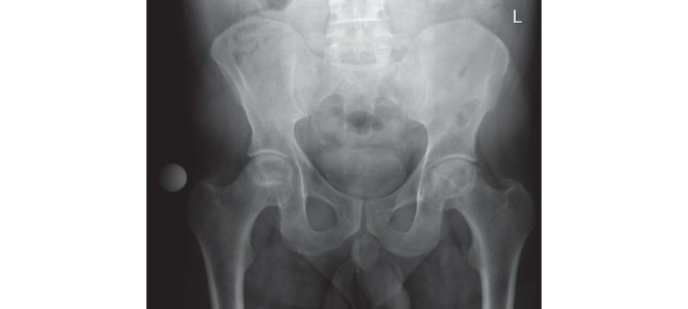
Osteonecrosis occurs due to interruption of the blood supply to the femoral head leading to ischaemia and cellular death. The term osteonecrosis is preferred to commonly used AVN because it best describes the histopathological processes involved and does not imply a specific aetiology .
What is the aetiology of osteonecrosis?
A number of conditions are associated with osteonecrosis. The most common cause is trauma secondary to fracture and/or dislocation of the femoral head. Other conditions include: Corticosteroid use. Alcohol abuse. Smoking. Coagulopathies. Sickle cell anaemia. Caisson disease. Hypercholesterolaemia.
Organ transplantation.
Systemic lupuserythematosus.
Gaucher disease.
Hypertriglyceridaemia.
Intramedullary haemorrhages.
Chronic pancreatis.
Figure 3.5a Anteroposterior (AP) radiograph of bilateral osteonecrosis.
AS IT GRIPS 3Cs (mnemonic)
Alcohol
Steroids
Idiopathic
Trauma
Gout Gauchers
Rheumatoid/radiation
Infection/incr eased lipids/inflammatory arthritis
Pancreatis/pregnancy
SLE/sickle cell/smoking
CRF/chemotherapy/Cassion’s disease
In approximately 10–20% of cases no cause can be identified.
What is the pathophysiology of osteonecrosis?
Aetiologic factors in osteonecrosis are usually related to underlying pathologic conditions that alter blood flow, leading to cellular necrosis and ultimately to collapse of the femoral head. This damage can occur in one of five vascular areas around the femoral head: arterial extraosseous, arterial intraosseous, venous intraosseous, venous extraosseous and extravascular extraosseous. 1. Extraosseous arterial factors are the most important. The femoral head is at increased risk because the blood supply is an end-organ system with poor collateral development. Blood supply can be interrupted by trauma, vasculitis (Ra ynaud disease), or vasospasm (decompression sickness). 2. Intraosseous arterial factors may block the microcirculation of the femoral head through circulating micro emboli. These can occur in sickle cell disease (SCD), fat embolization or air embolization from dysbaric phenomena. 3. Intraosseous venous factors affect the femoral head by reducing venous blood flow and causing stasis. These factors may accompany conditions such as Caisson disease, SC Dor enlargement of intramedullary fat cells. 4. Intraosseous extravascular factors affect the hip by increasing the pressure, resulting in a femoral head compartment syndrome. For example: fat cell hypertrophy after steroid administration or abnormal cells, such as Gaucher and inflammatory cells, can encroach on intraosseous capillaries, reducing intramedullary circulation and contributing to compartment syndrome.
5. Extraosseus extravascular (capsular) factors involve the tamponade of the lateral epiphyseal vessels located within the synovial membrane, through increased intracapsular pressure. This occurs after trauma, infection and arthritis causing hip effusion that may affect the blood supply to the epiphysis.
Specifically, how do steroids cause osteonecrosis?33
The mechanism postulated for steroid-induced ON is still unclear . Johnson proposed that fat cell hypertrophy within the bone marrow increases femoral head pressure resulting in sinusoidal vascular collapse and necrosis of the femoral head.34 The exact mechanism of fat cell hypertrophy remains obscure, but a disorder in fat metabolism is implicated. Jaffe et al. believe patients undergoing steroid treatment are in a hyperlipidaemic state, which can increase the fat content within the femoral head and raise intracortical pressure producing sinusoidal collapse and finally necrosis.35 Other investigators have proposed that this hyperlipidaemic state leads to fat embolism occluding the femoral head microvasculature, which initiates the pathophysiologic process.36 A recent study in rabbits suggests that the use of steroids can also damage endothelial and smooth muscle cells within the vasculature. This may result in interruption of the venous drainage from the femoral head, leading to blood stasis, an increase in intraosseous pressure, and osteonecrosis.37 Other studies suggest primary osteocyte cell death without any other features. This is seen with steroid use, in transplant patients and those who consume significant amounts of alcohol.
How common is steroids as a cause of osteonecrosis?
High-dose corticosteroids are the most common cause of non-traumatic osteonecrosis accounting for 10–30% of cases. However, only 10% of patients exposed to corticosteroids may develop osteonecrosis. Dosage is typically steroids > 2 g of prednisone, or its equivalent, within a 2– 3-month period. The period from the start of corticosteroid treatment to the diagnosis of osteonecrosis ranges from 1 to 16 months (mean 5.3 months), and the majority of patients are diagnosed within 1 year.
You mentioned the crescent line, what is its significance?
Therapeutic interventions are less likely to halt progression of the disease once this sign appears.
How does osteonecrosis of the hip present?
Although osteonecrosis can be clinically silent, typically apa tien t complains of pain, usually localized to the groin area but occasionally to the ipsilateral buft ock and knee. It is usually a deep intermift ent, throbbing pain, with an insidious onset that eventually occurs at rest and may be presentor even worsen at night. Physical examination reveals pain with both active and passive range of motion, especially with passive internal rotation. Range of motion is important as this helps determine the extent of the disease. In general, more limited flexion and abduction indicates more extensive articular damage whereas limited rotation alone may indicate less destruction. A careful examination of the contralateral hip should always be undertaken, as osteonecrosis is bilateral in 40–80% of cases.
How is osteonecrosis classified?
Several classification systems for osteonecrosis exist. Ficat and Arle tis the most commonly known and consists of four stages.38 Hungerford and Lennox later added a fit ihs tage (Stage 0) when MRI became available.39 Stage 0 (preclinical). Suspected disease in the contralateral hip when the index joint has definitive findings. No clinical symptoms. MRI non-diagnostic. Stage I (preradiological). Normal findings on radiographs and positive findings on MRI or bone scan. The MRI shows a double-line sign, consistent with a necrotic process. Stage II (pre-collapse). Osteopaenia, demineralization sclerosis or cysts. A late finding is the crescent sign, a linear subcortical lucency, situated immediately beneath the subcortical bone, representing a fracture line and impending femoral head collapse. Stage III (collapse). The femoral head is flattened and collapsed with the presence of sequestration manifested by a break in the articular margin without acetabular involvement. Stage IV (progressive degenerate disease). Severe collapse and destruction of the femoral head, acetabular osteophytes. Osteoarthritis superimposed on a deformed femoral head.
Any other classification systems?
Steinberg (Table 3.8) expanded the staging system into seven stages and quantified the amount of involvement of the femoral head into mild (< 15%), moderate (15–30%) and severe (> 30%), based on radiographs.40 It is considered more useful than Ficat because it grades the severity and extent of the involvement, both of which are thought to affect prognosis.
Any others?
Other classification systems include the ARCO (Association Research Circulation Osseous) classification University of Pennsylvania system and the Mitchell MRI classification.
What is the Kerboull necrotic angle and its importance?
The Kerboull necrotic angle is used to calculate the size of the necrotic segment. It is the sum of the angle of the necrotic seg mentas measured on both the anteroposterior and frog-lateral radiographs. Patients with a Kerboull angle > 200° more commonly have poor results with certain bone-preserving procedures.
How are you going to manage this patient?
I would perform bilateral core decompression. The osteonecrosis is still a t an early stage where it may be successful (Ficat stages I and II osteonecrosis). The procedure has no role in the management of Ficat stages III or IV disease. Results have been satisfactory when core decompression is combined with either non-vascularized or vascularized fibula gratis in patients with Ficat stage II lesions.
What are the pre-requisites for performing a free vascularized fibular graft (VF G)?
VFG for AVN is a major operative procedure with a long rehabilitation time and therefore patient selection to minimize the potential for an unsuccessful operation is critical. McKee from Toronto suggests the operation should be limited topa tien ts [scoring 7–8]:41 1. With 2 mm orless of femoral head collapse as measured on plain radiographs. 2. Who are 45 years of age or younger (and have a reasonable life expectancy). 3. Have had withdrawal of an identified ae tiologic agent. 4. Have no contractures about the hip. 5. Have a supple joint. These are obviously general guidelines that may be adjusted somewhat depending on the individual patient.
What are the advantages of performing a free VFG?
The advantages of vascularized fibular gratiing include: Being able to perform a core decompression of the femoral head. The ability to perform curettage and removal of the osteonecrotic focus. Impaction of autogenous cancelleous graft to fill the defect created by removal of the osteonecrotic bone. The structural support of the subchondral surface provided by the fibular graft. The addition of vascularized bone and blood supply to the area of osteonecrosis enhances there vascularization process.
What complications can occur with a free vascularized fibular graft?
Ga skillet al. from a tertiary centre in North Carolina performing a large volume of VFG reported a 16.9% complications rate, 4.3% of complications require reoperation or chronic pain management.42,43 Donor site morbidity:
Great-toe flexion contracture (4.3%). Majority asymptomatic, noticeable only on clinical examination with the ankle fully dorsi flexed. Occasionally requires z-lengthening of the FHL tendon at the level of the medial malleolus. Flexion contracture of the second and third toes may coexist in a small number of patients.
Persistent weakness in the operated extremity (0.6%) either long toe flexors or peroneal group.
Mild persistent pain and tenderness at the ankle or distal osteotomy site (4.1%) usually after prolonged standing or moderate activity such as jogging.
Sensory deficits (1.7%). The sensory deficit was not always consistent with peripheral nerve or dermatomal distributions.
Superficial infection.
Table 3.8 Staging system of Steinberg et al.
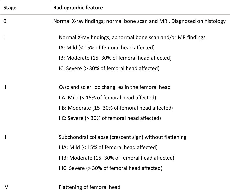
IVA: Mild (< 15% of surface and < 2 mm depression)
IVB: Moderate (15–30% of surface or 2–4 mm depression)
IVC: Severe (30% of surface)
Stage Radiographic feature
V Joint narrowing and/or acetabular changes (this stage can be graded according to severity)
VI Advanced degenerative changes
Graft site complication
Symptomatic lateral pin migration (2.4%). A Kir schner wire was used routinely to secure the fibular graft in its final position after placement in the femoral head.
Symptomatic heterotopic ossification (1.4%).
Femoral fracture (0.7%). All occurred in the intertrochanteric and subtrochanteric region after a fall.
Superficial infection (4%).
Deep infection (4%).
Haematoma (1%).
Trochanteric bursitis (1%).
What are the other techniques that can be used to manage osteonecrosis of the hip?
The trapdoor procedure is performed with an arthrotomy to dislocate the hip anteriorly, followed by curettage of the necrotic segment of the head and packing of the defect with iliac crest bone graft through a cartilage window in the femoral head. This can be used for Ficat stage III and early Ficat stage IV and reasonable results have been reported.
You have to be more specific than that; what do you mean by reasonable results?44
Michael Mont reported on a series of 30 hips Ficat stage III/IV at 5 years with 73% having good to excellent results.45
Any other options?
Osteotomy has been used to treat Ficat stage III and IV disease, but results have been variable because it is difficult to rotate the necrotic segment out from the weight bearing area, especially when the lesion is large. Sugioka et al. reported good to excellent results at 3–16 years of follow-up in 78% of 229 hips treated with the transtrochanteric anterior rotational os teotomy.46 Their results with this technically demanding procedure have not been reproduced by others. A success rate of approximately 30% at 5 years is common, with the best results reported in patients whose lesions do not result from trauma and who haveless than 30% of the head involved.
Any new technique that has emerged in the last 2 or 3 years?
Stem cells have been used to manage ON.
Go on – do you know about the technique or results?
Two techniques are being promoted. One is a three-stage procedure and the other is a single-stage procedure. The first method is by stem cell culture in the lab to multiply the number of cells several millionfold. These cultured stem cells arere injected into a previous core decompression site.
In the second method, bone marrow obtained from the pelvis is centrifuged in the operating room to yield a bone marrow concentrate rich in stem cells. The patient is supine on a traction t able with a
C-arm image intensifier. Percutaneous core decompression drilling with a Kirschner wire (diameter
2.7 mm) is performed to perforate the interface between the necrotic lesion and healthy bone.
Following this, concentrated autologous bone marrow aspirate is slowly transplanted into the necrotic area under fluoroscopic control. This is still an experimental procedure, but early results seem promising for early disease.
The patient had surgery on both hips. These are his postoperative radiographs (Figure 3.5b).
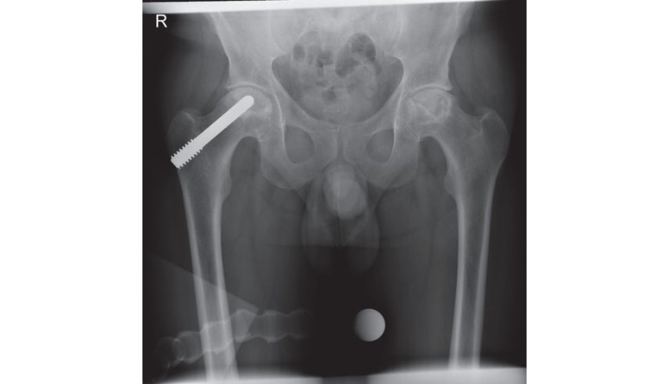
The AP radiograph demonstrates a metal core rod in the right hip.
What do we call this?
The patient has had a tantalum rod inserted into the femoral head. The implant achieves decompression, supports the subchondral plate of the necrotic areas and probably induces bone regeneration.
Anything else?
The use of a trabecular metal ‘AVN rod’ has a number of atir active theoretical advantages, including no donor site morbidity, improved rehabilitation structural support of the femoral head and the potential for ‘osseointegration ’ of the biologically friendly material.
The patient had core decompression performed on the left hip and a c ore decompression with tantulum rod inserted in the right hip. He initially got good pain relief from the procedures for about a year or so, but he returns to the orthopaedic clinic complaining both hips are now painful. The left side is worse than the right. What do you think of the radiographs?
The AP radiograph suggests osteonecrosis has progressed.
What will you do?
I would offer him bilateral hip arthroplasty, the left one being more symptomatic first.
What type of hip replacement would you use?
In view of his relatively young age I would perform an uncemented THA with a ceramic on HCLPE bearing surface.
What are the results like at 10 years for this bearing surface? What will you tell the patient about how long his hip will last?
Sorry, I am not sure, I think it is around 95%. The latest NJR report (15th, 2018) has shown a 5.33% (3.77% to 7.50%) RR at 14 years for males aged under 55 with an uncemented THA CoP surface.
Are there any other bearing surface options a vailable?
A ceramic-on-ceramic (CoC) bearing surface.
What are the advantages of using a ceramic bearing surface?
The advantages of using a ceramic bearing surface include superior lubrication, friction and wear properties compared with other bearing surfaces in clinical use. Specifically, it is an extremely hard material very resistant to wear, and has a low coefficient of friction excellent abrasive resistance and excellent wettability properties for improved lubrication. It is presumed that the lower wear rates lead to a lower rate of aseptic loosening and the need for revision surgery. Disadvantages include potential for catastrophic fracture, squeaking, and cost.
What is the incidence of squeaking?
The reported incidence of squeaking with alumina ceramic bearings varies widely from 0.45% in a series of 2716 ceramic implants to 7.0% in a series of 159 ceramic implants. Most reported series note that squeaking is rare and without clinical significance; however, on rare occasions, major squeaking has led to revision surgery.47
What are the results like for ceramic-on-ceramic hips?
UK NJR data report a 6.43% (5.17–7.98%) revision rate at 14 years for males aged under 55 with an uncemented THA CoC surface. Latest data from the 15th NJR report have shown a steady decline since 2011 in CoCT HAuse due to the concerns with squeaking, catastrophic failure and high cost. The use of a CoP bearing surface is steadily increasing and if used with a second-generation HCLPE becomes a highly atir active option in a young patient Better NJR survival figures in males under 55 at 14 years are reported for CoP compared to CoC. Definitely avoid mentioning MoM resurfacing as an option in a viva scenario unless specifically brought up in the discussion by the examiners.48 Ceramic on X3 poly is becoming the preferred bearing option for young patients with most hip arthroplasty surgeons moving away now from ceramic-on-ceramic use.
Will there be any special issues removing the tantulum rod and performing THA?
I would contact the manufacturers of the implant as there is a special implant removal kit. Otherwise not using the removal kit makes the surgery more difficult. I would use a Gigli and reciprocating saw to section the head, implant removal corer to take out the tantulum rod and then perform a conventional uncemented THA.
Are there any worries with tantalum material?
Studies suggesta trend towards an inferior outcome inpatients following conversion of tantalum rod to THA.49 There is also concern of residual tantalum metal within the joint space found in the majority of conversions. Although there is no catastrophic wear seen in studies there is the potential for accelerated joint damage in the medium to long term.
These are his radiographs (Figure 3.5c). He had a large jumbo MoM performed on the left side and a large ceramic jumbo head THA performed on the right side. Do you have any worries?
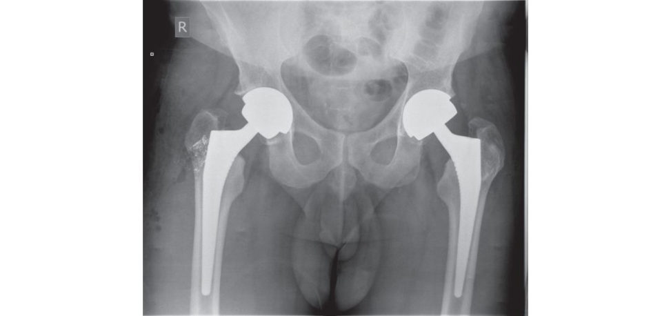
Following the BHS meeting in Manchester 2012 a statement was released advising that stemmed large-diameter MoM total hip replacements using bearing of 36 mm or above should no longer be performed. A higher than anticipated early failure rate for large jumbo head MoM hips had been reported. Concern was expressed regarding the trunnion at the ‘Morse’ taper where the large diameter metal head attaches to the stem with damage occurring from either wear or corrosion or both resulting in either loosening of the acetabular component, loosening of the femoral component or a metal reaction with necrosis and soft tissue damage Excluding the ASR implant these devices have a reported revision rate of 22.14% at 13 years (14th NJR data).
What about followup?
This should be as per recent MHRA and BHS guidelines for MoM bearing surfaces, yearly for the first 5 years and continuing on for life. Pain in this group of patients should betaken seriously and investigated appropriately with cobalt chromium levels, a MARS MRI scan of the hip and OHS. This patient should be considered at high risk for implant failure as he has a stemmed implant with a femoral head ≥ 36mm. I would review this patient at least yearly as ARMD may occur in symptomatic and asymptomatic patients and earlier detection should give a better revision outcome if needed. Although each patient needs to be assessed individually I would have a low threshold for obtaining blood level ion measurements and MARS MRI even for asymptomatic patients if I had concern about ARMD.
What would you look for in the MARS MR Iof the hip?
I would look for any soft -tissue local reaction or masses (pseudo tumour), abductor muscle detachment, the presence and extent of any osteolysis and any periprosthetic fluid collections. A fluid collection by itself around the joint in an asymptomatic patient, unless very large can be safely observed with interval scanning. MARS MRI scan is more important in the decision-making process to revise a MoM hip replacement than elevated cobalt/chromium levels. I would have significant concerns in any patients with surrounding muscle/bone damage visualized on MARS MRI. Solid lesions seen on MARS MR Iare more worrying than cystic Synovial thickness has a high sensitivity and specificity for ALVAL.
Is there a role for US of the hip?
Ultrasonography is a good screening tool, is cheap and has no radiation hazard. However, the detection of small or deep lesions is difficult and the use of ultrasound is highly dependent on the operator’s experience.
What about the other ceramic hip?
There are some worries again regarding the trunnion where the large ceramic head attaches to the stem, which may be the source of excessive wear or corrosion leading again to early failure, although the evidence isn’t as strong.
Why choose a Delta ceramic head over a cobalt chrome head?
It was originally thought that this would result in a lower revision rate with decreased clinically relevant wear rates. The use of a large ceramic head provides an advantageous head–neck ratio that theoretically reduces the risk of impingement and subluxation and provides for an increased ROM. And while a large femoral head may potentially cause elevated trunnion stresses, trunnion corrosion is likely to be multifactorial with taper design, contact area, preparation of the t aper, impact force, head–neck junction alloy composition also playing a part.
Figure 3.5b Anteroposterior (AP) radiograph of pelvis with tantulum rod inserted into the right hip.
Figure 3.5c Anteroposterior (AP) radiograph left MoM hip and right ceramic large jumbo hip arthroplasty.
Structured oral examination question 6#
This is an anteroposterior (AP) radiograph of a 73-year-old male who had a cemented THA performed 14 years ago (Figure 3.6).
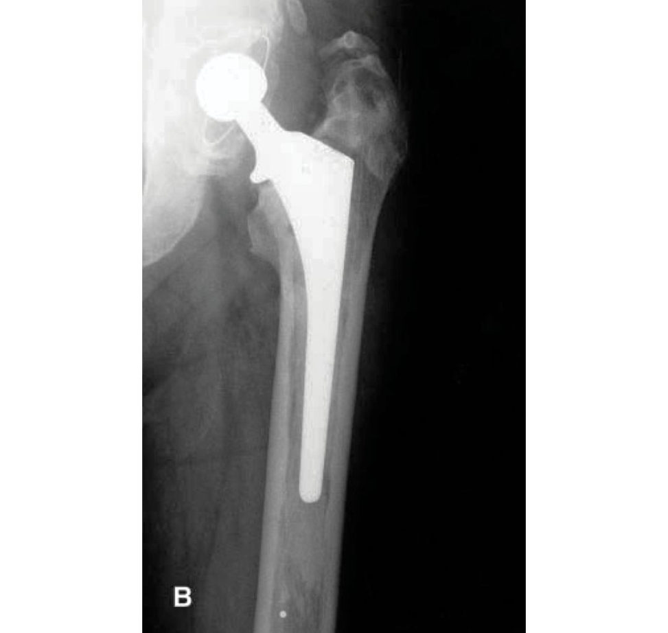
The AP radiograph demonstrates a cemented THA. I am unfamiliar with the implant, but both the cup and femoral stem have been cemented and most likely a 28-mm head size has been used. The cup does not appear excessively worn or loose. There are no significant lucencies in any of the DeLee and Charnley acetabular zones. However, there is a continuous radiolucency at the femoral cement–bone interface in all seven Gruen zones suggestive of gross femoral stem loosening.
What areG ruen zones?
This is a widely used system in which the femoral component interface is considered in seven zones. These allow the location of cement fractures and of lucent lines either at the cement– bone or the cement–prosthesis interface. It is the progressive changes that are seen in serial radiographs that are important in diagnosing osteolysis and femoral stem loosening.
What mode of cemented femoral stem failure has occurred?
This is Gruen mode 1b failure. Pistoning subsidence of stem and cement within bone.
Briefly, what are the other modes of failure?
There is mode 1, apis toning subsidence of stem within cement mantle; mode 2, medial midstem pivot; mode 3, calcar pivot and bending cantilever fatigue (distal pivot).
OK, what will you see radiographically with each mode of failure?
There is one mode of failure like a car windscreen wiper, but I am not sure which one, I think it may be medial stem pivot mode 2, no sorry I think it is mode 4. Far better for the candidate to say they aren’t sure and leave it at that rather than guessing and geting all mixed up.
What do we mean by the term wear?
Wear is defined as a progressive loss of bearing surface from a material as a result of chemical (corrosive) or mechanical action Types of mechanical wear include adhesive, abrasive and fatigue.
What do you mean by abrasive and adhesive wear?
Abrasive wear occurs when two surfaces with microscopic irregularities or asperities slide past oneanother while in intimate contact. The interaction generates particles mainly from the softer material.
Adhesive wear occurs when two opposing materials bond under contact load. Actual transfer of material from one surface to the other may occur, forming transfer films. When motion resumes between the two surfaces, particles may be broken free from one or both surfaces. These new particles then further contribute to wear from third-body abrasive wear.
The wear of UHMWPE in THA is mainly adhesive and abrasive.
What is freting w ear?
Freting occurs with small cyclic motions of one surface relative to another.
What are the wear sources in joint replacement surgery?
Wear sources include the primary articulation surface, secondary articulation surfaces, cement/prosthesis micromotion, cemen t/bone or prosthesis/bone micromotion and thir d-body wear.
What are the modes of wear in joint replacement surgery?
There are four modes of wear. 1. Mode 1 is the generation of w ear debris that occurs with motion between the two bearing surfaces as intended by the designers. 2. Mode 2 refers to a primary bearing surface rubbing against a secondary surface in a manner not intended by the designers (for example, a femoral head articulating with an acetabular shell following wear-through of the polyethylene). 3. Mode 3 refers to two primary bearing surfaces with interposed third-body particles (such as bone, cement, metal and so on). 4. Mode 4 refers to two non-bearing surfaces rubbing together (such as back-sided wear of an acetabular liner, freting of the Morse taper, stem–cement freting). While several modes of wear often occur simultaneously, mode 1 accounts for the majority of wear in well-functioning hip or knee replacements.
What do we mean by effective joint space?
Schmalzreid et al. coined the term ‘effective joint space’ to refer to all periprosthetic regions to which joint fluid, and hence wear debris, can gain access.50 In the acetabulum, wear debris can reach the interface through unfilled screw holes or via non-ingrown areas of the shell. On the femoral side, use of circumferential porous coating has reduced the incidence of diaphyseal osteolysis by blocking access of wear particles.
What is osteolysis?
Osteolysis is a biological phenomenon that can result in the loosening of the implant principally caused by the UHMWPE wear particles. Me tal or ceramic wear particles that are produced at the articulating surfaces of a hip prosthesis are also implicated but to a much lesser degree.
Osteolysis is influenced by the size and morphology of the UHMWPE particles Macrophages actively phagocytose (engulf) wear debris at the bone–implant interface. These cells release various enzymes and osteolytic mediators such as interleukin, tumour necrosis factor (TNF-α) and prostaglandin.
These cytokines cause inflammation and trigger bone dissolution or resorption around the implanted region.
What factors influence osteolysis (wear)?
Osteolysis (wear) is a multifactorial process dependent on surgical factors, implant design, patient factors and material composition. Implant-specific factors that affect wear performance of THA (and TKA) include (see Table 3.9):51,52 Surgical factors (e.g. component position, soft -tissue balancing) that affect joint loads and kinematics influence wear performance of THA (and TKA) (see Table 3.10). Patien t-specific factors that affect wear performance of THA (and TKA) include (see Table 3.11).
What do you know about osteoblastic regulators?
Three osteoblastic regulators (RANK, RANK Land OP Gare involved in bone resorption. This is linked to TNF-α, a cytokine responsible for encouraging osteolysis through the facilitation and augmentation of osteoclast differentiation and activation of pr e-existing osteoclasts.
Figure 3.6 Anteroposterior (AP) radiograph of a loose cemented left THA .
Table 3.9 Implant-specific factors affecting joint wear.
Implant design choices
Modularity versus monoblock
UHMWPE component thickness
Bearing couple conformity
Fixation (cemented versus ingrowth)
Implant constraint
Implant impingement
Material
Metallic alloy (Co–Cr–Mo alloy versus titanium alloy)
Ceramic (alumina, zirconia, oxidized zirconium alloy)
UHMWPE (highly cross-linked versus conventional)
Bearing couple
Metal-on-UHMWPE
Ceramic-on-UHMWPE
Metal-on-metal
Ceramic-on-ceramic
Quality control
Lot-to-lot variability
Shelf-life and packaging of UHMWPE components
Sterilization process (radiation versus ethylene oxide)
Table 3.10 Surgical factors affecting joint wear.
- Surgical approach
- Component position
- Restoration of appropriate mechanical and rotational axes
- Initial stability and method of component fixation
- Soft -tissue balance laxity versus overconstraint)
- Subluxation or dislocation
- Third-body wear
- Surgeon experience
Table 3.11 Patien t-specific factors affecting joint wear.
- Activity le vel (activities of daily living , pivot-shift activities). Patients with active lifestyles often return to recreational activities that markedly increase joint-loading conditions (eg. running, jumping, pivoting , stair climbing)
- Body mass index and bodyweight. Increased bodyweight can be associated with increased magnitude of force and altered kinematics, although the detrimental effects of excessive weight can be counterbalanced by decreased activity levels and loading cycles that accompany a sedentary lifestyle
- Gait mechanics (level and stairs)
- Limb alignment
- Implant time in situ
- Preoperative diagnosis post-traumatic arthritis and AVN have been associated with higher prosthesis failure rates as usually arthroplasty is performed in younger, more active patients
- Comorbidities. A CL and meniscal injuries predispose to osteoarthritis in a young age group
- Special cultural demands (e.g. kneeling in Middle Eastern and Asian populations). Deep flexion for kneeling, load implants beyond current design characteristics (TKA)
- Revision versus primary surgery
Gold medal candidates#
Periprosthetic osteolysis is the loss of bone surrounding an artificial implant. The formation of a periprosthetic interfacial membrane between the bone and the implant is implicated in bone resorption.
The interfacial membrane is composed primarily of two cell types, the macrophage and the fibroblast.
Aseptic osteolysis is thought to occur through a mechanism involving expression of bone resorptive cytokines such as interleukin-1β (IL-1β), interleukin-6 (IL-6), tumour necrosis factor-α (TNF-α), platelet-derived growth factor (PDGF), and receptor activator of nuclear factor-κ B ligand (RANKL).
RANK Lisa potent bone resorptive cytokine present on the membranes of bone marrow stromal cells, osteoblasts in bone, as well as onT cells, and as a soluble molecule secreted into the bone microenvironment by these cells. Receptor activator of nuclear factor-κ B (RANK), a RANKL receptor, is expressed on the cell surface of preosteoclasts.
Macrophages express RANK and, when exposed to RANK Lin the presence of macrophage colony-stimulating factor (M-CSF), have been shown to differentiate into mature osteoclasts capable of bone resorption. Osteoprotegerin (OPG) acts as a decoy receptor for RANKL by binding to RANK Land preventing the functional interaction of RANKL with RANK, thereby blocking the osteoclast formation and the bone resorptive effects of RANKL. Osteoclast activation is thus blocked.
What factors affect PE cup wear in THA?
Implant factors associated with an increased wear rate include non-cross-linked PE, longer shelf-life for liners γ-irradiated in air, thickness of PE. Patient factors include younger age due to higher activity levels, obesity due to increased joint loading. Surgeon factors include position of the cup relative toKo hler’s line, increase in cup abduction angle.
What is the current thinking about UHMWPE?
Three approaches are currently being investigate din an attempt to modify highly cross- linked UHMWPE so that the increased wear resistance provided by cross-linking can be maintained without the reduced fracture resistance that accompanies cross-linking:53 1. Stabilization of free radicals through the impregnation of irradiated ultra-high molecular weight polyethylene with vitaminE. VitaminE protects polyethylene against oxidation, which renders the melting step that normally follows cross-linking with radiation unnecessary VitaminE also quenches free radicals. 2. A second approach involves sequentially irradiating and annealing polyethylene. Irradiation is conducted in three steps with interspersed annealing processes that together improve oxidative stability compared with that resulting from a single large dose of irradiation followed by annealing.
3. The third approach involves the photo-induced graft polymerization of 2-me thacryloyloxyethyl phosphorylcholine (MP Conto cross-linked polyethylene (CLPE). The concept is to create a hydrophilic layer with better wettability than a conventional polyethylene surface, thus increasing the chance for lubrication.
Structured oral examination question 7#
This is a radiograph of a 68-year-old woman who has been referred up to the orthopaedic clinic by the physiotherapist-led musculoskeletal clinic with an 18-month history of left hip pain and difficulty walking (Figure 3.7).
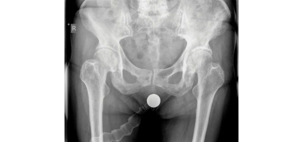
This is an anteroposterior (AP) radiograph of the pelvis demonstrating a coarsened trabecular pattern of the left hip, a thickened left cortex compared to the opposite hip, and increased density of the left hip compared to the right side. Both iliopectineal (Brim sign) and ilioischa tic lines are thickened. There is sclerosis involving the left pelvis (ileum, ischium and pubic r ami), left femora and lower lumbar spine. The radiograph is highly suspicious of Paget’s disease. Differential diagnosis would include other causes of increased and disorganized bone turnover such as sclerotic bony metastasis (prostatic carcinoma), renal osteodystrophy, fibrous dysplasia, multiple myeloma, lymphoma, osteopetrosis and hyperparathyroidism.
What is Paget’s disease?
Paget’s disease is a metabolic bone disorder of unknown aetiology characterized by a disorganized increase in osteoclastic bone resorption and compensatory osteoblastic new bone formation. The reis accelerated but chaotic bone remodelling in which the bone is biomechanically weak and prone to deformity and fracture. The disease can be divided into three major phases, lytic, mixed lytic/ sclerotic and sclerotic, each of which is associated with distinctive clinical, radiological and pathological features.
What causes Paget’s disease? What is the pathophysiology of Paget’s disease?
The primary abnormality of Paget’s disease is an intense focal resorption of normal bone by abnormal osteoclasts. These osteoclasts are abnormal in size, activity and quantity . The abnormal osteoclasts make large resorption cavities in the bone matrix. In response to the osteoclast resorption osteoblasts are recruited, resulting in bone formation. The osteoblast activity is rapid such that the newly formed bone is not organized and remains irregular and woven in nature, less- resistant and more elastic than typical lamellar bone; prone to deformity and fracture.
What are the other radiographic features of Paget’s disease?
Radiographic features of Paget’s include: Advanced disease in the long bones is characterized by coarsened trabecula, bony sclerosis, bony enlargement, and deformity. A ‘candle flame’ or ‘blade of grass’ sign represents a wedge- or V- shaped pattern of advancing lysis in the diaphysis of long bones. The femur develops a lateral curvature while the tibia develops an anterior curvature that may result in fracture. Fine cracks may appear (stress fractures) which resemble Looser zones but occur on the convex bone surface.
Lateral radiographs of the lumbar spine demonstrate a ‘picture-frame’ vertebral body that is secondary to severe osteoporosis centrally and a thickened, sclerotic cortex.
The skull is involved in 29–65% of cases. Inner and outer table involvement leads to diploic widening. Osteoporosis circumscript ais a well-defined lysis, most commonly involving the frontal bone producing well-defined geographic lytic lesions in the sk ull. It is seen in the early or lytic phase when osteoclastic resorption overwhelms bone production. A t a later stage a ‘coft on wool appearance’ represents mixed lytic and blas tic pattern of thickened calvarium.
Protrusio deformity of the pelvis is a common occurrence with advanced Paget’s disease.
What are the current theories regarding the aetiology of P aget’s disease?
The aetiology of P aget’s disease is still unknown. Proposed theories include viral, genetic and environmental causes. Paramyxoviruses such as measles virus, respiratory syncytial virus and canine distemper virus have been implicated. Electron microscopy has shown virus-like structures that resemble the paramyxovirus in osteoclast nuclei and cytoplasm of cells affected byP aget’s disease. However, more recent studies have been unable to confirm the presence of specific viral antibodies in patients with Paget’s disease. Environmental factors implicated include high levels of arsenic and an uncertain association with cats and dogs. Genetically, 5–40% of patients have first- degree relatives with the disease.
That’s fine. I am, however, a bit sceptical about the cats and dogs theory. Moving on – what are the complications of P aget’s disease?
Complications of P aget’s disease include: Compression fractures of the vertebral body (commonest complication of spinal P aget’s). Pagetic spinal stenosis, defined as compression of the spinal cord, caudae quina or spinal nerves by expanded Pagetic bony tissue of the spine. Most common in the lumbar region and typically single level, causing cord or nerveroot compression. An enlarged and deformed skull can lead to increased intra-cranial pressure, hydrocephalus or cranial nerve deficits such as facial palsy (narrowing of neural foramina), hearing loss or blindness (pressure on optic nerve). High cardiac output secondary to increased bone vascularity (rare). Insufficiency fractures. Osteosarcoma, chondrosarcoma, malignant fibrous histiocy toma and giant cell tumours all have been reported with Paget’s disease.
What are the indications for THA in Paget’s disease?
The indications are similar to non-Pagetoid disease. It is important to make sure that the pain is arising from the joint surface and not the bone. Bone pain with active Page tis suggested by an increased alkaline phosphatase value. It is also important to exclude insufficiency fractures, neurological compression in the spine or Paget’s sarcoma as a cause of pain.
How do you assess disease activity?
Patients with active Paget’s disease have raised alkaline phosphatase (AlkPhos) and urine hydroxyproline values. The higher the level the more active the disease is. Patients with very high AlkPhos levels are thought to be at higher risk of bleeding and heterotrophic ossification formation.
If the Paget’s disease is active what will you do?
I would refer him to one of my rheumatoid colleagues for a Pamidronate (Aredia) injection. This is a bisphosphonate, which is a potent inhibitor of osteoclastic activity and hence bone resorption. This reduces bone vascularity and bleeding and possibly the incidence of heterotopic ossification. The other option is the use of bis phosphonates or calcitonin to reduce bone-related pain, reduce postoperative bone resorption and decrease bleeding should surgery be required.
What are the technical issues of performing THA in Paget’s disease?
There is a tendency for excessive bleeding at surgery due to increased vascularity. Blood should ideally be cross-matched or at the least available from a group and saved within 10 minutes. Bone can be very hard and sclerotic making it difficult to ream and broach. Burrs may be needed to enter the bone prior to reaming and/or broaching. Varus deformity of the proximal end of the femur predisposes to varus placement of the femoral component. Protrusio, as we have mentioned, is a common finding and I would consider using bone graft medially to compensate. Some surgeons use lateral offset liners and antipr otrusio cages, although this complicates surgery. AsP aget’s bone is britile the reis a higher risk of both intraoperative and postoperative fracture. There is some controversy as to whether there is an increased risk of heterotopic ossification occurring from the abnormalities of osteogenic differentiation in P aget patients Some surgeons routinely give prophylaxis to reduce the risk of HO [Score 6].
There is a bit more than that when planning THA.
As bone pain is common in Paget’s disease and does not necessarily improve with THA a diagnostic local anaesthetic injection to rule out concurrent bone pathology may be indicated. It is also important to exclude referred pain from spinal stenosis or radiculopathy and other causes of musculoskeletal pain. Good-quality, full-length radiographs to assess the degree of deformity and the extent of bone involvement. Radiographs should be scrutinized for the presence of a stress fracture that could account for hip pain. The fractures may be in the region of the femoral neck, intertrochanteric area or femoral shaft. The y usually present as incomplete or fissure fractures on the tension side of the bone. Unrelenting hip pain and radiographic bone destruction suggests sarcomatous change.
Consider using cell salvage, hypotensive anaesthesia and predonation of autologous blood if intraoperative blood loss is anticipated to be high with active disease. Concurrent osteotomy may be needed if component alignment is difficult.
Marked protrusio can make hip dislocation very difficult.
You mentioned osteotomy, how often do you perform osteotomy when you perform THA for Paget’s disease?
In the majority of patients with Paget’s THA can be performed without need for osteotomy. However, if deformity is severe, precluding implantation with a standard stem, then planning for reduction osteotomy to correct the deformity and/or the use of modular stems must be done preoperatively.
What type of hip replacement would you use?
Although there has been a trend in recent years to use uncemented components in Paget’s disease, in this patient I would use a cemented THA. She is 68 and hasP aget’s disease and I think it is a reasonable option in this situation. If the patient is younger, then the choice becomes more controversial. Although previous studies have recommended the use of cementin the last 20 years there has been a trend to use uncemented components. The worry that the altered morphology of pagetoid bone adversely influences ingrowth into cementless implants has not been borne out in practice. In addition, previous concerns for the problem with osseointegration are mostly unfounded. The biology of bone ingrowth for initial fixation of uncemented components depends, in parton the ability of bone to proceed through the early phase of fracture healing. Patients with Paget’s disease are not known to have compromised ability for fracture healing and these patients progress through the biological process of fracture healing at normal speed. Parvizi et al. reported on 21 cementless THA implanted against pagetoid bone; all were stable and demonstrated radiographic evidence of ingrowth at 7-year follow-up.54 Lusty et al. from Sydney, Australia reported medium-term results of 23 uncemented THA at 6.7 years follow up.55 There were three revisions, one stem for aseptic loosening and two stems after periprosthetic fracture. Some surgeons prefer cementless components especially when bone is very sclerotic, or a concurrent osteotomy is done. Extremely sclerotic bleeding bone will make interdigitation of cement difficult and cement extravasation in to the fracture gaps may occur after osteotomy. If using a cementless cup the use of adjuvant acetabular screws is recommended.
Any special complications that can occur postoperatively?
There is a reported greater incidence of heterotopic ossification.
Anything else?
Dislocation.
No, I am not aware of an increased risk of dislocation.
Several studies have documented osteolysis following THA inpatients with Paget’s disease.56 This is thought to be related to the increased metabolic turnover of the pathologic bone. Other authors have reported that osteolysis is not a problem following THA in Paget’s disease.57 Other complications include periprosthetic fracture around total hip implants; and the continuation of bone pain following arthroplasty, microfractures, and malignant transformation to osteosarcoma.
Figure 3.7 Anteroposterior (AP) radiograph demonstrating P aget’s disease of the left hemipelvis.
Gold medal candidates
What causes have been identified for the increased number and activity of Page tic osteoclasts?
Causes identified include: 1. Osteoclastic precursors are hypersensitive to calcitriol (1,25(OH) 2D3). 2. Osteoclasts are hyper-responsive to RANK ligand (RANKL), the osteoclast stimulatory factor that mediates the effects of most osteotropic factors on osteoclast formation. 3. Marrow stromal cells from Pagetic lesions have increased RANKL expression. 4. Osteoclast precursor recruitment is increased by interleukin (IL)-6, which is increased in the blood of patients with active Paget’s disease and is over-expressed in pagetic osteoclasts. 5. The antiapop totic onc ogene Bcl-2 in Pagetic bone iso ver-expressed. 6. Expression of the proto-oncogene c-fos, which increases osteoclastic activity , is increased. 7. Numerous osteoblasts are recruited to active resorption sites and produce large amounts of new bone matrix. As a result, bone turnover is high and bone mass is normal or increased, not reduced.
Structured oral examination question 8#
DD His one of the most common hip viva questions that regularly gets asked in the oral viva examination.
We think this is because it is a fairly common hip condition with a lot to talk about. The story can go in many different directions.
These are the anteroposterior (AP) radiographs of a 66-year-old woman with bilateral hip pain (Figure 3.8). Would you like topass comment on them? CANDIDATE 1 : The AP radiograph demonstrates a severely dysplastic hip on the right side with secondary OA changes. On the left side again, there is dysplasia but to a lesser degree with again secondary OA changes present. [Score 5] CANDIDATE 2 : This is an AP radiograph of the hips and pelvis of a 66-year-old woman taken on the 16/5/11, which demonstrates severe bilateral dysplasia.58 There is a high dislocation on the right side, Crowe IV or Hartofilakidis III hip. There is no contact between the true and false acetabulum. The femoral head appears poorly developed, probably absent, with the femoral neck articulating against the iliac crest. The view of the proximal portion of the femoral canal on the right side suggests a very narrow medullary canal. On the left sid ether e is a Crowe III hip or Hartofilakidis II hip. There is a low dislocation and secondary osteoarthritis. 59 [Score 6–7] COMMENT60: The left side is a Hart ofilakidis I hip as the femoral head is still contained within the original acetabulum. With a low dislocation the femoral head is in contact, at least in part, with the true acetabulum and in this situation, this is the most severe deformity. In high dislocation, the femoral head and acetabulum make no contact and the head has migrated superiorly and posteriorly. Often in this situation the true acetabulum is reasonably well-preserved, although underdeveloped and osteoporotic. What do you mean by dysplasia?
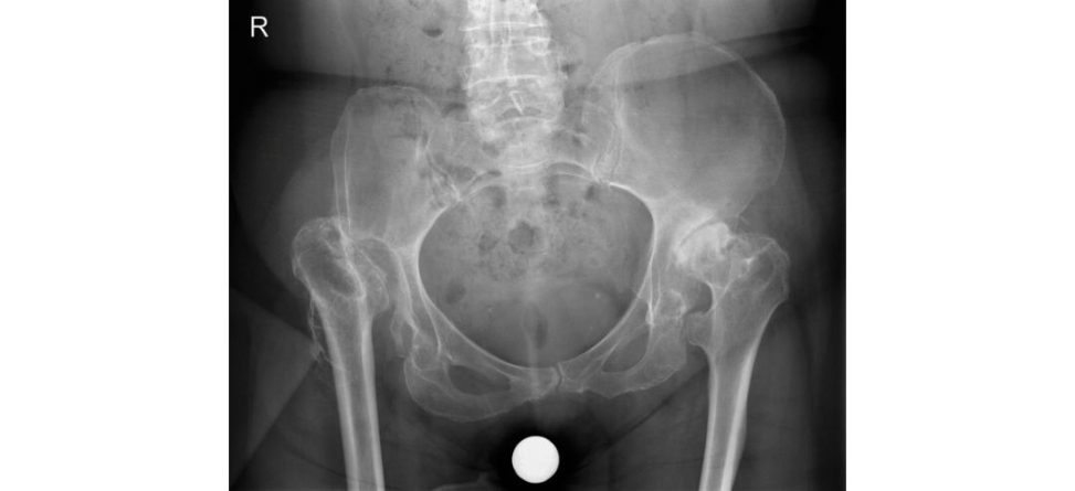
Dysplasia is lack of coverage of the femoral head, whether it is subluxed or dislocated.
How do you classify dysplasia?
Crowe classified dysplasia radiographically into four categories based on the proximal migration of the femoral head. The migration is calculated on an AP radiograph by measuring the vertical distance between the inter-teardrop line and the junction between the femoral head and medial edge of the neck. Cro weI isless than 50% subluxation, Cr owe II hips have between 50% and 75% subluxation.
[Interrupting] That’s fine. That’s OK. Any other classification systems that you know?
[Sharp intake of breath, shaking of head and then silence.] No.
Have you heard of the Hartofilakidis classification?
I have, but I can’t remember the specifics.
The Hartofilakidis classification system, which divides DD Hin adults into three types: dysplasia, low dislocation and high dislocation. Many surgeons prefer this system, as it is more practical and simpler to use. What are the anatomical issues associated with DDH?
The anatomical differences are divided into acetabular, femur and soft -tissue issues. The acetabulum is shallow and anteverted; the femur has a small deformed head and short anteverted valgus neck.
That’s not all the differences. There are some you have missed. Do you know any more?
Muscles around the hip are usually shortened ander, er ...
The greater trochanter is small and posteriorly displaced, the femoral canal narrow, the acetabulum is usually small with poor bone quality, hip capsule elongated and redundant, psoas tendon hypertrophied, and abductors orientated transversely as a result of the superior migration of the femoral head. The femoral and sciatic nerves may be shortened and therefore more vulnerable to injury during arthroplasty surgery. What is the role of a CT scan in planning an operation for DDH?61 CANDIDATE 1: CT scans can be used to determine the available acetabular coverage and to estimate the degree of femoral anteversion. CANDIDATE 2: CT scans are useful in assessing available bone stock, and the morphology, dimensions and orientation of both the acetabulum and femur. Any leg length discrepancy can be precisely evaluated and allow for design of custom femoral implants. Various measurements include: femoral neck shaft angle anteversion of the femoral neck, medial head offset, position of the isthmus and height. The AP size of the acetabulum as measured by CT is often different from the supero-inferior size evaluated on plain radiographs. Proximal femoral anteversion is calculated by measuring the angle between the posterior bicondylar axis and the mediolateral dimensions of the medullary canal 20 mm above the lesser trochanter. These measurements are useful to know but how are they actually going to help you to plan surgery?
In the acetabulum following the abnormal anatomy too closely might lead to anterior instability if the cup is overanteverted. It is important to recognize that a substantial amount of acetabular anteversion and deformity can be present with a relatively normal-looking AP pelvic radiograph.
In addition femoral anteversion may be difficult to recognize. Even in normal-looking AP radiographs a significant amount of anteversion may be present. Attempting to implant an uncemented stem in a deformed anteverted femur may result in a proximal femoral fracture.
What are the technical difficulties in performing a THA in a DDH patient?
Crowe type II and III hips have a marked superolateral rim deficiency and anterior wall defect. Bulk autogratiing of the superolateral acetabulum with bone from the femoral head can be used to increase the cover and stability of the acetabular component. The graft and its bed need careful preparations table fixation and precise positioning. Gr aft resorption can occur, leading to cup migration and loosening. Although it is technically difficult for anatomic placement of the acetabular component the forces on the THA are significantly reduced. Linde et al. found a 42% rate of loosening of cemented Charnley components after a mean of 9 years if the component was positioned outside the true acetabulum compared to 13% if placed inside.62,63
Any other options to deal with deficient superior coverage of the cup?
A small, uncemented cup can be placed in a high hip centre location. In this position the cup is completely covered with host bone and avoids the need for gratiing. Disadvantages include decreased polyethylene thickness associated with a small acetabular component, difficulties with correction of leg length inequality and altered hip biomechanics. Hip instability is increased due to the use of a small femoral head component along with the risk of femoral–pelvic impingement either inflexion or extension.
What do we mean by cotyloplasty?
Cotyloplasty involves a deliberate fracture of the medial wall of the acetabulum in order to place the acetabular component within the available iliac bone. The acetabulum is advanced medially by the creation of a controlled comminuted fracture of the medial acetabular wall. Mixed results have been reported, but there is a worry that future revisions may be difficult because issues with restoration of bones tock have not been addressed.64
How do you preoperatively plan for DDH surgery?
On the acetabulum side the position of the true acetabulum should be identified, and a decision made whether to restore the acetabulum to its true position or not. The degree of anteversion of the acetabulum should be defined as well as the adequacy of bone stock for satisfactory cup fixation and coverage. Preoperative planning would also include an estimation of the acetabular component size, the preferred method of fixation (cemen t/uncemented) and need for bone graft.
On the femoral side the size of the femoral canal and the need for special or custom implants should be assessed.
The need for femoral shortening should be made preoperatively. The method and amount of femoral shortening need to be worked out beforehand.
Preoperative planning should also include the surgical approach to be used, solutions to deal with the hypoplastic acetabulum and femur, management of LL Dand restoration of abductor function.
What is the effect of anteversion of the femoral stem on THA?65
When there is more than 40° of anteversion, a corrective rotational osteotomy or a modular implant in which the version of the femoral neck can be varied may be necessary.
That’s not really the question I asked.
A large amount of femoral anteversion increases the risk of dislocation.
That’s correct, but not the whole story. You have already partly answered the question earlier on.
I am sorry, I don’t understand.66 Attempting to implant an uncemented stem in a deformed femur may result in a proximal femoral fracture. In this situation you may want to use either a cemented or modular stem that allows control of anteversion. Also, excessive anteversion of the femoral component can lead to internal rotational contracture of the hip.
How do you correct length inequality in DDH?
With Crowe type III and IV hips, if the cup is placed in the anatomic position femoral shortening is required. Without femoral shortening it is very difficult to reduce the prosthetic head into the acetabular component because of soft -tissue contractures. If one attempts to fully correct a significant leg length discrepancy a sciatic nerve palsy may occur. If permanent this can be a disabling complication from surgery and which patients are less willing to accept these days. The exact amount of lengthening that results in sciatic nerve palsy is not known. Acute limb lengthening of more than 2–4 cm during arthroplasty is associated with an increased risk of neural injury. Therefore, as a general rule the hip should be lengthened the minimum amount required to re-establish reasonable function and hip stability. Any lengthening more than 4 cm becomes very risky for a sciatic nerve injury and is generally not advised. Shortening is performed either by sequential resecting of the proximal femur or by performing a shortening subtrochanteric osteotomy. Sequential proximal resection results in a small, straight femoral tube with a small metaphyseal flare which is usually unsuitable for an uncemented femoral implant. Typically, a small cemented
DDH stem needs to be used with a straight proximal medial geometry and without a metaphyseal flare.
Advantages of a subtrochanteric shortening osteotomy include preservation of the metaphyseal femoral region (which provides most of the rotational stability of the implant) and allowing concomitant correction of angular and anteversion deformities. It is technically difficult and there is a risk of non-union.
How do you reduce the risk of non-union?
Different subtrochanteric osteotomy geometries can be used. These include transverse, oblique, stepcut and double Chevron osteotomies. A transverse osteotomy is simplest and the resected bone can be used as an onlay graft. Avoiding the use of a cemented stem prevents the risk of the cement interfering with healing of the osteotomy site. A press fit achieves distal fixation of the prosthesis. Strut allograft and cir clage cables may also be needed for support.
What are the principles of revision hip surgery with DDH?
Two major concerns are deficient acetabular bone stock and the position of the acetabular cup, particularly if the centre of the hip has not been restored during the primary procedure. Several surgeons have advocated the use of a high hip centre in order to take advantage of the remaining bone stock and to avoid the use of a structured graft. However, a high hip centre does not correct leg length discrepancy, does not provide good bone stock for revision hip surgery and is associated with early acetabular loosening and a higher rate of dislocation because of ischial impingement. The pattern of bone loss associated with DD His a reduced AP diameter combined with poor superior support. This loss is further increased by surgical bone loss at the time of the index operation migration of the cup and osteolysis. Bone graft would need to be ordered along with special equipment such as universal screwdrivers, screw extractors, high-speed burrs and metal cuft ers.
Figure 3.8 Anteroposterior (AP) radiograph of the pelvis of severe bilateral DDH.
Structured oral examination question 9#
Periprosthetic fracture (PPF) around the hip
This is a 70-year-old man who was admift ed to the orthopaedic ward last night after a fall. He is generally fit and healthy although is on Warfarin for a mitral value replacement. These are his radiographs that were presented at the trauma meeting in the morning ( Figure 3.9a). What do you see?
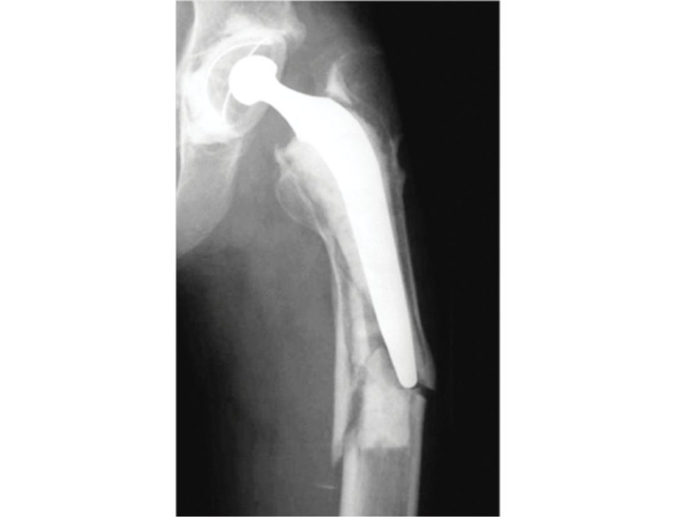
This is an AP radiograph showing a left Vancouver B2 periprosthetic fracture around a Charnley cemented total hip replacement.
What makes you state that this is a Vancouver B2 periprosthetic fracture?
A B2 fracture is around the distal stem or tip with significant communition and a looses tem with adequate bone stock.
Any difficulties with the Vancouver classification ( Figure 3.9b)?
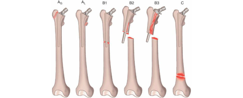
There are some difficulties differentiating between type B1 and type B2 on plain radiographs. What you think is a well-fixed stem may in fact be loose. If you haven’t thought through a plan B to deal with the possibility of a loose stem, then this will significantly increase the complexities and difficulties of managing the case intra operatively.
Are you aware of any more recent classification system?
The Vancouver group have recently published their Unified Classification System (UCS) for periprosthetic fractures around a hip or knee arthroplasty in 2014.67 Fractures are categorized A to F. Type A fractures involve the apophysis, e.g. greater trochanter fracture around a THA. Type B fractures involve the ‘bed’ of the implant, e.g. femoral shaft fracture around a stem, and are still graded B1, B2 and B3 as per the original Vancouver classification system. Type C fractures are ‘clear’ from the implant, e.g. distal to the stem but within the same bone. TypeD is a PF in a bone between two joint replacements, e.g. femoral shaft fracture between a hip and knee replacement TypeE is a fracture in which two bones support one joint replacement, e.g. an acetabular and femoral fracture around a THA. Type F fractures involve a joint surface that has not been replaced or resurfaced, e.g. acetabular fracture around a hip hemiarthroplasty.
What advantages does this classification system have over the Vancouver classification system?
The Vancouver classification system was based on the key principles of management of periprosthetic fractures. It has demonstrated good inter- and intra-observer reliability and has become widely accepted.
However, there was felt to be a need to expand the Vancouver classification system to include three other types of fracture that may occur in combination or in isolation, and to deal with the pelvis as a whole (not just the acetabulum).
This is actually a B3 fracture as there is poor bone stock around the stem, which is loose. This patient is a 70-year-old male who had a left THA 20 years ago and had a fall sustaining this PPF. He has had a previous mitral valve replacement and is on warfarin. He is a diet-controlled type 2 diabetic. He is otherwise quite fit, well, is fully active and independent and still drives a car. What will be your management plan?
This is a difficult and complex case. The review article by Schwarzkopf provides a good treatment algorithm for dealing with periprosthetic fr actures68 (Figure 3.9c). Firstly, I would like to get details of the original operation and implants if at all possible, although in this case, it may prove impossible given the length of time since the original hip replacement. I would also seek advice from other senior colleagues who were experienced hip surgeons or discuss this case in a regional revision hip network. I would need to consider the following: whether to just deal with the femoral stem or also deal with the acetabular component. I would additionally need to take into account the patient ability to cope physiologically with a complex and prolonged procedure. This patient is still very active and healthy with few comorbidities, and this will influence my surgical plan. His Warfarin will need to be stopped but he must be covered with an alternative anticoagulant because of his MVR. I would use a posterior approach as this is an extensile approach and provides excellent acetabular and femoral exposure. I can also extend the incision distally if I needed to, along the length of the femur. Assuming the patient is fit enough, I would plan to also revise the socket as the radiographs show acetabular component loosening. This is a B3 fracture, which necessitates revision of the femoral stem, and I would bypass the fracture using a long-stem, titanium, modular fluted revision hip such as the ZMR or ARCOS system (which I am familiar with) (Figure 3.9d) in order to get distal fixation and provide rotational and axial stability. I would ensure that I had templated preoperatively the different stem options a vailable. Using such a stem helps deal with the issues of poor proximal bone quality, bone loss and stress shielding. I would also use cerclage cables or wires distally and around the diaphysis of the femur to help maintain control of the femoral tube and prevent fracture propagation distally. The modular stem allows for greater flexibility in controlling for leg length, offset and version. I would also consider doing an ORIF for the fracture with or without an allograft to help maintain the integrity and stability of the femoral tube. For the acetabular component, I would use an uncemented socket (such as a trabecular metal (TM) backed socket) with supplementary screws, but have augments available in case there are significant acetabular bone defects present (Figure 3.9e). A preoperative CT scan of the hip would be useful in helping identify significant acetabular bone defects, although from what I can determine on the radiograph shown to me, there doesn’t appear to be significant bone loss or any protrusio. My bearing surfaces of choice would be a ceramic head (36 mm ideally) with a highly crosslinked lipped polyethylene liner to help minimize the risk of a posterior dislocation.
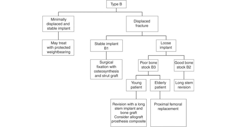
Why not just fix the fracture with a long femoral plate and leave the stem in situ?
The evidence suggests that reoperation rates are higher when B3 fractures only have an ORIF performed and are not revised. The systematic review in the BJJ 2017 by Khan et al.69 from Notingham showed almost 30% reoperation rate compared to around 15% for revision ± ORIF.
Are there any other surgical options a vailable?
A distally locked, uncemented, hydroxyapatit e-coated long femoral stem prosthesis such as the Cannulok hip could be used. The paper by El-Bakoury in 201770 looking at B2 and B3 fractures in 28 patients aged 75 and over showed a 95% fracture union rate and 100% survivorship of the revised hip at a mean 4 years follow-up. The Exeter group have reported on their experience using impaction bone gratiing , supported by a plate to contain the graft, with a long cemented femoral stem for situations with extensive femoral bone loss including B2 and 3 periprosthetic fr actures.71
Are there any other options a vailable that you might need to consider if the patient was very frail and the bone quality very poor?
Another potential option would be to do a proximal femoral replacement (PFR). The study from Parvizi’s group72 looking at 21 patients with a B3 PPF showed a good outcome using a PFR. They advised using a constrained liner in such patients if the hip was unstable intraoperative lyas is often the case because the abductor mechanism is often disrupted in such patients and the risk of dislocation is high). The y also advised trying to maintain as much of the proximal femoral bone as possible even if the bone quality is very poor to help maintain bone stock andre approximate the proximal bone onto the PFR.
You perform the operation revising the femoral and acetabular components using an uncemented acetabular socket, a long uncemented femoral fluted titanium stem and a ceramic-on- polyethylene bearing couple. All went well during the operation. Postoperatively, on Day 1, he appeared quite comfortable and had no neurovascular complications. On Day 2, you see him and he has developed a foot drop on the left (operated side. What will you do?
This patient has developed a foot drop secondary to some form of sciatic nerve injury. He was well immediately postoperatively, and this implies that the sciatic nerve must have been intact and working then. It is therefore unlikely that the nerve was injured iatrogenically intra operatively. I would firstly establish exactly when the patient started to get neurological symptoms and signs. I would look at the incision site. Is there excessive bruising or swelling? Are the distal pulses intact? I would review the operation notes and anaesthetic notes. What type of anaesthesia was used (spinal/epidural, etc.) although, again, there was no foot drop initially postoperatively.
The patient has noticed that the thigh has swollen significantly and there is quite a lot of bruising and the soft tissues f eel tense.
I note that the patient was on warfarin. Has this been restarted? I would get an urgent IN Rand cloting screen. My concern is that the patient has an evolving large haematoma, and this could be causing a sciatic nerve neuropraxia leading to a foot drop 48 hours after surgery. The difficulty is balancing the need to protect his mitral valve replacement and minimize the risk of get inga haematoma that is now causing a foot drop. I would also seek urgent advice from the cardiologists and haematologists as to whether we can stop the warfarin and put the patient on an alternative anticoagulant such as IV heparin or LMWH. I would prepare to take this patient to theatre and evacuate the haematoma urgently. An MRI might be helpful preoperatively to identify the location of any significant haematoma. An urgent CT angiogram may also help identify any significant bleeding vessels which may be amenable to embolisation.
Figure 3.9a Vancouver B3 periprosthetic fracture left THA .
Figure 3.9b Vancouver classification of PP Far ound the hip.
Figure 3.9c Treatment algorithm B3 PF.
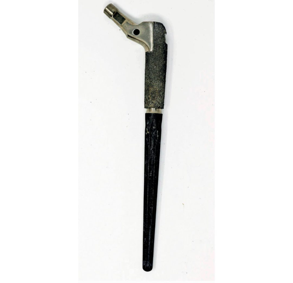
Figure 3.9d ARCOS uncemented revision femoral stem. Grit-blasted, anatomic bow.
Figure 3.9e TM revision shell with augment.
Structured oral examination question 10#
What does the radiograph show (Figure 3.10)?
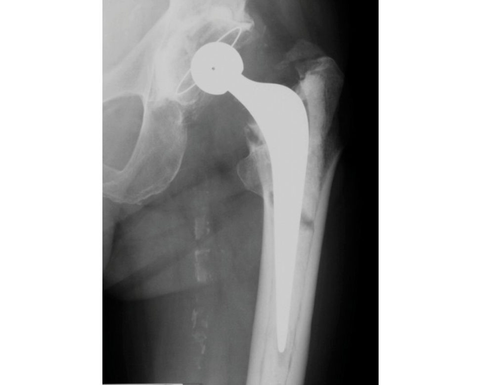
This is an AP radiograph of a cemented Exeter THA. The acetabular cup and stem are well fixed with an adequate cement mantle thickness which is greater than 2 mm.
Anything else?
The stem is in slight varus, but the cup is well aligned with no suggestion of excessive anteversion or retroversion or being too closed or open.
Anything else?
His femoral artery has a large amount of calcification present in its wall.
Anything else?
Not sure.
Look here; what do you think this lucency is?
This is a cement fracture.
What are the reasons for a broken cement fracture?
Cement mantle fractures are worrying as they are associated with early loosening and the need for revision surgery. The early development of stem–cement interface debonding (separation) and subsequent cement fracture are thought to be the initiating events of aseptic loosening. Studies suggest that the initiating events that result in cement failure are due to stresses. Listen to the question the examiner asks you. Trying to waffle through a question isnt going to score you any marks. Studies suggest that the initiating events that result in cement failure are due to stresses experienced at the cement mantle that exceed the fatigue endurance limit of both the stem–cement interface and the cement material itself. It is important to reduce cement stresses so as to minimize the risk of cement debonding and fracture.
So how can high cement stresses be avoided?
By the creation of an optimally thick symmetric, and homogeneous cement mantle.
So how do we achieve this?
Stresses experienced in the cement mantle have been shown to be highest at the stem tip and secondarily at the proximal–medial cement mantle. Stem malalignment produces non-uniform cement mantle thickness in key areas. Defects or voids in the cement mantle reduce bulk cement thickness and have a substantial effect on cement stresses. Variations in implant geometry (e.g. diameter and contour) and material have also been shown to affect stresses experienced in the cement mantle.
Can you be more specific?73
A varus femoral stem is associated with higher incidence of aseptic loosening. This results in thin or non-existent cement mantle in the proximal medial and distal lateral zones. Large voids up to 5 mm in diameter are detrimental. Location of voids is important. Small voids in areas of the cement mantle known to experience high strains may result in premature fixation failure. A proximal–medial cement mantle greater than 10 mm orless than 2 mm in thickness is associated with a significant increase in cement fracture, radiolucent lines at the prosthesis–cement and progressive component loosening when compared to proximal–medial cement mantles that measure 2–5 mm in thickness.74 An asymmetrical distal cement mantle significantly increases the risk of implant failure. Inadequate centralization of the s tem or malrotation will result in excessively thinned areas of distal cement, increased cement strains and prosthesis bone contact. Best results for femoral components allow for 2–5 mm proximal medial thickness of cement mantle, less than 2 mm of proximal medial thickness of cancellous bone, a stem that fills more than half the distal part of the meduallary canal and a stem in neutral orientation. Worst results for femoral components occur with a cement mantle thickness >10 mm or in a femur with more than 2 mm proximal medial cancellous bone, those that filled half orless of the medullary canal and those in varus orientation. In general, too thin a layer of cement will occur if there is lack of removal of proximal medial cancellous bone whilst too much removal of proximal medial cancellous bone results in poor cement interdigitation and fixation with bone, as the bone surface is mostly cortical.
Figure 3.10 Anteroposterior (AP) radiograph cemented Exeter hip with broken cement mantle.
What is the optimal cement mantle thickness?
The femoral stems of hips that have a 2–5 mm thick cement mantle in the proximal medial region have a better outcome than stems implanted with a thicker (>10 mm) or thinner (<2 mm) cement mantle.
What about the cement, how can this be improved?
Improvements in the inherent properties of the cement increased strength, reduced britileness, improved interface adherence) to increase strain resistance and thus retard early debonding and microfractures will aid in the quality and long-term success of the cement.
What measures can betaken intra-operativ ely to improve the quality of the cement mantle?
1. Canal preparation Use of correctly sized broaches that allow a mantle of adequate thickness, pulsatile lavage, and brushing and drying of the prepared canal before and during insertion (clean dry bone).
Packing of the femoral canal with adrenaline soaked swabs, hypotensive analgesia to reduce bleeding, suction catheter and avoidance of blood/cement occlusions.
2. Cement preparation
Centrifugation or vacuum mixing to minimize pore formation and timing of cement injection to achieve optimal viscosity during insertion improves the cement mantle quality.
Occlusion of the canal using a distal plug, retrograde filling of the canal and cement gun pressurization of the cement column with a tight proximal seal are essential in achieving an interdigitating , uniform, and homogeneous cement mantle.
Have you heard of boneloc bone cement?
This is a bone cement that was withdrawn quite soon after introduction because of unacceptable revision rates with its use. Mean fracture toughness and mean tensile strengths were significantly lower than other conventional bone cements.
What is the function of a centraliser?
Femoral stem centralizers were originally designed for double tapered, straight stems. A stem centralizer guides the femoral prosthesis to a neutral position within the cement and guarantees an even cement layer between the bone and prosthesis. Use of a centralizer improves the quality of the distal cement mantle as well as improves stem position central within mantle). Means the stem is “Non-end bearing”, the void below the stem allows a degree of stem subsidence without directly bearing onto the cement thus preventing cement cracking and deterioration. This subsidence also seals off the stem-cement interface to prevent any fluid flow that may lead to loosening (effective joint space). Stem subsidence into an air filled centralizer leads to low shear stresses, high compressive stresses and almost no tensile stress
What is the ideal cement mantle thickness?
I would aim for a cement mantle thickness greater than 2 mm as any less than this increases the risk of cement mantle fracture.
Have you heard of the French paradox?
No, I am sorry I haven’t.
The ideal cement mantle thickness is still uncertain Two philosophies about cement mantle thickness exist. Int heUK and USA, the first technique aims to produce a complete cement mantle of at least 2 mm in thickness and without ‘windows’. It is believed that ‘windows’ may allow debris to reach the interface and that thin cement mantles will be highly stressed and may fracture. The second technique used in France is the use of a thinner cement mantle in which the possibility of windows is accepted. This has been called the French paradox in which implantation of a canal filling femoral component in a line to line manner is associated with a thin cement mantle75. The reason for good results is thought to be the fact that a thin cement mantle in conjunction with a canal filling stem was supported mainly by cortical bone and subjected to low stresses. This is Score 8 material not expected for the average Score 6 pass candidate. The discussion could move on to any number of topics related to cement use in arthroplasty surgery depending on how the viva is progressing. Barracks grading of cement.76,77 Generations of cementing technique. Categories of loosening of cemented stems (Harris). Exeter vs. Charnley stem design.
Structured oral examination question 11#
A 52-year-old man has been referred to the orthpaedic clinic with a 6-week history of (right)78 hip pain. The pain was unrelated to trauma and was a severe, deep aching groin pain worse at night. The patient has a limp with pain on weight bearing and a positive Trendelenburg sign. These are his radiographs (Figure 3.11a). What do you see?
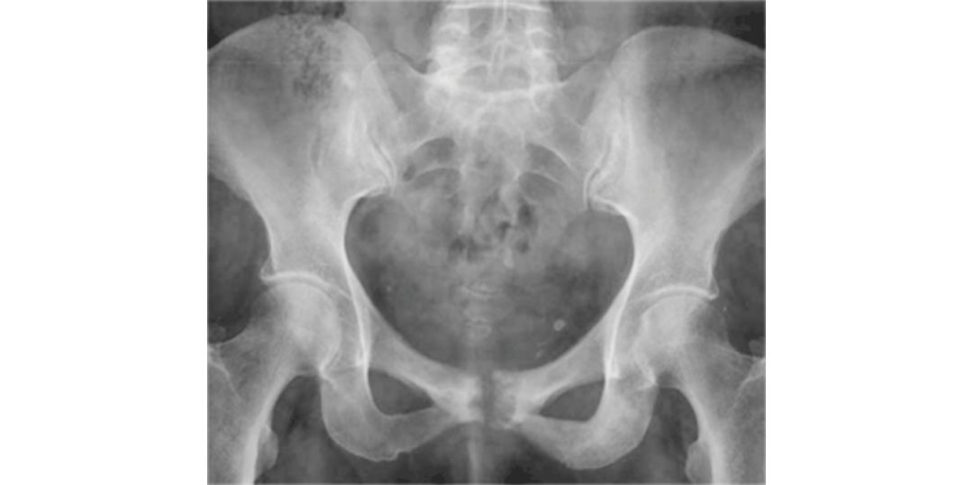
The anteroposterior (AP) pelvis radiograph shows no significant abnormality. At most there is a suggestion that the right femoral head may be more opaque than the left. 79 OR The anteroposterior (AP) pelvis radiograph reveals diffuse osteopenia of the right femoral neck and head. There is no joint space narrowing, scalloping or fracture noted. The left hip appears normal. Do we have a lateral radiograph of the right hip?
That’s all we have got. What further investigations would be appropriate?
I would start by taking a full history and examining the patien t.80
That’s all been done. He has severe hip pain keeping him off work.
I would like to perform bloodtests (FB CUE, bone profile, LFT, cloting , ESR, CRP, PSA and serum electrophoresis). My first choice investigation would be an MR Iof the hips and pelvis. Other investigations may involve a nuclear bone scan which would provide information about other possible skeletal abnormalities. A CT scan is another option that would provide more information about the bone architecture.
This is his bone scan (Figure 3.11b). What does it show?
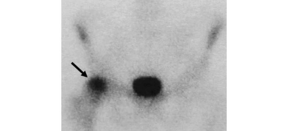
The bone scan shows heterogeneous intense uptake of isotope in the femoral head (mainly anterosuperior) and neck region.
These are his MRI hip images (Figure 3.11c). What do you see?
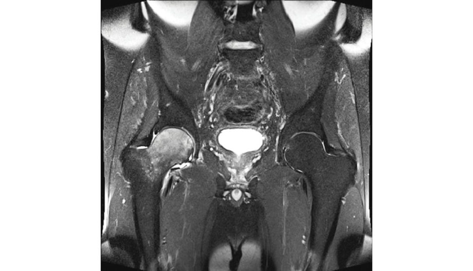
This is a coronal MR image of both hips short-tau inversion recovery (STIR sequence) characterized by a heterogeneous bone marrow oedema pattern in the right femoral head and neck regions. In the superior aspect of the head in the subchondral area, there are areas of low signal surrounded by rims of high signal. MRI images of the hips demonstrate decreased marrow signal on T1 images of the right hip with striking T2 hyperintensity in the same area. No masses are noted.
What is the differential diagnosis?
The differential diagnosis is duet o other conditions that cause bone marrow oedema on MRI that include infection (septic arthritis), osteonecrosis , osteochondromatosis and reflex sympathetic dystrophy. Other conditions that should be considered are: malignancy (primary and secondary), stress fracture of the femoral neck, osteoarthritis and inflammatory arthritis. Benign tumours including osteoid osteoma, osteoblastoma, chondroblastoma and malignant lesions such as leukaemia, osteosarcoma, Ewing’s sarcoma and chondrosarcoma have been reported to be associated with bone marrow oedema. In this case the history and imaging would suggest either ITO Hor osteonecrosis. The radiographs are unremarkable (sometimes may show osteopenia). The reduced range of movement on examination can beseen with either osteonecrosis orB MES. In BMES one would usually find diffuse oedema but no focal defects or subchondral changes on T2 MR IOne would see low-signal intensity on T1-weighted images, high-intensity signal on T2-weighted images and short-tau inversion recovery (STIR) images. These changes reflect increased intracellular and extracellular fluid of the bone marrow resulting from the inflammatory process. The bone scan in BMES is sensitive for early disease typically showing homogeneous increased uptake in the head and neck and sometimes the trochanteric region. In ON the isotope uptake is more localized and on MRI there are corresponding focal lesions in the same area (double line sign and subchondral changes), typically seen in the anterosuperior femoral head.
What other joints are commonly affected?
Regional migratory osteoporosis can affect several joints. The hip is the most frequently affected joint (75%), followed by the knee, ankle, foot and tibial shaft. Rare cases affecting the upper extremities and the spine have also been reported.
What are the features of this condition?
The syndrome is characterized by acute disabling pain in the hip and functional disability without a history of previous trauma. It is a rare condition. Men are more commonly affected than women (3:1). Two distinct groups are seen: BMES rarely presents in women other than in the third trimester of pregnancy. Middle-aged men. Most cases are unilateral. Up to 40% of patients may show involvement of other joints. The aetiology is largely unknown. The ESR may be raised. There is thought to be a relationship with cloting abnormalities and impaired venous return with marrow oedema and increased intramedullary pressure. There is controversy as to whether BMES may represent a very early reversible stage of osteonecrosis.
What is the natural history of this condition?
The disease is usually self-limiting and will resolve over a period of 6–12 months, restoration of bone density and normal MR Iare seen by 6–12 months. Turner et al. reported five patient sin which the condition evolved into ON with head collapse; however, these patients may have actually had ON at the time of initial presentation . 81 Patients should be followed-up with an MRI at around 2 months, with the focus on reducing oedema.
What are the treatment options?
Treatment is usually symptomatic consisting of analgesia, protected weight-bearing, rest and physiotherapy to help maintain strength and mobility of the hip. Various studies have reported that bisphosphonates improve the symptoms. Other options are prostacycline infusion and there is limited evidence for hyperbaric oxygen. Some may consider operative treatment in the form of core decompression for intractable pain which may allow for a faster recovery than in the conservatively treated group of patients.
Figure 3.11a AP radiograph of pelvis.
Figure 3.11b Nuclear bone scan.
Figure 3.11c The likely diagnosis is bone marrow oedema syndrome (BMES) or idiopathic transient osteoporosis of the hip (ITOH). The main differentiating feature from osteonecrosis is the lack of focal lesions typically seen in osteonecrosis.
Key points
Around 10,000–20,000 new cases of osteonecrosis (ON) are reported each year in the United
States. Bone marrow oedema syndrome (BMES) is a relatively rare disease.
ON and BMES similarly present in young and middle-aged patients with hip or groin pain.
Magnetic resonance imaging (MRI) is the most sensitive and specific diagnostic tool for both ON and BMES.
ON progresses to end-stage arthritis in as many as 80–90% of patients. BMES has an excellent prognosis, typically resolving within 2–9 months.
BMES should be treated non-operativ ely with protected weight-bearing and analgesics.
References
1. Ber bariE, Mabry T, Tsar asG et al. Inflammatory blood laboratory levels as markers of prosthetic joint infection: a systematic review and meta-analysis. J Bone Joint Surg Am. 2010;92(11):2102–2109.
2. This is teaching a candidate not examining them, and is no longer allowed by the ICB.
3. Spangehl MJ, Masri BA, O’Connell JX, et al. Prospective analysis of preoperative and intraoperative investigations for the diagnosis of infection a t the sites of two hundred and two revision total hip arthroplasties . J Bone Joint Surg Am. 1999;81:672–682.
4. Wyatt MC, Beswick AD, Kunutsor SK, Wilson MJ, Whitehouse MR, Blo mAW. The alpha-defensin immunoassay and leukocyte esterase colorimetric strip test for the diagnosis of periprosthetic infection: a systematic review and meta-analysis. JBJS. 2016;98(12):992–1000.
5. It may be enough just to mention the uncertainties with nuclear imaging or you may have to quantify your answer a bit more fully. It is a judgement decision, but don’t persist with your answer if the examiners want to move on.
Technetium-99 bone sc ans are sensitive but not specific. Some investigators have found that a negative scan rules out infection, while others have reported that a scan can occasionally be negative in the presence of infection if the reis inadequate blood supply to the bone. A technetium-99m bone sc an identifies areas of increased bone activity through preferential up take of the diphosphonate by metabolically active bone. Increased uptake occurs with loosening, infection heterotopic bone formation, P aget’s disease, stress fractures, modulus mismatch of a large uncemented stem, neoplasm, reflex sympathetic dystrophy and other metabolic conditions. In the uncomplicated THA, uptake around the lesser trichinae and shaft is usually insignificant by 6 months, but in 10% of cases, uptake may persist at the greater trochanter, prosthesis tip and acetabulum for more than 2 years. The pattern of uptake has not been found to consistently reflect the presence or absence of infection. Gallium imaging likewise has a poor sensitivity and accuracy. The use of leukocyte scans is generally preferred, having a higher sensitivity (88 –92%) and specificity (73–100%), but their usefulness for the diagnosis of infection continues to be debated. FDG-PET is expensive and limited to a few institutions, and although very sensitive does not allow differentiation between an inflamed aseptically loosened prosthesis and an infected one.
6. This is sometimes referred to as Gustilo ’s classification.
7. Tsukayama DT, Estrada R, Gustilo RB. Infection after total hip arthroplasty. A study of one hundred and six infections. J Bone Joint Surg Am. 1996;78:512–523.
8. This is sometimes referred to as Gustilo ’s classification.
9. McPherson EJ, Woodson CHol tom P, Roid isN, Shufelt C, Patzakis M. Periprosthetic total hip infection. Outcomes using a staging system. Clin Orthop Relat Res. 2002;403:8–15.
10. Lötscher PO Clauss M, Sen diP, Kessler B, Graber P, Zimmerli W. Debridement and implant retention in the management of hip periprosthetic joint infection: outcomes following guided and rapid treatment at a single centre. Bone Joint J. 2017;99(3):330.
11. Grammatopoulos G, Bolduc MEAt kins BL et al. Functional out come of debridement, antibiotics and implant retention in periprosthetic joint infection involving the hip. Bone Joint J. 2017;99(5):614–622.
12. Buchholz HW, Elson RA, Engelbrecht E et al. Management of deep infection of total hip replacement. J Bone Joint Surg [Br]. 1981;63B:342–353.
13. Raut VV, Siney PD, Wroblewski BM. One-stage revision of infected total hip replacements with discharging sinuses. J Bone Joint Surg [Br]. 1994;76B:721–724.
14. With due respect, although Ra utis the first author, I think ‘Wroblewski from Wrighting ton has shown’ is easier to remember. There is enough to learn already without making things difficult for yourself!
15. Hanssen AD, Rand JA. Evaluation and treatment of infection a t the site of a total hip or knee arthroplasty. J Bone Joint Surg [Am]. 1998;80A:910–922.
16. StrangeS, Whitehouse MR, Beswick AD et al. One-stage or two-stage revision surgery for prosthetic hip joint infection – the INFORM trial: a study protocol for a randomised controlled trial. Trials. 2016;17(1):90.
17. Taylor GJS, Bannister GC. Infection and interposition between ultraclean air source and wound. J Bone Joint Surg Br. 1993;75:503–504.
18. Madhavan P, Blom A, Karagkevrakis B, Pradeep M, Huma H, Newman JH. Deterioration of theatre discipline during total joint replacement – have theatre protocols been abandoned? Ann R Coll Surg Engl. 1999;81:262–265.
19. Lidwell OM, Lowbury EJ, Why teW et al. Effect of ultraclean air in operating rooms on deep sepsis in the joint after total hip or knee replacement: a randomised study. BMJ. 1982;285:10–14.
20. Occasionally if an examiner doesn’t know what a candidate is discussing they will enquire further. Equally the examiner may let it pass so as not to reveal their own knowledge gap. Skilful wily candidates may be able to bait and tempt the examiner into asking for clarification so ast o then appear very studious and knowledgeable. Be careful, however, as there is a very real danger you may irritate the examiners by coming across as a ‘know-all’.
21. Campbell P, Ebramzadeh E, NelsonS, Takamura K, DeS met K, Amstutz HC. Histological features of pseudotumor-like tissues from metal-on-metal hips. Clin Orthop Relat Res. 2010;468:2321–2327.
22. Deva nePA, Horne JG, Ashmore AMu timer J, Kim W, Stanley J. Highly cross-linked polyethylene reduces wear and revision rates in total hip arthroplasty: a 10-year double-blinded randomized controlled trial. JBJS. 2017;99(20):1703–1714.
23. Ideally, they should have published their resurfacing results in peer-reviewed journals and perform enough cases to justify continuing on with this procedure (GIRFT – geting it right first time).
24. This is an old-style viva as this type of going on background case discussionis no longer allowed.
25. Huo MH, Stockton KG, Mont MA, Parvizi J. What’s new in total hip arthroplasty. J Bone Joint Surg Am. 2010;92(18):2959–2972.
26. Barrack RL, Mulroy RD Jr, Harris WH. Improved cementing techniques and femoral component loosening in young patients with hip arthroplasty: a 12-year radiographic review. J Bone Joint Surg Br. 1992;74:385–389.
27. Real-life working in the NHS understanding inherent practical difficulties rather than reading facts from a book. Examiners score candidates higher if they manage to include the ‘NHS working environment’ into their answer.
28. Lewthwaite SC, Squires B, Gie GA, Timperley AJ, Ling RS. The ExeterTM universal hip inpatients 50 years or younger at 10–17 years’ follow-up. Clin Orthop Rel Res. 2008;466(2):324–331.
29. Petheram TG, Whitehouse SL, Kazi HA, et al. The Exeter Universal cemented femoral stem at 20 to 25 years: a report of 382 hips. Bone Joint J. 2016;98B:1441–1449.
30. Examiners should not teach candidates in the exam. Their role is to score candidates on their knowledge. They should not use up candidates’ precious scoring opportunities by talking too much!
31. Know the biochemical differences between ageing and osteoarthritis inc artilag e as your examiners may want candidates to continue answering the question. If all else f ails, with ageing the cartilage dries out’.
32. If you initially miss a subtle AVN spot diagnosis it is difficult to recover the viva past a bare 6 pass, especially if the candidates before and after you spot it without prompting.
33. Take your pick. On the day steroids, but you may be asked about alcohol, smoking, Caisson disease, sickle cell anaemia and transplant recipients, etc. A few buzzwords may be sufficient to bluff your way through, although it is more likely the examiner will want a more detailed explanation.
34. Johnson LC. Histiog enesis of avascular necrosis. Presented at the Conference on Aseptic Necrosis of the Femoral Head, St Louis, 1964.
35. Jaffe WL, Epstein M, Hey manN, Mankin HJ. The effect of cortisone on femoral and humeral heads in rabbits. An experimental study. Clin Orthop Relat Res. 1972;82:221–228.
36. Jones JP Jr. Fat embolism, intravascular coagulation, and osteonecrosis. Clin Orthop Relat Res. 1993;292:294–308.
37. Nishimura T, Matsumoto T, Nishino M, Tomita K. Histopathologic study of veins in steroid treated rabbits. Clin Orthop Relat Res. 1997;334:37–42.
38. Fica tRP. Idiopathic bone necrosis of the femoral head. Early diagnosis and treatment. J Bone Joint Surg (Br). 1985;67(1):3–9.
39. Hungerford DS, Lennox DW. The importance of increased intraosseous pressure in the development of osteonecrosis of the femoral head: implications for treatment. Orthop Clin North Am. 1985;16(4):635–654.
40. Steinberg ME, Hayken GD, Steinberg DR. A quantitative system for staging avascular necrosis. J Bone Joint Surg (Br). 1995;77:34–41. (Level 2/3 evidence.)
41. McKee MD, Waddell JP, Kudo PA, Schemitsch EH Richards RR. Osteonecrosis of the femoral head in men following short-course corticosteroid therapy: a report of 15 cases. Canadian Med Assoc J. 2001;164:205–206.
42. Gaskill TR, Urbaniak JR, Aldridge JM 3rd. Free vascularized fibular transfer for femoral head osteonecrosis: donor and graft site morbidity. J Bone Joint Surg Am. 2009;91(8):1861–1867.
43. Standard protocol is that Gaskill should be mentioned as the first author when quoting papers. Rules sometimes need to be bent and as Urbaniak is a recognized world expert in VFG the examiners may be more familiar with his research and therefore mentioning him as the lead author may be tactically more astute. There were 215 complications (a 16.9% rate) at the time off ollow-up, at an average of 8.3 years, after the 1270 procedures. Quote papers and results but be sensible about it.
44. Sometimes you will getaway with this type of general statement regarding results; other times the examiners will press you.
45. Mont MA, Einhorn TA, Sponseller PD, Hungerford DS. The trapdoor procedure using autogenous cortical and cancellous bone gratis for osteonecrosis of the femoral head. J Bone Joint Surg Br. 1998;80:56–62.
46. Sugi okaY, Hotokebuchi T, Tsutsui H. Transtrochanteric anterior rotational osteotomy for idiopathic and steroid-induced necrosis of the femoral head: indications and long-t erm results. Clin Orthop. 1992;277:111–120.
47. Jarrett CA, Ranawat A, Bruzzone M, Yossef B, Rodriguez J, Ranawat C. The squeaking hip: a phenomenon of ceramic-on-ceramic total hip arthroplasty. J Bone Joint Surg Am. 2009;91:1344–1349.
48. Less risk of viva meltdown.
49. Lewis POl sen O, Mckee M, Waddell J, Schemitsch E. Total Hip Arthroplasty Following Failure of Core Decompression and Tantalum Rod Insertion for Femoral Head Avascular Necrosis. 11th Congress Effort E poster content, 2–5 June 2010, Madrid, Spain.
50. Schmalzreid TP, Jasty M, Harris WH. Periprosthetic bone loss in total hip arthroplasty: polyethylene wear debris and the concept of the effective joint space. J Bone Joint Surg [Am]. 1992;74A:849–863.
51. Tsao AK, Jones LC, Lewallen DC. What patient and surgical factors contribute to implant wear and osteolysis in total joint arthroplasty? J Am Acad Orthop Surg. 2008;16:S7–13.
52. For ease of learning and memorizing we have provided the information int able form. Be aware of the need to carefully apply this knowledge into an appropriate usable answer in the exam. If the radiograph demonstrates amal aligned THA (cup open or stem in varus, etc.) tell this to the examiners as a probable cause of accelerated wear and then followup with other surgeon-related factors. Be proactive and mention this sooner rather than later on, especially if the topic is travelling down the wear rather than revision route. If the patient is young mention pa tien t-related factors associated with wear such as activity or diagnosis osteonecrosis).
53. Ramage SC, Urban NH, Jiranek WA, Maift A, Beckman MJ. Expression of RANK Lin osteolytic membranes: association with fibroblastic cell markers. J Bone Joint Surg Am. 2007;89(4):841–848.
54. Parvizi J, Schall DM, Lewallen DG, Sim FH. Outcome of uncemented hip arthroplasty components inpatients with Paget’s disease. Clin Orthop Relat Res. 2002;403:127–134.
55. Lusty PJ, Walter WL, Walter WK, Zicat B. Cementless hip arthroplasty in Paget’s disease at mediumterm follow-up (average of 6.7 years). J Arthroplasty. 2007;22(5):692–696.
56. Alexakis PG, Brown BA, Howl WM. Porous hip replacement in Paget’s disease: an 8–2/3-year followup. Clin Orthop Relat Res. 1998;350:138–142.
57. Ludkowski P, Wilson-MacDonald J. Total arthroplasty in Paget’s disease of the hip: a clinical review and review of the literature. Clin Orthop Relat Res. 1990;255:160–167.
58. It is not unreasonable to mention the patient’s age and when the radiograph was taken to the examiners with the first radiograph shown in the viva exam. Just like the trauma viva and the ‘I would initially manage the patient with the ATLS protocol’, if you keep repeating the catchphrase it will severely annoy the examiners. Once is reasonable to let the examiners know it is part of your standard practice. An y is more irritating and wastes time.
59. The score is 6–7, as the candidate didn’t classify the left side correctly. If the candidate had correctly identified a Hart ofilakidis I hip it would be more towards the 7–8 mark. The candidate would have correctly used two classification systems to grade the severity of the DDH. He/she has already preempted questions on DDH classification.
60. Examiners aren’t allowed to teach. See first edition viva book.
61. This is probably one of the pre-agreed oral viva questions that the examiners need to ask. The examiners have a set standard answer with various bullet points provided so as to be able to mark candidates accordingly.
62. Lin deF, Jensen J, Pilgaard S. Charnley arthroplasty in osteoarthritis secondary to congenital dislocation or subluxation of the hip . Clin Orthop. 1988;227:164–171.
63. The candidate’s answer isn’t particularly well structured.
64. Candidates can either volunteer this extra information or perhap s wait for the examiners to ask it!
65. Technically, the candidate hasn’t really answered the question.
66. The candidate is not quite appreciating what the examiner wants and has just gone a bit blank in the stress of the moment.
67. Duncan C. The Unified Classification System (UCS): improving our understanding of periprosthetic fractures. Bone Joint J. 2014;96B:713–716.
68. Schwarzkopf R, Oni JK, Marwin SE. Total hip arthroplasty periprosthetic femoral fractures: a review of classification and current treatment. Bull Hosp Jt Dis. 2013;71(1):68–78.
69. Khan T, Grindlay D, Ollivere BJ, Scammell BE, Manktelow AR, Pearson RG. A systematic review of Vancouver B2 and B3 periprosthetic femoral fractures. Bone Joint J. 2017;4(Suppl B):17–25.
70. El-Bakoury AHo sny H, Williams M, Keenan J, Yarlagadda R. Management of Vancouver B2 and B3 periprosthetic proximal femoral fractures by distal locking femoral stem (Cannulok) inpatients 75 years and older. J Arthroplasty. 2017;32(2):541–545.
71. Tsiridis E, Amin MS, Charity J, Narvani AA, Timperley J, Gie GA. Impaction allogr atiing revision for B3 periprosthetic femoral fractures using a Mennen plate to contain the graft: a technical report. Acta Orthop Belg. 2007;73:332–338.
72. Klein GR, Parvizi J, Rapuri V, et al. Proximal femoral replacement for the treatment of periprosthetic fractures. J Bone Joint Surg Am. 2005;87(8):1777–1781.
73. Dennis DA, Lynch CB. Optimizing the femoral component cement mantle in total hip arthroplasty. Orthopedics. 2005;28(8):S867–871.
74. Ebramzadeh E, Sarmiento A, McKellop HA, et al. The cement mantle in total hip arthroplasty: analysis of long-term radiographic results. J Bone Joint Surg Am. 1994;76:77–87.
75. El Masri F, et al. Is the so-called ‘French paradox’a reality? Bone Joint J. 2010;92(3):342–348.
76. Barrack RL, Mulroy R, Harris WH. Improved cementing techniques and femoral component loosening in young patients with hip arthroplasty. A 12-year radiographic review.Bone Joint J. 1992;74(3):385–389.
77. Banaszkiewicz PA. Improved cementing techniques and femoral component loosening in young patients with hip arthroplasty: a 12-year radiographic review. In PA Banaszkiewicz, DF Kader (Eds.), Classic Papers in Orthopaedics. London: Springer; 2014:31–34.
78. The examiners may not specify a particular side, which will make the radiograph slightly more difficult to interpret.
79. Although the radiograph doesn’t show anything significant, try to avoid the terms ‘there is no obvious abnormality of bone’ or ‘there may be perhaps slightly more opacity in the right femoral head’.
Be definite in your answer.
80. You have to play safe and default to this standard reply for your first couple of viva questions, but it can start to irritate the examiners by your fitih viva question.
81. Turner DA, Templeton AC, Selzer PM, Rosenberg AG, Petasnick JP. Femoral capital osteonecrosis: MR finding of diffuse marrow abnormalities without focal lesions. Radiology. 1989;171(1):135–140.