Chapter 16 Paediatric trauma
Structured oral examination question 1#
This 10-year-old boy was hit by a car while crossing the road and sustained a closed head injury with GCS 8/15. He has been intubated because he is combative. A secondary survey has revealed this associated limb injury (Figure 16.1).
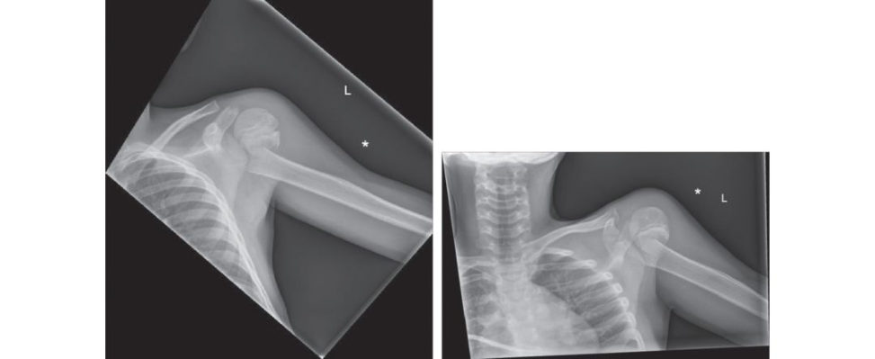
I can see a shoulder trauma series with an AP and an attempted shoot-through or trans- scapular view. It’s not quite a lateral Y-view I’m suspecting as the radiographer couldn’t get the correct projection Therea transverse fracture of the metaphysis, which is angulated medially due to the pull of the pectoralis major. If he was conscious I would specifically look at the axillary nerve function. I ’d also document the vascular status of the arm.
Assume that there is no associated neurovascular deficit. How are you going to manage this patient?
My concern is that the fracture fragments are completely translated with no bony contact and the distal fragment is being pulled medially into the axilla. As he has a closed head injury of unknown prognosis, he may not be able to be nursed upright for some time. I would consider surgical intervention in this child for ease of nursing. When the child was stable, I would plan to take him to theatre after appropriately counselling the parents, marking the child and the WHO checklist. I would screen with the image intensifier to check the shoulder is in joint and realign the fracture with gentle traction.
What if it doesn’t reduce closed?
I would ask the anaesthetist to give muscle relaxant for the MUA. If the fracture doesn’t reduce closed I would do a limited open reduction through a deltopectoral approach (internervous plane: axillary and the medial and lateral pectoral nerves). In terms of the fracture, I would stabilize this with retrograde K-wires from the lateral side through a mini-incision. The longhead of the biceps can sometimes get interposed in the fracture, blocking reduction. The major structure at risk from percutaneous K-wires is the axillary nerve. Its surface landmark is 5 cm from the lateral edge of the acromion. The nerve is a branch of the posterior cord, and travels through the quadrangular space with the posterior circumflex vessels before going anterior around the surgical neck, underneath the deltoid.
What do you think about this postop radiograph (Figure 16.1c)? I thought these fractures were usually managed conservatively?
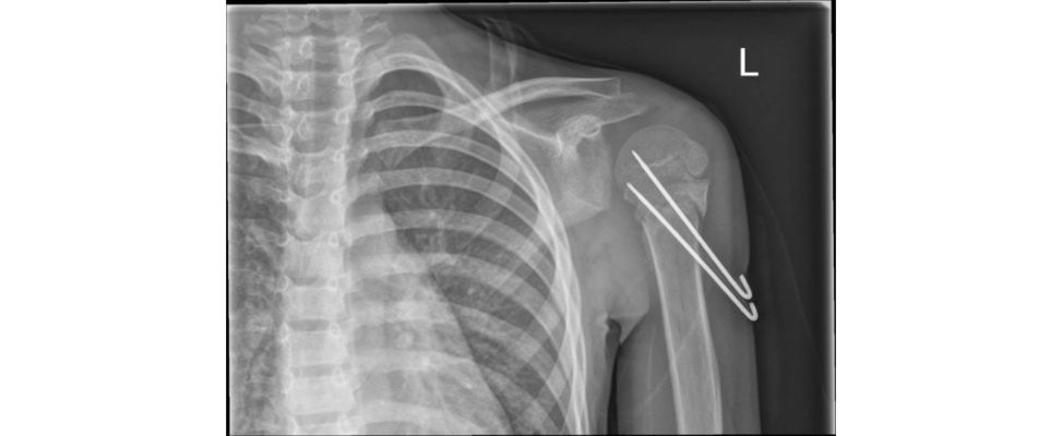
I can see the fracture is reduced and held with K-wires from the lateral side obliquely into the medial metaphysis. One of them is crossing the growth plate, and on this view, there’s no penetration of the surface of the head. The reduction is in slight varus, but overall, it’s well aligned. I would be careful about allowing the patient to mobilize before the wires come out at 4 weeks; I’d anticipate there might be limited hold in the metaphysis. Taking the wires out will need another GA as they are threaded, and you could screen it at the timet o check it was united.
For proximal humerus fractures in young children, almost any level of deformity is acceptable, for instance, bayonet apposition, as the remodelling potential is so gr eat. Typically, these are managed conservatively in a sling for a short time.
In children older than 13 with litile growth remaining there is an argument for accepting less deformity, such as < 30° angulation or < 50% translation, although the reno consensus in the literature about what level of deformity is an absolute indication for fixation, and the reis a higher incidence of stiffness compared topa tien ts treated non-operativ ely [1].
Figure 16.1a and 16.1b Displaced proximal humerus fracture.
Figure 16.1c Anteroposterior (AP) radiograph, shoulder following K-wire fixation.
Structured oral examination question 2#
This 7-year-old child fell from swings in the park sustaining a closed injury (Figure 16.2a and 16.2b).
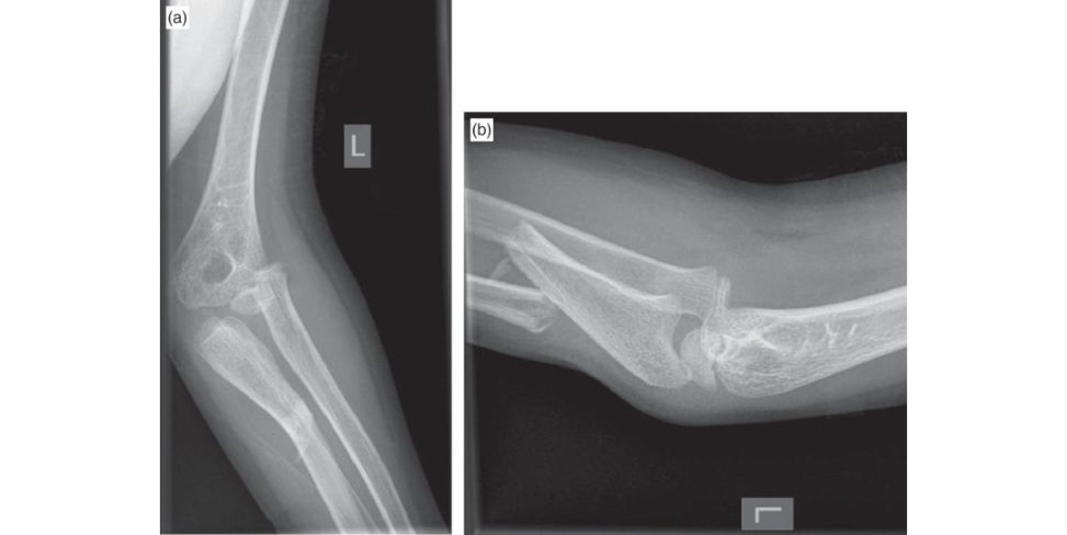
I can see AP and lateral radiographic views of the left elbow. I’m assuming this is an isolated injury and I would like to get additional views of the wrist. My initial priority ist o assess the neurovascular status and splint the limb. This is a Monteggia injury where there is a displaced and volarly angulated ulna fracture in addition to an anterior dislocated radial head.
Let’s assume there is available timet o do this on the trauma list and you are taking the child to theatre. What are the principles of treating this injury What would be your surgical plan?
By restoring the ulna length this should reduce the radial head. I would initially attempt a closed reduction by traction. If length, alignment and rotation were correct and with restoration of the radiocapitellar alignment, I would stabilize the ulna with an elastic nail. If I had an y doubts about the reduction, I would plate it.
How do you perform an elastic nailing? What problems do you anticipate arising if you try to use a nail for this?
Patient is positioned supine, arm board, bipolar (diathermy), tourniquet and image intensifier screen positioned within unrestricted view. Initially I ’d screen (with the image intensifier) and mark the physis and the fracture with a skin marker. The nail is sized according to one-third diameter of the isthmus. By pre-bending, the nail has elastic recoil, which exerts a force in the intramedullary canal to reduce the fracture. Elastic nails are best suited to diaphyseal fractures. This is metaphyseal and near the entry point for the nail, so I’d have a low threshold for open reduction and plate fixation with a one-thir d tubular plate or LCP.
Let’s say the nail doesn’t work. Even when you plate the ulna, the radial head does not reduce. What do you do?
I’d recheck I had properly reduced the fracture. In some cases, the ulna is plastically deformed, so you have to osteotomize it, but I don’t think that’s happening here. In this case I would suspect that the annular ligament is either torn or interposed, blocking reduction of the radial head. I’d open the radiocapitellar joint via aKo cher’s approach (between the anconeus and ECU). It is possible to reconstruct the annular ligament with a fascial sling. This fascia can be obtained from a number of sites within the upper limb, e.g. triceps fascia.
Figure 16.2a and 16.2b Anteroposterior (AP) and lateral radiographs, left elbow.
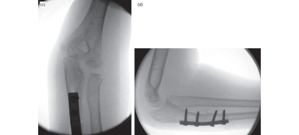
Figure 16.2c and 16.2d Intraoperative image intensifier anteroposterior (AP) and lateral radiographs, left proximal ulna.
Structured oral examination question 3#
This 6-year-old child is a new referral to the fracture clinic, please look at and describe these radiographs (Figures 16.3a and 16.3b).
These are AP and lateral radiographic views in plaster. There is a displaced lateral condyle fracture which is easiest to see on the AP. This looks like a Milch type 2 and the fracture line extends from the trochlear groove into the metaphysis. Because it’s extending from the medial side of the lateral ridge of the sulcus, the ulnohumeral articulation is unstable. A Milch type 1 is lateral to the ulnohumeral articulation: going through the capitellar physis into the metaphysis. This is the equivalent of a Salter Harris type IV. A more recent and practical classification by Jakob describes three types. Type 1: under 2 mm of displacement, indicating the presence of a cartilaginous hinge. Type 2: where there is between 2 and 4 mm of displacement with intact intra-articular cartilage on arthrogram. Type 3: greater than 4 mm of intra-articular displacement.
What problems can you foresee with this fracture?
These are intra-articular fractures and have a higher incidence of specific complications: Non-union due to synovial interposition from the joint surface, causing persistent elbow instability. Fishtail deformity from a fracture gap between the condyles, central area of avascular necrosis or physeal bar. Apparent cubitus varus due to lateral periosteal overgrowth, after the fracture heals, causing a cosmetic deformity. Cubitus valgus occurs from physeal arrest of the lateral capitellar physis, which can cause a tardy ulna nerve palsy. In undisplaced fractures, assessing the intra-articular component of the fracture can be difficult on plain radiographs, as the distal humerus is cartilaginous. In these cases, an MRI or EUA and arthrogram may be useful to assess the fracture further.
How do you surgically manage displaced fractures?
The aim is to restore articular congruity. For those fractures where the arthrogram demonstrates an articular hinge or mildly displaced (< 2 mm) fractures, the fracture can be stabilized with percutaneous divergent wires. For those fractures that are displaced, visualizing the articular surface is required. This can be achieved with a lateral approach to the elbow. Avoid dissecting around the posterior aspect of the capitellar fragment as this can devascularize the fragment. Reduce the fracture and hold with at least two divergent K-wires. These can either be buried or left outside of the skin.
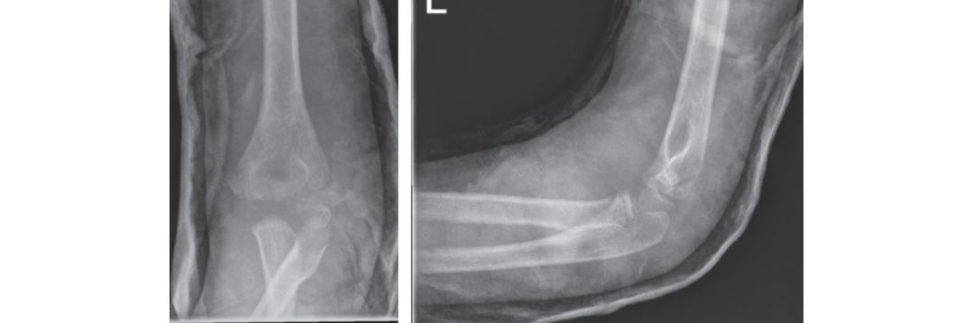
Figure 16.3a and 16.3b Anteroposterior (AP) and lateral radiographs, left elbow.
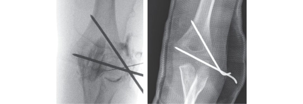
Figure 16.3c and Figure 16.3d Intraoperative arthrogram of left elbow and postop radiograph incast.
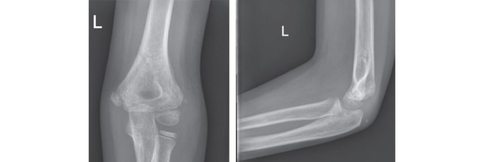
Figure 16.3e and 16.3f Anteroposterior (AP) and lateral radiographs, left elbow after healing.
Structured oral examination question 4#
This 7-year-old boy was waiting for his dinner and fell outside in the back garden (Figure 16.4a).
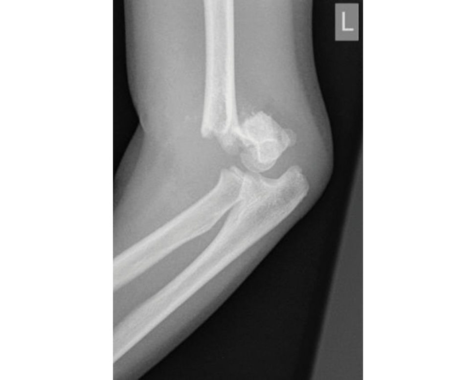
This is a lateral radiograph in a skeletally immature patient which demonstrates a displaced supracondylar fracture. Typically, the mechanism is a fall with the elbow extended and axial loading. My first priority is assessment of the child and limb. I’m assuming this is an isolated injury?[Yes.] I would check vascularity by checking the radial and ulna pulse, capillary refill and ensuring the hand is warm and well perfused. I would check the motor and sensory function of the median radial and ulna nerves. I would examine the antecubital fossa to see if the skin was under threat: such as a pucker sign due to buft onholing through brachialis. I would splint in a position of comfort after intranasal diamorphine.
The child has a pink hand that’s perfused but no palpable radial pulse. There is no motor or sensory deficit.
While the evidence is a pink, perfused hand without any neurological deficit, it can be elevated and observed [2,3]: if the child is fasted and I can get him to theatre at a reasonable time, my preference is to operate on him that evening.
He’s fasted, consented and ready. Explain what you are going to do.
I would warn the vascular surgeons. Set-up is key: the child is supine, with headring and shoulder at the level of the arm table extension. I would have the C-arm from the foot-end of the table: for access for AP and lateral views. The image intensifier screens are opposite me, on the other side of the table. I would prep and drape with a high tourniquet applied but not inflated. After the WHO checklist, I’d ask the anaesthetist to give muscle relaxant and antibiotics. I would reduce this by traction with the elbow in slight flexion. I would first check the AP to see I’ve got it out to length and correct for medial or lateral translation. I then flex the elbow with my thumb pushing on the olecranon, and depending on which best reduces the fracture, the forearm is either supinated or pronated. If the child’s fingers can touch their shoulder: this indirectly shows the fracture is reduced. I would check the medial and lateral columns are restored by taking oblique column views and a lateral X-ray. I put my lateral side wires in first. I do this by laying a 2.0-mm K-wire on the skin and marking with a skin marker. I make a small stab, place the wire free-hand on the entry point and tap it in a bit with a toffee hammer before driving it with the wire driver under image guidance. I aim for bi cortical fixation, engaging both distal and proximal fragments. Either lateral divergent wires or a crossed configuration is biomechanic ally stable, with maximal spread and avoiding crossing at the fracture site. On the medial side, I would extend the elbow to take the ulna nerve away from the epicondyle, then make a mini-incision to see the bone before placing and driving the medial wire in. I would screen the fixed construct under real-time imaging to check stability.
I’d recheck pulse and circulation before applying abacks lab. If there are any doubts about the pulse, a hand-held Doppler can be used. If the hand remained pink, it may take a few hours for the palpable radial pulse to return.
You find the hand is white when you recheck his circulation!
I’d reduce flexion on the elbow to see if that restored circulation. If it didnt, I’d screen to see if there was a gap at the fracture site, indicating the brachial artery was interposed or tethered. If I thought this was the case I’d remove the wires, place him on straight longitudinal traction and c all the vascular surgeon, who would do an on-table angiogram and explore the brachial artery through an anterior approach.
Figure 16.4a Lateral radiograph, left elbow.
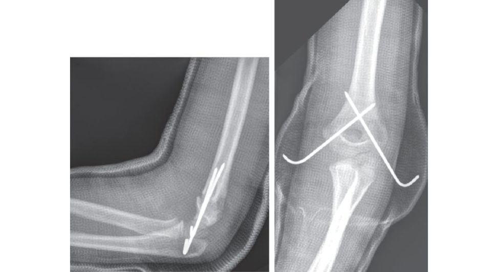
Figure 16.4b and 16.4c Lateral and anteroposterior (AP) radiographs incast.
Structured oral examination question 5#
This 13-year-old boy had a previous forearm injury which healed, and he was due to have an operation to remove the metalwork. He was involved in exuberant play and pushed over (Figures 16.5a and 16.5b). The injury is closed and neurovascularly intact.
I can see an initial radiograph and a repeat view, probably in A&E resus, after procedural sedation with his forearm reduced into a straighter position. The reis an elastic nail in the ulna with a double bend, with mid-shaft r e-fractures of both bones.
Could he betaken to theatre and have it manipulated, and a cast putback on it?
As he’s 13 years old, there isn’t much remaining growth. The mid-shaft position of the fracture also implies that this will have poor remodelling potential. A this age, I would not accept any deformity with this fracture pattern. I think trying to manipulate his forearm closed and achieve an adequate reduction of both bones is unlikely to work. My plan would be to either remove the ulna nail and then insert elastic nails in to both bones or open the fractures and plate them.
Are there any technical problems you anticipate while doing this?
Trying to remove the bent nail may be a problem, in which case I would open the fracture site and cut the nail. It’s then possible to capture each half of the nail with the pliers and pull it out.
Is it necessary to remove metalwork? Do you know any evidence?
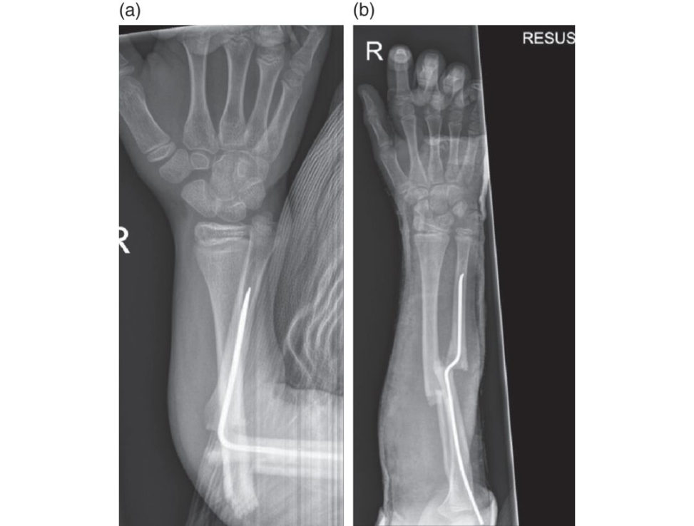
Figure 16.5a and 16.5b Anteroposterior (AP) radiographs, right forearm.
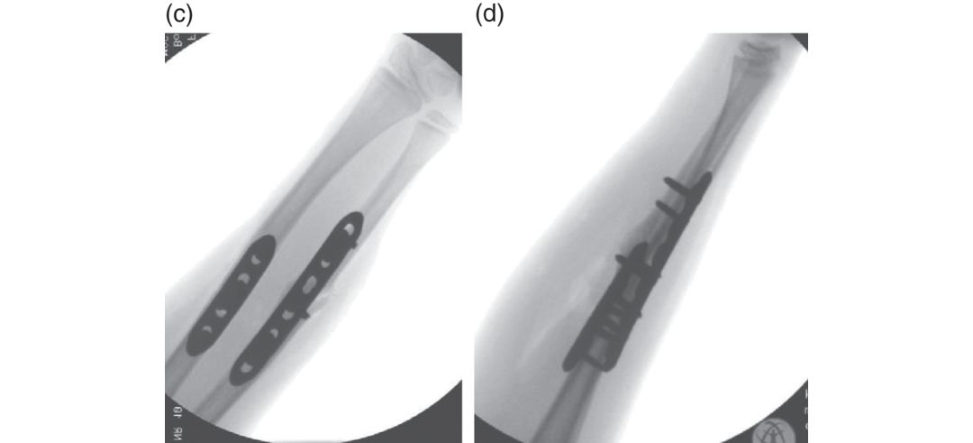
Figure 16.5c and 16.5d Anteroposterior (AP) and lateral intraoperative images.
There is divided opinion on the necessity for routine metalwork removal. Children have exuberant periosteal bone formation and thereby metalwork can get buried when placed at a young age. Assuming there are no further complications then they can be left in situ. With the risk of subsequent periprosthetic fracture and the difficulty of excavating metalwork, some surgeons have a low threshold to remove paediatric metalwork.
There is morbidity associated with metalwork removal, which includes a 40% complication rate when removing forearm plates that is related to the seniority of the surgeon doing the procedure [4].
Structured oral examination question 6#
This 8-year-old boy has had multiple previous fractures which have healed but left him with clinical deformities of the long bones ( Figure 16.6a). He had an innocuous fall which caused this injury (Figures 16.6b and 16.6c).
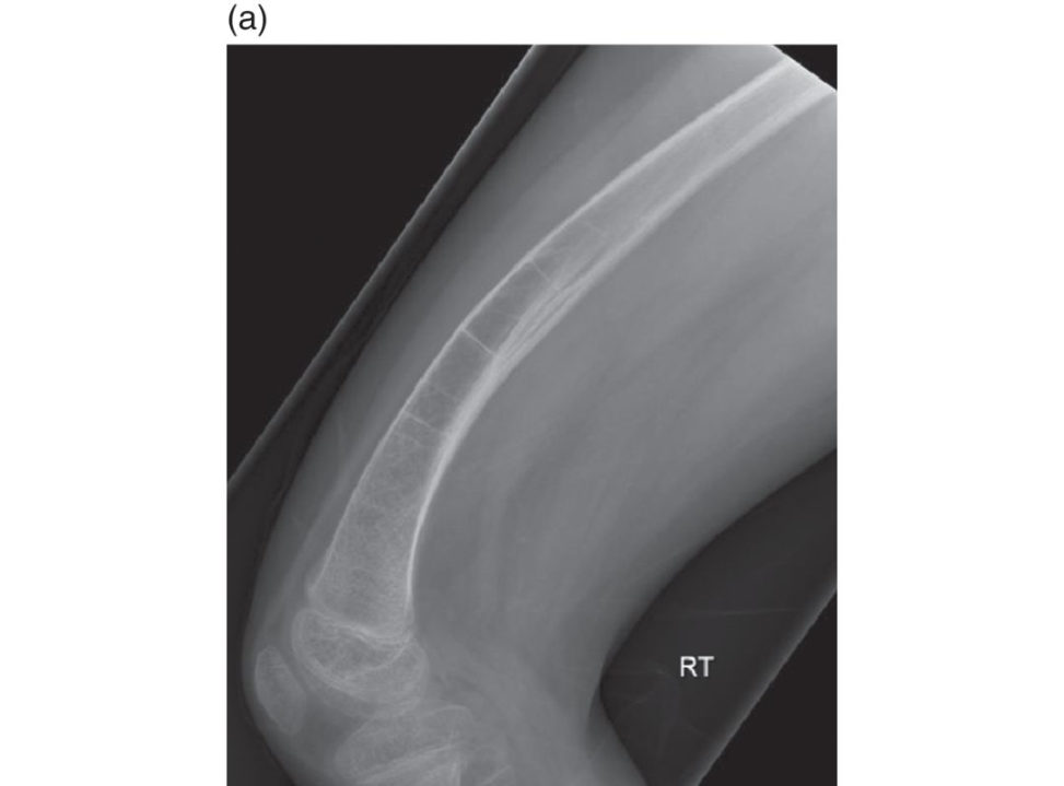
I can see a pre-injury radiograph of a right femur with a significant anterior bow and appearances of Park Harris growth arrest lines. On the right-hand side AP and lateral, there is an oblique fracture through the distal third of the femur with a Thomas splint. It’s translated and in varus, but the fracture is approximately out to length in his traction. The cortices are thin and bone quality is osteopenic. Does the child have an underlying metabolic bone disease? I would take a history and ask about inherited conditions in his family.
What are Park Harris growth arrest lines?
Park Harris lines are transverse sclerotic lines in the metaphysis which correspond to stressor an insult to the bone and then resumption of growth. A similar appearance can occur with bisphosphonate treatment.
Do you know of any paediatric orthopaedic conditions where we use bisphosphonates?
Osteogenesis imperfecta.
His sclera are blue and dad is affected. What type of OI does he have?
Likely Type 1 (according to Sillence): as they have blue sclera and the condition is autosomal dominant.
What is the treatment for this fracture? Are there any technical considerations?
I would discuss with a tertiary centre because of his pre-existing OI, but essentially the principles of management are to restore alignment and correct pre-existing deformity. The options are limited due to the anatomical abnormality and background osteopenia. An intramedullary device offers the strongest biomechanical option however, due to the age of the child this needs the capacity to grow with the child and not damage either the physis or the vascularity to the proximal femur. I am aware of the use of growing telescopic rod systems. The rods have a trochanteric entry point with male and female sliding components. The proximal and distal ends are threaded to anchor in the epiphysis. As the nails are straight, there may be a requirement for sequential osteotomies (Shish–Kebab/Sofield procedure) in pre-existing bony deformities to allow tension-free passage of the nails. Postoperatively I will liaise with his physician about the commencement of bisphosphonate therapy.
Figure 16.6a Lateral radiograph, femur.
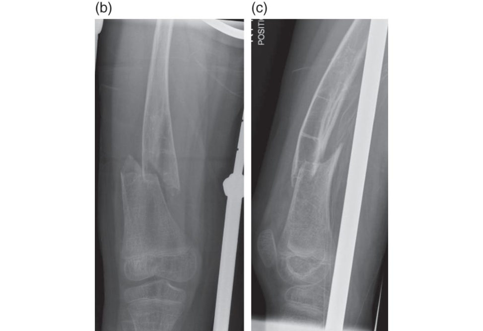
Figure 16.6b and 16.6c Anteroposterior (AP) and lateral radiographs, femur.
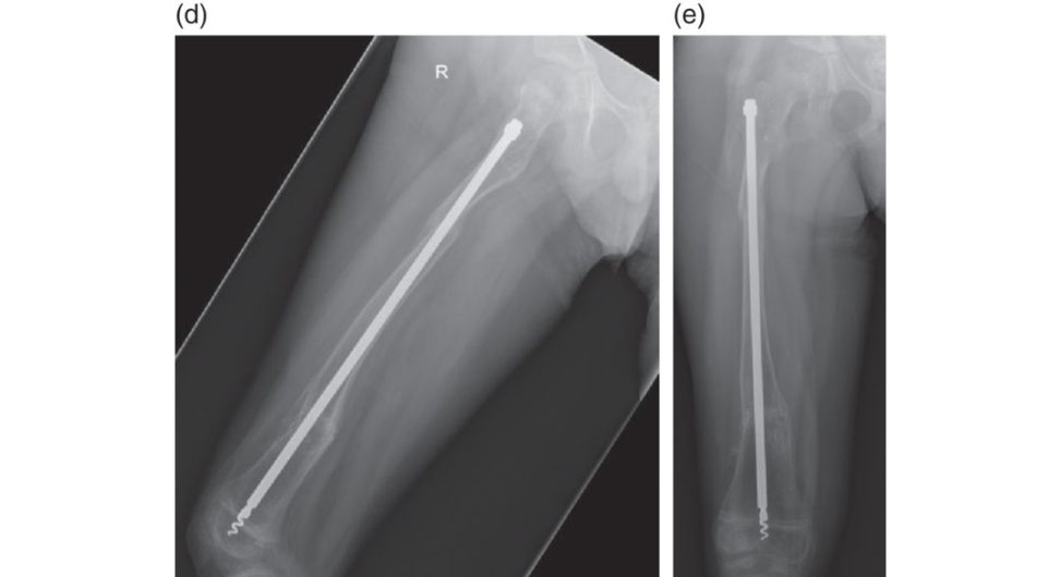
Figure 16.6d and 16.6e Lateral and anteroposterior (AP) postoperative radiographs, femur.
Structured oral examination question 7#
This 8-year-old girl sustained an injury walking home from school when she was hit by a car at low speed, pedestrian versus car (Figures 16.7a and 16.7b).
These are AP radiographs in a Kendrick traction splint used by paramedics at the scene as well as in a Thomas splint. I can see a short, oblique fracture of the femoral diaphysis, at the junction of the proximal and middle thirds. I would initially assess the patient according to ATLS protocol: prioritizing the resuscitation and treatment of life- and limb-threatening injuries. From the point of view of this injury: I would want to check the condition of the skin, ensure the foot is warm and well-perfused with intact pedal pulses, and the patient is moving the toes actively with no motor or sensory deficit. Is this a closed, isolated injury?
Yes. On secondary survey, you are happy this is an isolated injury and the patient is physiologically suitable for an operation, if you think it appropriate.
I would be inclined to treat this by elastic nails. This pattern of injury is length-stable. It’s a fracture in a paediatric femur with an appropriate size of canal for elastic nails: typically, a child between 5 and 10 years of age. The nails should each be one-third the diameter of the canal at the narrowest point. As a roughest ima te, I would measure canal diameter using the PACS system to check this. My set-up would be a Jackson table with my assistant applying traction on the leg. I would check alignment and rotation with fluoroscopy. I would use retrograde nails via medial and lateral entry points about 2.5 cm above the distal physis. The nails are pre-bent, so the apex is at the fracture site. The biomechanical principle is of ‘double arc secant inequilibrium’: the bending moments of two nails counteract each other with an equal and opposite force to create a stable construct.
Stable? What does that mean? Is it rigidly fixed? What type of bone healing occurs?
Fracture healing occurs by secondary bone healing with callus formation; this is a relative stability technique. The nails are far from rigid. They especially do not resist torsion: I’d be observant for rotational malalignment on follow-up in clinic. Loss of alignment is particularly seen with nails that have unequal curves, different size nails and when the fracture is comminuted. End caps have been advocated by some to increase the stiffness of titanium elastic nails, although the evidence for this is in biomechanical studies only as far as I’m aware. In children greater than 50 kg bodyweight, stainless steel flexible nails can be used to increase the nail strength and have been shown to have a lower rate of malunion compared to titanium elastic nails [ 5].
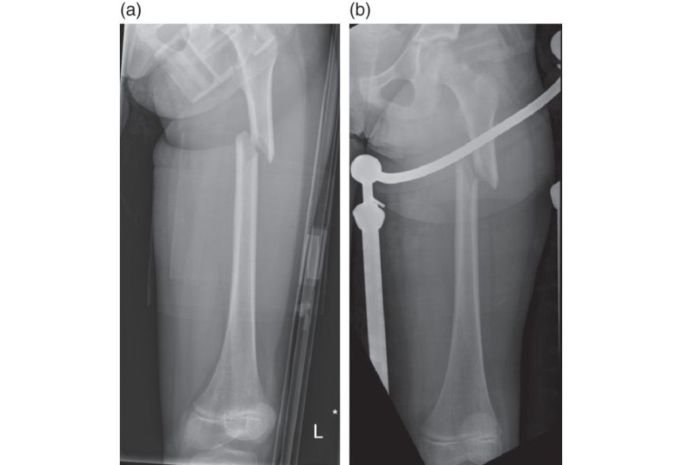
Figure 16.7a and 16.7b Anteroposterior (AP) radiographs, femur.
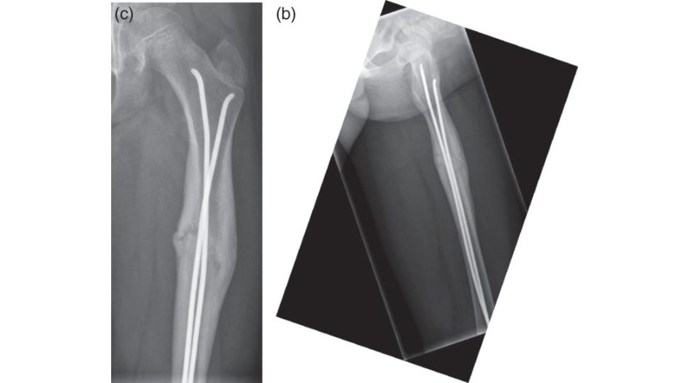
Figure 16.7c and 16.7d Anteroposterior (AP) and lateral postoperative radiographs, femur.
Structured oral examination question 8#
This 14-year-old boy was a trauma call from an RTA. He has sustained multiple rib fractures and a lung contusion on the same side as this closed injury to his leg. He’s had a pan-CT scan in A&E resus (Figure 16.8a) and is awake and stable on paediatric HDU. The general surgeons have said his liver laceration can be managed conservatively without the need for laparotomy and CT has excluded a pelvic injury.
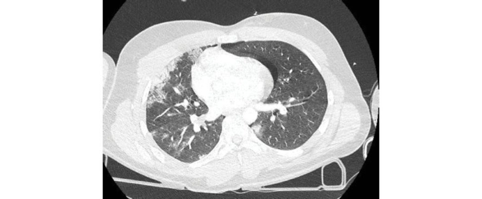
I’m assuming he’s had a primary survey according to ATLS protocol by the trauma team. My priority would be the secondary survey on HDU. I would check him head to toe for additional injuries. I would have a high index of suspicion for a spinal injury or an injury to the hip. I would speak to the ITU team to see if they have been happy with his monitoring, and to make a plan for theatre. His lactate on blood gas and urine output are indicators of organ perfusion and whether he is physiologically safe to proceed. From the point of view of assessment of the distal femur fracture: I would check pulses in his leg, the condition of the skin, neurological status and for signs of impending compartment syndrome.
The ITU team says you can proceed, what is your plan?
My plan would be to fix his femur using a distal femoral locking plate. He’s almost an adult, with an adult pattern of injury in terms of the buft erfly fragment and comminution. The capital femoral physis on his AP pelvis appears almost shutdown in terms of remaining growth. The distal physis is open. To control this would be difficult using a paediatric lateral entry nail, because it is at the junction of the diaphysis and metaphysis. A retrograde nail is not an option. Hes also had a significant lung injury and there are risks from reaming in terms of fat embolus. I would approach this using a lateral approach, being careful around the blood supply to the physis. It might be possible to do a submuscular technique and slide the plate up: I’d have a low threshold for opening it to check reduction was adequate in terms of length and rotation.
Do you foresee any problems after this has been fixed? What do you warn him and his parents?
An injury near the distal femoral physis has a potential for growth disturbance. Although this injury was in the metaphyseal region, I can see on later radiographs his physis is starting to shutdown. I would check to see if clinically he had signs of a significant leg length discrepancy and obtain standing alignment views. There is an incidence of valgus deformity associated with plating of femoral shaft fractures close to the physis and I would warn the parents we would monitor him for this during follow-up. As he’s almost at maturity I would not expect there to be a significant angular deformity or length discrepancy.
Figure 16.8a Axial CT, chest.
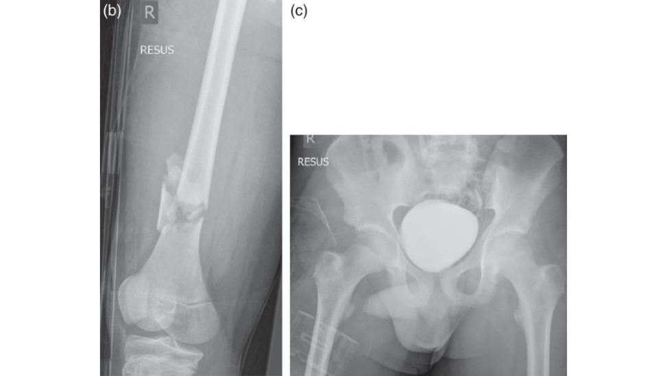
Figure 16.8b and 16.8c Anteroposterior (AP) radiographs, right femur and pelvis trauma series.
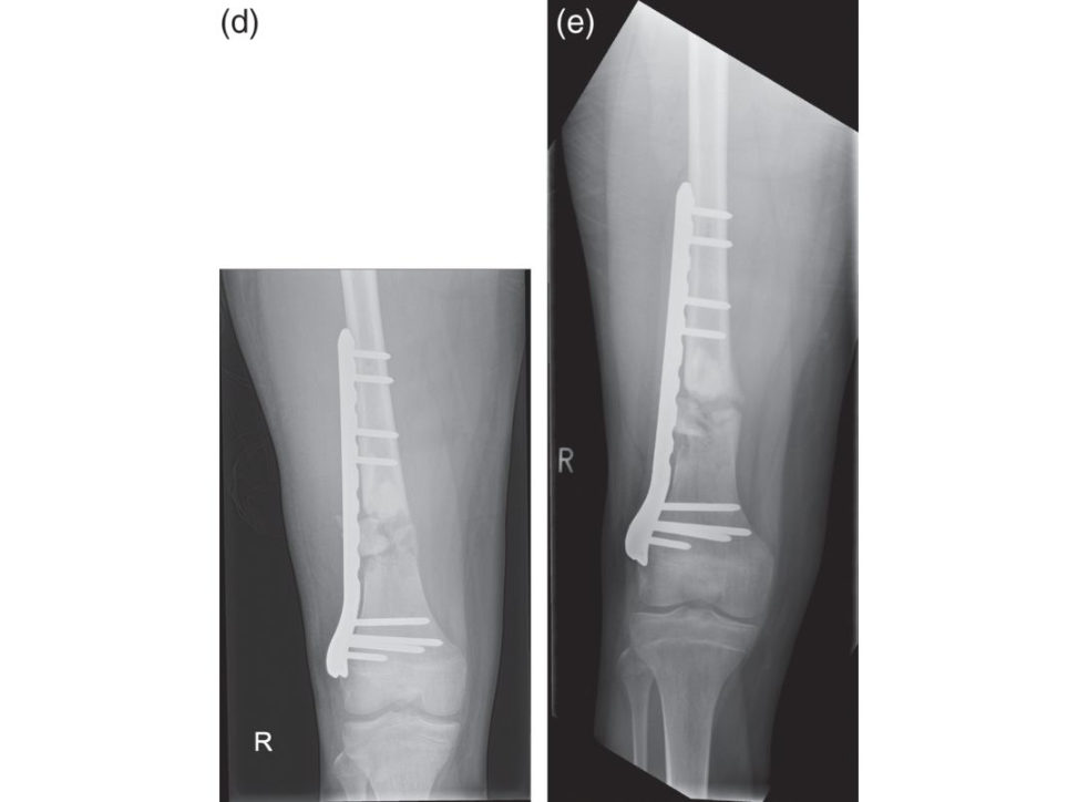
Figure 16.8d and 16.8e Anteroposterior (AP) postoperative radiographs, right femur.
Structured oral examination question 9#
This 13-year-old boy sustained this injury playing football (Figures 16.9a and 16.9b).
I can see an AP and lateral plain radiograph of an adolescent knee showing a comminuted tibial tubercle avulsion fracture. This is typically due to eccentric quadriceps contraction with a flexed knee, such as jumping and landing on a bent knee. On examination, I would expect to find swelling, bruising and the patient would be unable to straight-leg raise. I would check for impending compartment syndrome as there is a risk of injury to a branch of the recurrent anterior tibial artery, which bleeds into the anterior compartment. Initial management is splintage, admission for elevation and planned definitive fixation. The reis a Watson-Jones classification modified by Ogden. This looks like a comminuted Type II: where the apophysis is avulsed from the metaphysis.
Tell me about the Watson-Jones classification as modified by Ogden.
Type I is a fracture of the tibial tubercle apophysis (secondary ossification centre where the patella tendon inserts). It is distal to the junction of the ossification centres of the apophysis and epiphysis. IA is undisplaced. IB is a displaced avulsion, with the fragment hinged anteriorly. Type II is at the junction of the apophysis and epiphysis. II Ais undisplaced. II Bis comminuted and the apophysis is displaced by the pull of the patella tendon. Type III is above the junction , where the fracture extends into the knee joint through the epiphysis. III Ais where the apophysis and epiphysis are a ‘composite’ or single fragment. III Bis where they are comminuted and separated.
What is your management plan?
This needs operative fixation to restore the integrity of the extensor mechanism. My preference is to use 4-mm partially threaded cannulated screws. I would have a tourniquet and a sterile gown rolled up under the calf as a bolster to hyperextend the knee. I would make an incision anteriorly over the fracture: pullout any interposed periosteum and then pass my guidewires from anterior to posterior, while avoiding crossing the physis and being mindful of the neurovascular structures within the popliteal fossa. After passing the screws I would screen the knee to check the fixation was stable when flexing to 90°. Pos topI would mobilize the patient touch weight-bearing with a hinged brace initially locked inextension, and then allow progressively more flexion over 6 weeks.
This boy is an elite sports person. Are there any future problems you need to warn his parents about?
He may require removal of the metalwork after the fracture has united if it is prominent or causes a bursitis and anterior knee pain. There is a risk of genu recurvatum from decreased tibial slope after premature closure of the anterior physis in younger children. I’d anticipate, as he has litile remaining growth before his physes fuse, that this is not likely to be an issue, but I would follow him up to watch for this. If there were any signs this was starting to develop, I would do an epiphysiodesis to complete the growth shutdown and he may then require a corrective osteotomy.
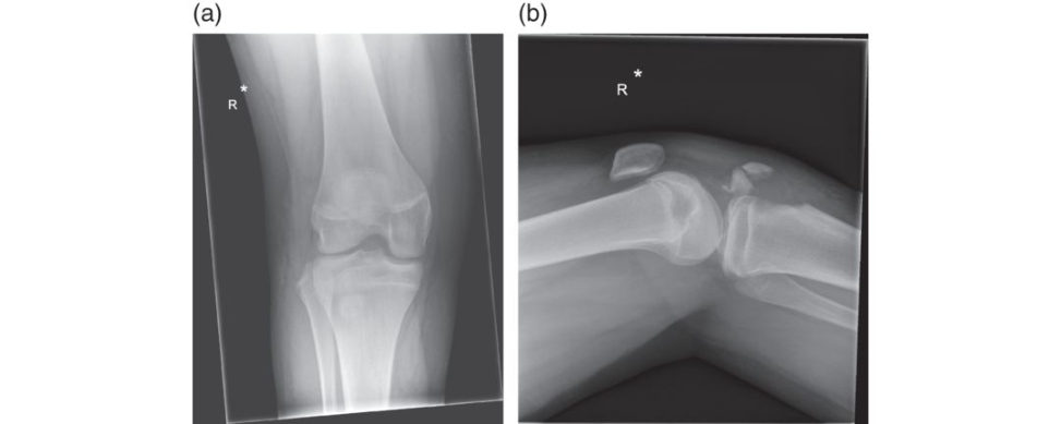
Figure 16.9a and 16.9b Anteroposterior (AP) and lateral radiographs, knee.
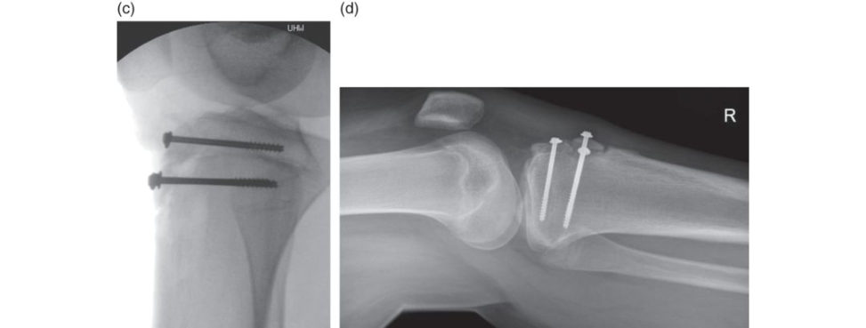
Figure 16.9c and 16.9d Intraoperative and postoperative lateral radiographs, right knee.
Structured oral examination question 10#
This 12-year-old girl attended A&E and was told she had a sprain after a trampoline injury (Figures 16.10a and 16.10b). She was allowed to weight bear with an ankle stirrup, although is struggling with pain when you see her in the fracture clinic.
I can see AP and lateral radiographs of a skeletally immature patient There’s evidence of an effusion within the joint and I can see a fracture line in the distal tibial physis exiting in to the plafond. At this age, I’d be concerned about a triplane or transitional type of injury and obtain a CT scan.
What do you mean by transitional?
Closure of the distal tibial epiphysis occurs in adolescents at 12–15 years of age, over an 18- month period. This is an injury when they are transitioning to skeletal maturity. The growth plate closes asymmetrically: first centrally then anteromedially, posteromedially and finally the lateral distal tibial physis is the last to close.
Is this a triplane fracture (Figures 16.10c–16.10e)? What is the management?
A triplane injury involves fracture lines in the sagift al, transverse and coronal planes, traversing the physis and entering the ankle joint. On the lateral X-ray typically, there is a Salter Harris II and a Salter Harris III on the AP, corresponding to the displaced anterolateral part of the physis. The CT confirms a triplane pattern of injury. The principles of management are to restore the articular surface by anatomical reduction, and fixation using absolute stability. Any displacement > 2 mm at the articular surface is an indication for surgical intervention. In this case, I would see if I could obtain a closed reduction and percutaneously fix it with a partially threaded 4-mm cannulated screw from a lateral to medial direction. I would use a large reduction clamp to aid reduction if necessary. If there was movement of the Thurston Holland fragment on screening, I would also use metaphyseal screws in an anterior to posterior direction. If I could not obtain reduction by closed means, I would openly reduce the articular surface with an incision over the fracture line to remove any potential soft -tissue interposition.
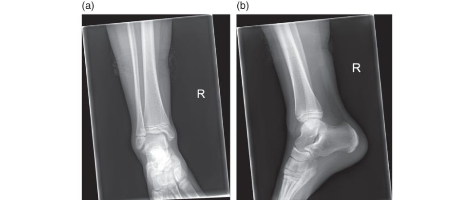
Figure 16.10a and 16.10b Anteroposterior (AP) and lateral radiographs, right ankle.
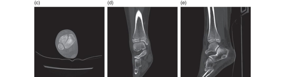
Figure 16.10c–16.10e Sagift al, coronal and axial CT slices respectively, right ankle.
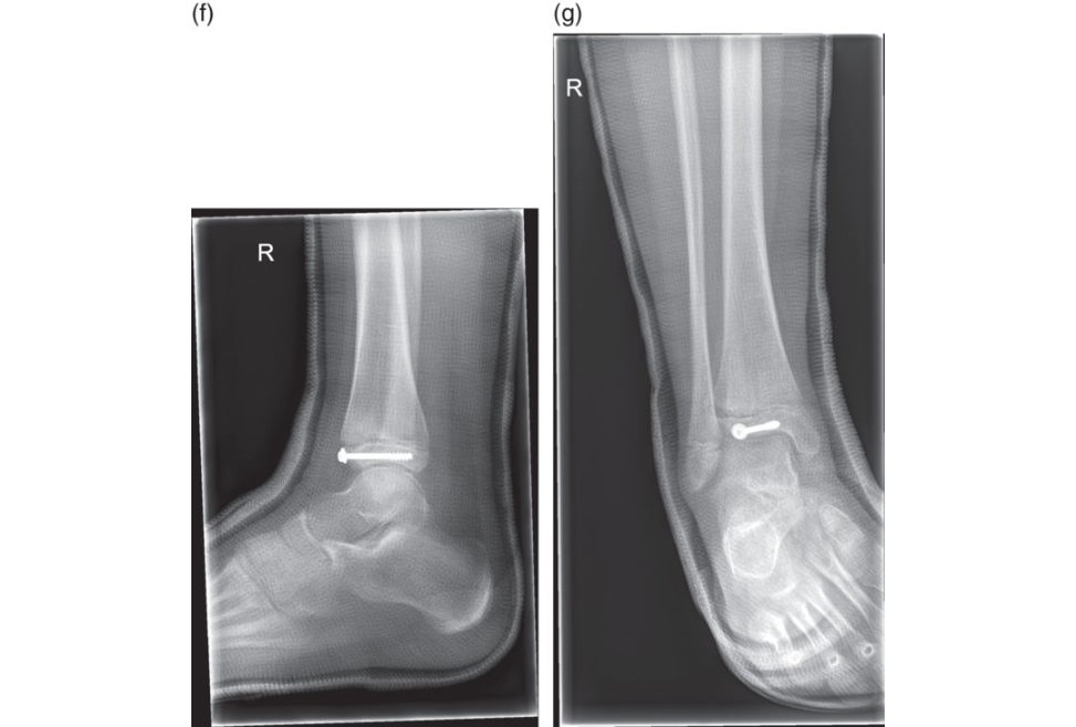
Figure 16.10f and 16.10g Postoperative lateral and mortise/ anteroposterior (AP) radiographs, right ankle incast.
References
1. Pahlavan S, Baldwin K, et al. Proximal humerus fractures in the pediatric population: a systematic review. J Child Orthop. 2011;5:187–194.
2. Mangat KS, Martin AG, Bache CE. The ‘pulseless pink’ hand after supracondylar fracture in children: the predictive value of nerve palsy. J Bone Joint Surg Br. 2009;91(11):1521–1525.
3. Scannell BP, Jackson JB, et al. The perfused, pulseless supracondylar humeral fracture: intermediate-term follow-up of vascular status and function . J Bone Joint Surg Am. 2013;95(21):1913–1919.
4. Langkamer VG, Ackroyd CE. Removal of forearm plates. A review of the complications. J Bone Joint Surg
Br. 1990;72(4):601–604.
5. Wall EJ, Jain V, et al. Complications of titanium and stainless steel elastic nail fixation of pediatric femoral fractures. J Bone Joint Surg Am. 2008;90(6):1305–1313.
Section 4