Chapter 20 Structure and function of connective tissue
Paul A. Banaszkiewicz
Introduction#
Section 2 of the basic science (Tr & Orth) syllabus is a large topic, difficult to grasp at face value as it appears quite removed from the average orthopaedic surgeon’s practice. However, it pervades many aspects of clinical practice and therefore must be understood.
It contains large sections of A-list topics that just need to be learnt as well as possible, otherwise marks will be thrown away.
Candidates may be asked very general questions or questions in more detail, so you need to cover both bases. Esoteric or un-Googleable questions are also fairly common in this section.
Candidates will need to double time on this section to both understand the topics and then work through viva practice sessions tohome in on the target answers.
This section was generally well received in the first edition viva book and we have kept and updated the majority of previous viva questions. As such, incertain sections there is a rough marking scheme, so you can judge your level.
1. Bone structure and function#
This is an A-list topic. The topic is large with a number of subtopics within the main topic that the examiners can easily focus in on.
Structured oral examination question 1#
What is bone?
Bone is a dynamic composite form of specialized connective tissue composed of cells (10%) and matrix (90%). The matrix has inorganic (60%) and organic (40%) components ... The viva could start off awkwardly with a definition that may catch the unsuspecting candidate off-guard. Go for an uncomplicated, non-controversial answer that allows you to continue talking if you feel confidently able to do so. Or Bone is an organ.
What is an organ?
[Pause] I am not sure. Make sure you know the definition of an or gan.1 Try to avoid giving an answer that will lead you up a blind alley. An organ is composed of multiple tissue types. F or bone these include: Bone tissue (a.k.a. osseous tissue). Fibrous connective tissue. Cartilage. Vascular tissue. Lymphatic tissue. Adipose tissue. Nervous tissue.
What are the functions of bone?
The three main functions of bone are: 1. As a reservoir for calcium. 2. As a source of haematopoietic cells such as erythrocytes leucocytes and platelets. 3. A mechanical role in supporting the bodys tissues providing attachment for muscles and protecting internal organs. There are other minor functions, but again, keep it simple.
Describe the structure of bone.
There are two main macroscopic types of bone, either (1) lamellar or (2) woven. The structure of lamellar bone can be either cortical compactor cancellous trabecular bone. Woven bone can be either immature (fracture callus) or pathological. Candidates may be pressed in a bit more detail about the differences between woven and lamellar bone, especially at the beginning of a viva, because it is basic information candidates would be expected to know. Woven bone Has a random arrangement of collagen, there are no lamellae, it is weaker and more flexible than lamellar bone. More cellular (×8 lamellar bone). More metabolically active with increased turnover. Variable irregular mineral content. It is found in the embryonic skeleton and the metaphyseal region of growing bones. It is also present in early hard callus and found in pathological bone such as tumours, osteogenesis imperfecta and pagetic bone. It is found where bone needs to be laid down rapidly. Lamellar bone This is mature adult bone that is subdivided into cortical or cancellous bone. Osteoblasts laydown collagen matrix in microscopic thin, layered sheets called lamellae. Collagen fibres are orientated more parallel to each other. Low number of cells. Slow deposition/turno ver. Stronger and less flexible than woven bone. Anisotropic. Table 20.1 Lamellar bone versus woven bone.
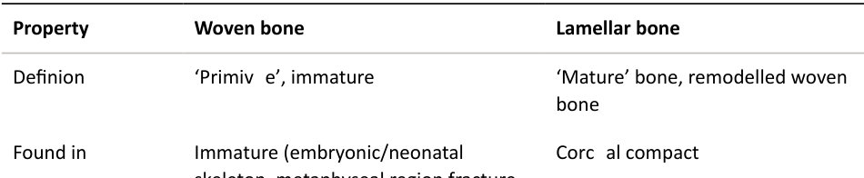
Found in Immature (embryonic/neonatal skeleton, metaphyseal region fracture healing)
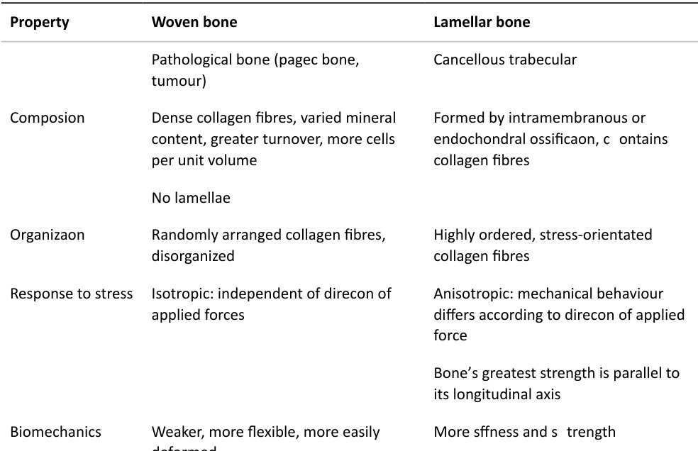
High-yield orthopaedics: Differentiating features of woven vs. lamellar bone.
No lamellae, isotropic.
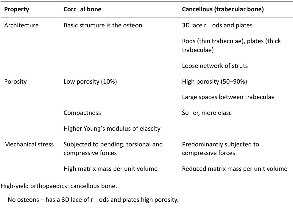
What is the difference between cortical and cancellous bone (Table 20.2)?
Cortical bone is compact with a high matrix mass per unit volume, low porosity and is subjected to bending, torsional and compressive forces. It is usually found in the diaphysis of long bones. The basic structure is the osteon or Haversian system. Cancellous bone is found in the metaphysis or epiphysis of long bones. It has an architecture of 3D latice rods and plates, high porosity with large spaces between trabeculae and predominantly subjected to compressive loads.
What do we mean by the term isotropic?
Isotropic refers to uniform properties in all directions, independent of the direction of load application.
So, which type of bone is isotropic?
Woven.
So, what about lamellar bone. Is this isotropic?
No.
Why?
I am not sure. These are fairly straighfoorward questions but may catch the unprepared candidate out. Lamellar bone has stress-orientated collagen fibres and has anisotropic features. The mechanical behaviour differs according to the direction of applied force: the bone’s greatest strength is parallel to the longitudinal axis.
What is the structure of bone?
The main structural unit within cortical bone is the Haversian system osteon) with its central neurovascular channels enclosed within concentric lamellae. Lying in between intact osteons separated by cement lines are incomplete lamellae called interstial lamellae. These fill the gaps between osteons and are remnants of bone remodelling. Neither collagen nor canaliculi cross cement lines, forming areas of relative weakness. Each osteon or Haversian system consists of five to seven concentric layers (lamellae) of bone matrix. Volkmann’s canals run perpendicular to the long bone axis carrying blood vessels to and from the Haversian systems to the outer surfaces of the bone. The viva may just begin by candidates being shown a diagram of the bone Haversian system and being asked to talk through the diagram. Less likely but still possible is for candidates to be asked to drawout the bone Haversian system. Bone compromises cells (10%) and extracellular matrix (90%). The cells include osteoblasts, osteocytes, osteoclasts and bone lining cells. The matrix has organic (collagens, mainly type 1) and inorganic (calcium phosphate, osteocalcium phosphate) constituents.
What are the main types of bone cells and what are their functions?
The main types of bones cells, and their function, areas follows: 1. Osteoblast: bone-forming cells. 2. Osteoclast: bone-resorbing cells, multinuclear irregular giant cells. 3. Osteocyte: maintains bone, important for calcium and phosphate homeostasis. 4. Osteoprogenitor cell: precursors to osteoblasts that line the Haversian system and can be stimulated to differentiate into osteoblasts and form new bone. 5. Bone lining cells: inactive osteoblasts. Some textbooks mention only three or four types of bone cells, usually omiting osteoprogenitor cells and sometimes bone lining cells.
The osteoprogenitor cells originate from mesenchymal stem cells and line the Haversian canals, endosteum and periosteum. They can quickly differentiate into osteoblasts if needed.
How do osteoblasts and osteoclasts differ?
Osteoblasts are derived from undifferentiated mesenchymal cells; they are bone-forming and laydown osteoid (type 1 collagen) as well as activating osteoclasts to resorb bone via the receptor activator of nuclear factor κβ (RANK) and its ligand (RANKL) system. RANK Lis expressed by macrophages and osteoblasts, and functions as an activator of RANK. RANK, expressed on osteoclast precursors, is a key regulator of osteoclastogenesis. These processes are controlled by cytokines, growth factors and bone morphogenic protein (BMP). Osteoclasts are derived from a haemopoietic monocyte cell lineage. They are multinucleated giant cells that resorb bone and are characterized by a cytoplasm that has a homogeneous foamy appearance due to a high concentration of vesicles and vacuoles. These vacuoles include lysosomes filled with acid phosphatase. They can sit in small pits called Howship’s lacunae, on the bone surface, or lead cuting c ones that tunnel through the bone. Under their ruffled brush border, with an increased surface area, they create a low pH microenvironment that dissolves inorganic apatite crystals. Enzymes are released (tartrate resistance acid phosphatase, TRAP) and proteases then breakdown the organic matrix components. This process is controlled via the RANKL system (inhibited by osteoprotegerin) of activated osteoblasts. Osteocytes are osteoblasts that have become trapped in bone matrix (comprising up to 90% of the cells in bone). They have an important role in the homoeostasis of calcium and phosphate metabolism.
What is Wolf flaw?
Wolf flaw states that bone will adapt to the loads placed through or across it. It is the result of the close coupling within bone remodelling units consisting of osteoblasts, osteoclasts and supporting stromal tissues. If loading on a particular bone increases, the bone will remodel itself over timet o become stronger to resist that sort of loading. Bone models and remodels in response to the mechanical stresses it experiences, resulting in a minimal-weight structure that is adapted to its applied stresses.
Can you give me an example of Wolf flaw?
The racket-holding arms of tennis players are stronger than the other arm. There is thicker cortical bone alongside hypertrophy of the muscle attachment sites. The arm is about a third bigger in size (35%). The femoral neck width of obese people changes to accommodate the added weight. In this case the width of the femoral neck has increased to dissipate weight throughout the bony area by increasing surface area and strength through redistribution of bone.
The reverse is true: if loading on a bone decreases, the bone will become less dense and weaker due to the lack of stimulus required for continued remodelling.
What is the Hueter–Volkman law?
Remodelling occurs in small packets of cells known as basic multicellular units (BMUs). Compressive forces inhibit longitudinal growth; tension stimulates it. This law suggests mechanical factors influence longitudinal growth, bone remodelling and fracture repair. The underlying mechanisms remain unclear.
What controls the differentiation of osteoblasts?
Two transcription factors are important for osteoblastic differentiation. 1. Osterix (Os xis an osteoblast-specific transcription factor essential for osteoblast differentiation and bone formation. 2. Runx2 is a multifunctional transcription factor that induces differentiation of multipotent mesenchymal cells into immature osteoblasts.
What are transcription factors?
Transcription factors are proteins involved in the process of converting , or transcribing, DN Aint oRNA. Transcription factors include a wide number of proteins, excluding RNA polymerase, that initiate and regulate the transcription of genes.
What is this cell line?
A picture was shown of an osteoclast in Howship’s lacuna (Figure 20.1).
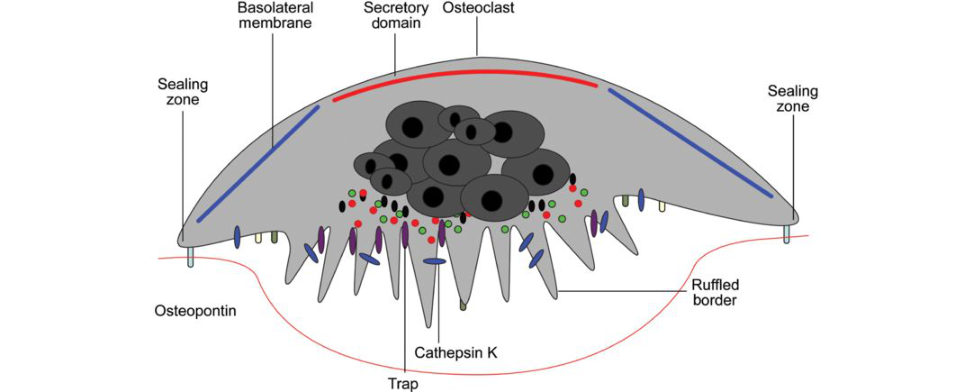
Figure 20.1 Activated osteoclast. The osteoclast plasma membrane is divided into multiple domains. A t the ruffled border, the osteoclast secretes acid and lysosomal enzymes that digest the mineral and protein components of the underlying bone. The degradation products of collagen and other matrix components are endocytosed, transported through the cell and exocytosed through a functional secretory domain.
This is an activated osteoclast.
How do osteoclasts resorb bone?
Osteoclasts resorb bone by binding to the bone surface using integrin anchor proteins and secreting hydrogen ions into the sealed area produced with a carbonic anhydrase system allowing dissolution of hydroxyapatite mineral matrix. They also secrete proteolytic lysosomal enzymes that hydrolyze the organic cellular components. The ruffled border greatly increases the surface area osteoclasts.
What do you understand by the term remodelling? Describe the process.
Remodelling is the process whereby the structure of bone is transformed from disorganized, haphazard immature bone to organized lamellar bone by osteoclast cuting c ones. The osteoclasts dissolve the inorganic matrix by acid secretion and as the y move forwards the resorption cavity is occupied by osteoblasts that laydown osteoid before it is calcified.
Can you please draw an osteoclastic cuting c one for me?
Learn to draw a cuting c one and be able to describe how this functions to remodel bone as you go along (Figures 20.2 and 20.3).
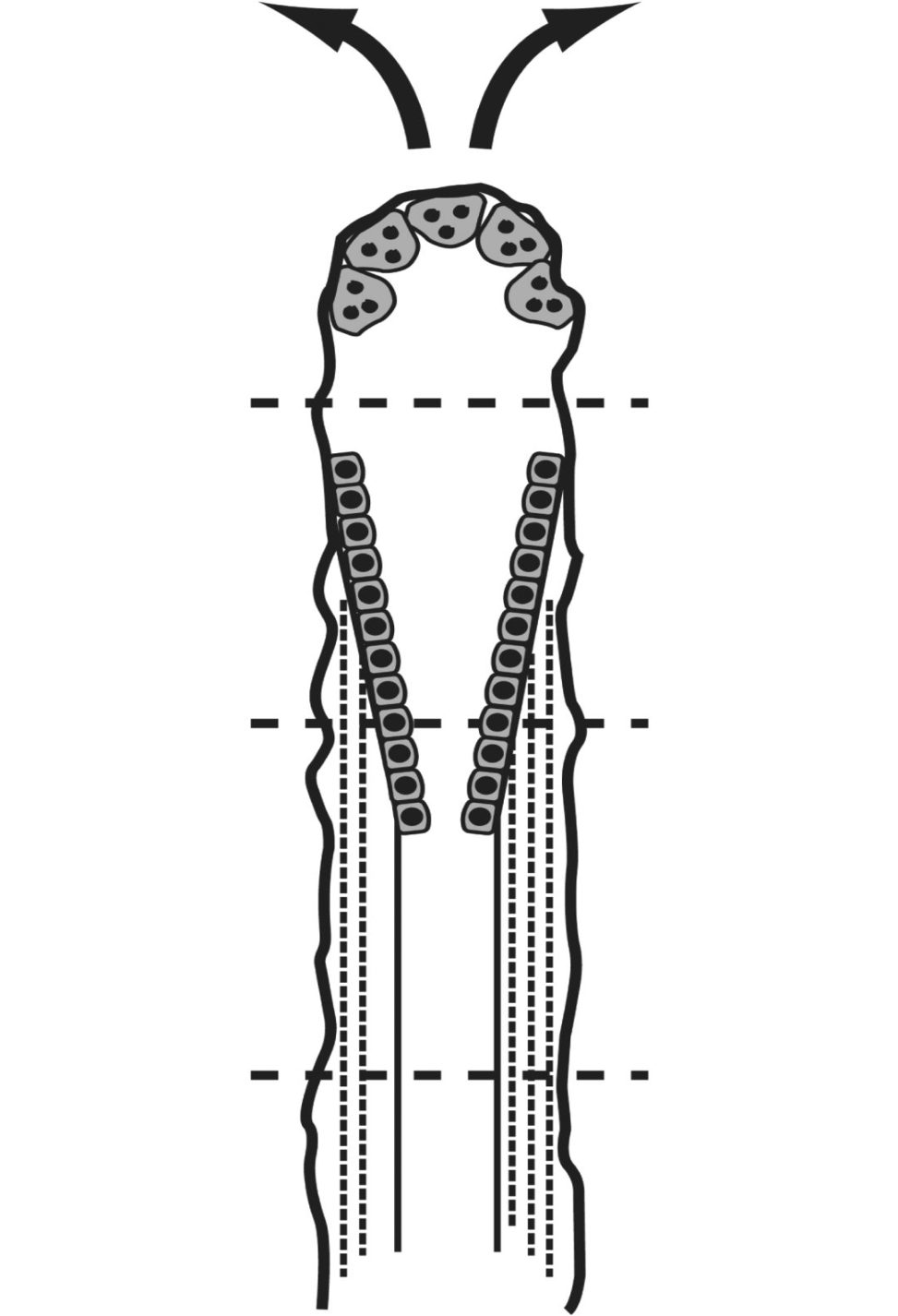
Figure 20.2 Osteoclastic cuting c one. Candidate drawing. The cuting filling c one has a head of osteoclasts that cut through the bone, and a tail of osteoblasts that form a new secondary osteon.
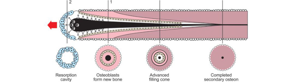
Figure 20.3 Osteoclastic cuting c one. At its tip osteoclastic resorption takes place while in the latter parts of the cone osteoblasts deposit osteoid with subsequent mineralization Reversal refers to a 1- to 2-week interval between completion of resorption and initiation of bone matrix formation. The structure terminates as a closing zone in which osteoblasts close the newly excavated osteon by adding centripetal layers of lamellar bone inward from the cement line boundary.
How and from where does bone derive its blood supply?
The blood supply to bone is derived from three sources. (a) High-pressure nutrient artery system. The nutrient arteries are branches of the systemic circulation and en ter the bony mid-diaphysis through the nutrient foramen passing to the medullary canal before branching into ascending and descending vessels and arteriolar branches supplying the inner two-thirds of the diaphyseal cortex (endosteal circulation). Remember that the end arterioles run in the Volkmann canals which drain into the Haversian system and finally drain back into the central venous sinus and out via the nutrient vein. (b) The low-pressure periosteal system circulation . This supplies the outer third of the bone cortex and consists of an extensive network of capillaries covering the length of the diaphysis. The normal direction is centrifugal; however, following endosteal damage this system is reversed to centripetal. This is the dominant system in children, which allows their circumferential growth. (c) Metaphyseal–epiphyseal system is the periarticular vascular complex that penetrates the cortex and supplies the metaphysis, physis and epiphysis with end arterioles.
What is the direction of blood flow within a long bone?
Arterial flow in mature bone is centrifugal (inside to outside), which is the net effect of the high-pressure nutrient artery system and the low-pressure periosteal system. When a fracture disrupts the nutrient artery system, the periosteal system pressure predominates, and blood flow is centripetal (outside to inside). Flow in immature, developing bone is centripetal because the highly vascularized periosteal system is the predominant component.
Venous flow in mature bone is centripetal.
Describe the structure of the periosteum.
The periosteum consists of an outer layer of fibroblasts and an inner layer of osteoblasts. The outer fibrous layer is structural, less cellular and continuous with the joint capsule, while the inner cambial layer is vascular, osteogenic and contributes to bone growth and fracture healing. With age, the periosteum thins and has less osteogenic capability.
What are the functions of the periosteum?
Functions of the periosteum include: Medium through which muscles, tendons and ligaments are attached. Forms a nutritive function. Can form bone when required. Forms a limiting membrane that prevents bone tissue from ‘spilling out’ into neighbouring tissues.
What is the structure of collagen in bone?
Collagen is type 1 in bone. The structural unit of type 1 collagen is called tropocollagen and is a trimer composed of three polypeptide chains. T wo chains are α1 chains and the third chain is α2. The three chains form a distinctive unit in which the polypeptide chains wraparound each other for most of their length, forming a tight triple helical braid. Each polypeptide chain is a le ft-handed helix, but the triple helix is a right-handed superhelix (i.e. the opposite way around) (Figure 20.4). The triple helical structure is not the same as the α helix that is formed by a single polypeptide chain and is the defining feature of all collagen. Collagen is secreted as an oversized molecule, procollagen, with specialized enzymes removing the N and C terminal propeptides leaving a triple helix with short non-triple helical stubs at the amino and carboxyl terminal chain ends. These non- helical regions are denoted telopeptides and the y play a role in the registering of collagen α chains and cross-linking. It is a fibril-forming collagen.
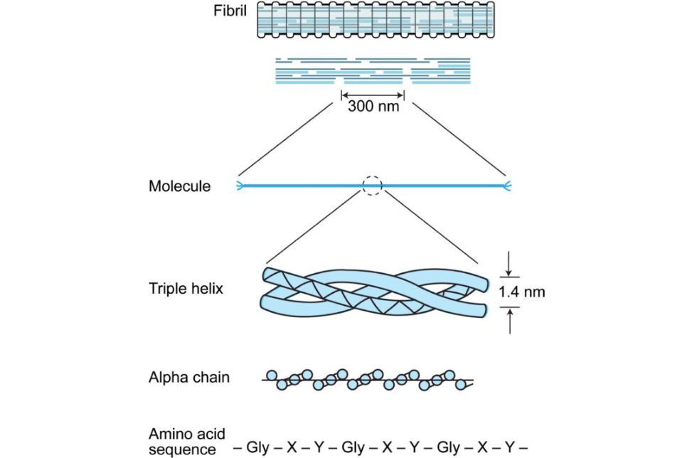
Figure 20.4 Several tropocollagen molecules are aggregated in an organized head-to-tail fashion into a structure called a collagen fibril. These collagen fibrils can beseen with an electron microscope and exhibit a 67-nm D-period banded appearance due to staggered gaps between the heads and tails of the molecules in each row.
How is collagen assembled?
Collagen biosynthesis and assembly is a complex process that involves several steps. Intracellular events include post-translational hydroxylation of proline and lysine and subsequent glycosylation. Pro collagen is secreted out of the cell. Extracellular events include terminal peptide cleavage and cross-linkage and self-assembly of collagen fibrils.
What do we mean by osteoclastogenesis?
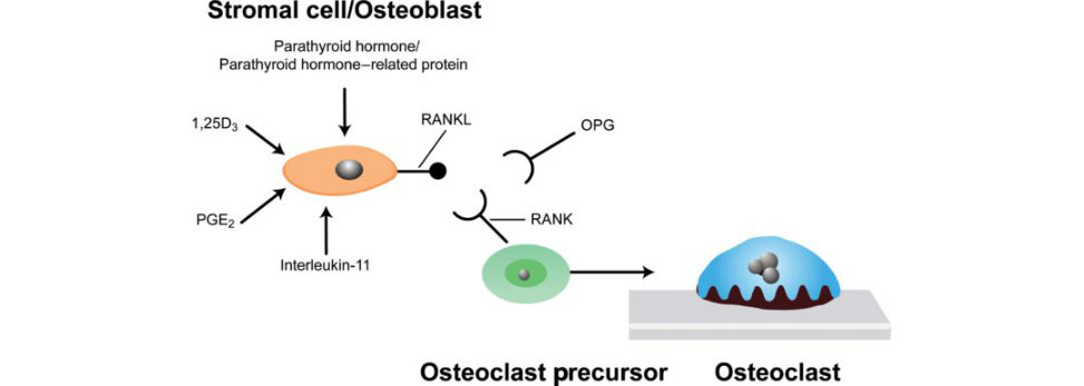
Osteoclastogenesis refers to the process of osteoclast differentiation and function. It is regulated by receptor activator of nuclear factor κB (RANK), RANK ligand (RANK Land osteoprotegerin (OP GRANK is expressed on the surface of osteoclast precursors and mature osteoclasts. Osteoblasts secrete receptor activator of nuclear factor κβ ligand (RANK Land macrophage colony- stimulating factor (M-CSF) to activate osteoclasts. RANK Lisa potent inducer of osteoclast formation. With RANK activation, specific genes are switched on and the osteoclast becomes programmed to resorb bone. OPG acts as a decoy receptor, blocking RANKL binding and subsequent activation of the
RANK system, thus inhibiting osteoclast differentiation and bone resorption.
What are osteotropic factors?
Osteotropic factors include1,25-dihydroxyvitamin D3, parathyroid hormone, prostaglandin E2, and interleukin 11. They induce the formation of osteoclasts by upregulating RANKL expression on the surface of marrow stromal cells and immature osteoblasts (see Figure 20.5).
What is an osteoclastic cuting c one?
Figure 20.5 Osteoblast/osteoclast coupling.
An osteoclastic cuting c one is a mechanism to remodel cortical bone by osteoclastic tunnelling (cuting c ones). Osteoclasts at the front of the cuting c one remove bone and are followed by layering of osteoblasts and successive deposition of layers of lamellae after the cement line has been laid down. The tunnel size narrows to a Haversian canal. The head of the cuting c one is made up of osteoclasts (which boreholes through hard cortical bone). Behind the osteoclast front are capillaries followed by osteoblasts (which laydown osteoid to fill the resorption cavity).
Can you draw a cuting c one out?
[Figure 20.3] Remember to keep the diagram simple.
Draw me the structure of bone.
Again, keep the diagram simple. Use the orthopaedic 20-second diagram. Candidates should discuss the structure of bone as they draw paying particular attention to Haversian systems (Figure 20.6).
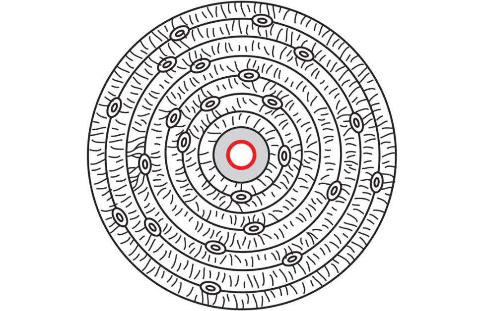
Figure 20.6 Structure of bone. Candidate drawing.
What is bone composed of?
Bone consists of cells (10%) and extracellular matrix (90%). The extracellular matrix has organic (40%) and inorganic (60%) components. Organic (40%). Collagen (type I) 90%. Proteoglycans. Matrix proteins (non-collagenous).
- ▪ Osteocalcin, osteonectin, os teopontin. ·
Growth factors and cytokines.
Inorganic (60%).
Primarily hydroxyapatite Ca5(PO4)3(OH)2.
What type of collagen is present in bone?
Type 1 [Remember – BONE].
Draw me some collagen.
Figure 20.7(a) focuses on the triple helix. Figure 20.7(b) is more complicated, but allows candidates more opportunity to focus on the hierarchical collagen arrangement.
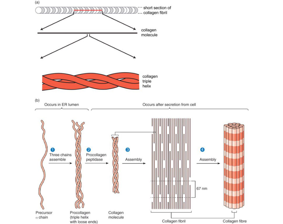
Figure 20. 7 (a) Candidate drawing. Structure of collagen. (b) More complex drawing of collagen assembly.
What do you know about the collagen structure in osteogenesis imperfecta (OI)?
Collagen is type 1 in bone. The structural unit of type 1 collagen is called tropocollagen and is a trimer composed of three polypeptide chains. T wo chains are α1 chains and the third chain is α2. The three chains form a distinctive unit in which the polypeptide chains wraparound each other for most of their length, forming a tight triple helical braid. The collagen triple helix forms because both the α1 and α2 chains contain repeat sequences of amino acids (-gly-X-Y), where gl yis glycine, X is proline andY is usually hydroxyproline. This arrangement results in a constrained structure that imparts a tight kink in the polypeptide chain with the glycine chain preventing steric hindrance that would otherwise impair wrapping of the helical band.
Wit hOI the majority of identified mutations are single nucleotide substitutions that result in alteration of glycine codons within the triple helical domain of either of the chains of type I procollagen (Figure 20.8). These substitutions for glycine within the triple helix are severely destabilizing and interrupt the triple helix. One-fitih of glycine substitutions in α2(I) are lethal, whereas nearly one-third of all glycine substitutions in α1(I) are lethal.
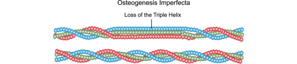
Figure 20.8 Collagen structure in osteogenesis imperfecta. Triple helix steric hindrance.
What is the gene coding fo rOI?
COL1A1 and COL1A2 are the genes that encode the two chains pro α1(I) and pro α2(I), respectively, of type I procollagen.
What about qualitative versus quantitative collagen deficiencies in osteogenesis imperfecta (OI)?
OI is a group of disorders with broad variations in clinical severity. Inheritance can be autosomal dominant or recessive. Quantitative defects are often heterozygous, with one copy not producing any collagen. Qualitative defects are usually errors in substitution or deletion leading to abnormal, less effectual collagen.
What about compression and tension of bone?
An eccentrically loaded bone has a compression and tension side. When bending occurs, one side of the bone is intension, the other side is in compression. Bone resists compression better than tension and the side with tension will fracture first. Whenever feasible, any internal or external fixation device should be applied to the tension side to provide maximum stability.
Draw me a longitudinal cross section of a long bone and t ell me the areas (Figure 20.9).
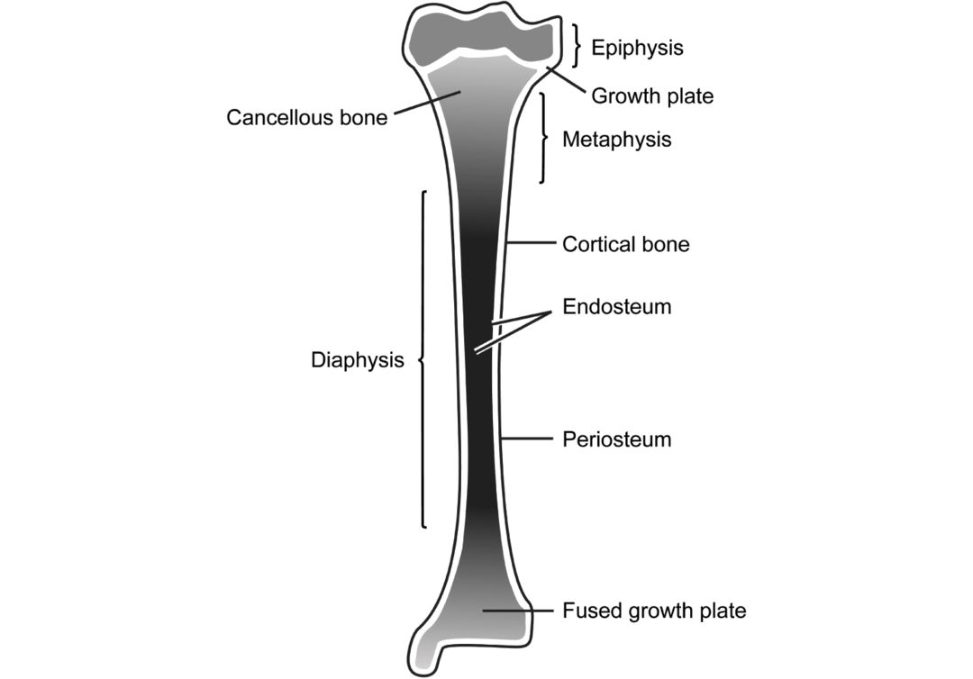
Figure 20.9 Drawing of a typical long bone.
Where are the cells? Where are the osteoblasts? Where are the osteocytes?
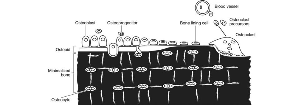
Osteoblasts are responsible for laying down new bone and are found in the growing portions of bone such as the periosteum and endosteum. Osteocytes occupy lacunae that are contained within the calcified matrix of bone between lamellae. Osteocytes are derived from osteoblasts and are essentially osteoblasts surrounded by the products they secrete (Figure 20.10).
What do you know about skeletal dysplasias?
Skeletal dysplasias are a large group of rare, complex, heterogeneous disorders that involve cartilage and bone. Rubin’s classification of bone dysplasia is based on the type of abnormality (hypo-/hyperplasia) and the site involved in the bone: (1) epiphyseal, (2) physeal, (3) metaphyseal, (4) diaphyseal location. Table 20.2 Cortical bone versus cancellous bone.

Structured oral examination question 2#
Draw me the structure of cortical bone (Figure 20.11a and 20.11b).
The cross-section of a long bone was quickly drawn, but we then almost immediately moved on to discussing the structure of compact bone.
What runs in a Haversian canal?
Haversian canals contain blood vessels lymphatics and nerves and are enclosed by closely packed concentric lamellae of bone. These canals branch into large transverse Volkmann canals that provide circulation to the cortical bone.
What is the Haversian system?
The Haversian system, or osteon, is the basic structural unit of cortical bone and lies parallel to the long axis of the bone. The osteon consists of a central Haversian canal that transmits a neurovascular bundle; surrounding this canal are at least five concentric lamellae of collagen. Within each lamella, collagen fibres lie in parallel but perpendicular to those fibres of adjacent lamellae. Volkmann’s canals run transversely to the bone’s long axis and permit communication between the outer vessels of the periosteum and the Haversian canals. Cement lines separate osteons.
Where do osteocytes originate from?
Osteocytes are trapped osteoblasts located within lacunae between lamellae, communicating with adjacent osteocytes via cytoplasmic processes that travel through canaliculi.
What do canaliculi do?
Canaliculi are the spaces or ‘canals’ occupied by osteocyte cell processes. They connect the lacunae together within the cortical bone. Canaliculi are thought to modulate the response of bone to mechanical stimuli. Simple basic questions on the structure of bone, but in the viva situation the y usually appear much more than straighfoorward.
How do bisphosphonates work?
Bisphosphonates are a class of anft-r esorptiv e agents used to treat diseases characterized by osteoclast-mediated bone resorption. There are two classes of bisphosphonates. 1. Nitrogen-containing bisphosphonates (etodronate). 2. Non-nitrogen-containing (alendrolate, zoledronate, risedronate).
They act differently to diminish bone resorption.
Non-nitrogen-containing BPNs are metabolized into non-functioning ATP analogues, which cause eventual osteoclast apoptosis.
Nitrogen-containing BPNs act by inhibiting farnesyl pyrophosphate synthase (FPPS), resulting in decreased prenylation of small GTPases. These small GTPases are signalling proteins that regulate a number of cell processes such as membrane ruffling, cytoskeletal organization and trafficking of vesicles, which are required for osteoclast function.
Can you tell me some clinical uses of bisphosphonates?
Clinical uses would include osteoporosis, hypercalcaemia of malignancy, Paget’s disease, solid tumours and metastatic bone disease AVN and stress fracture. With children they can be used in fibrous dysplasia, osteogenesis imperfecta and Perthes’ disease.
Structured oral examination question 3#
Bone healing, primary vs. secondary. Including cellular signalling pathways. Draw an osteonal cuting cone.
What do we mean by primary bone healing?
Primary bone healing is a direct attempt by the cortex to re-establish the Haversian structure after a fracture but without fracture callus formation. Primary bone healing is led by osteonal cuting c ones that consist of osteoclasts at the front of the cone that ream out a tunnel in the bone into which a blood vessel grows. This is followed by trailing osteoblasts that laydown new bone across the gaps to form a secondary osteon. Primary bone healing occurs only under low interfragmentary movement (rigid fixation). The reis direct osteonal remodelling and healing without external callous formation. Primary bone healing can be further divided into gap and contact healing.
What is contact healing?
Contact healing occurs if bone fragments have direct appositional contact and the gap between bone ends isless than 0.01 mm with an interfragmentary strain of less than 2%. Osteons are able to grow parallel to the long axis of the bone by tunnelling osteoclastic activity . Osteons traverse the fracture line.
And gap healing?
Gap healing occurs when a small stable fracture gapless than 1 mm is present between bony fragments. The fracture site is initially filled with transverse lamellar bone without intermediate fibrous or cartilage precursors. The bone is initially deposited perpendicular to the long axis of the bone prior to later osteonal remodelling along the lines of functionals tress. Contact healing – essentially no or minimal fracture gap. Direct osteonal remodelling. Absolute stability. Gap healing – small stable fracture gap.
What is secondary bone healing?
Secondary bone healing involves the formation of fracture callus. There is: 1. Haematoma formation with de vascularization of the bone ends. 2. An inflammatory phase with local accumulation of macrophages, MSC, cytokines. Delivery of osteoprogenitor cells from the periosteal cambium layer and osteoclasts starting to remove necrotic bone Coagulation and fibrin formation.
3. Primary soft callus formation. New blood vessels invade the haematoma, fibroblasts from the periosteum colonize the haematoma and produce collagen fibres. Granulation tissue gradually differentiates into fibrous tissue and afterwards fibrocartilage.
4. Callus mineralization (hard callus). Osteoblasts laydown woven bone at the periphery
(intramembranous ossification) and fibrocartilaginous callus bridges the fracture site, chondroid matrix calcifies, new woven bone is laid down (endochondral ossification).
5. Remodelling. According to Wolf fand Heuter–Volkmann’s laws: remodelling occurs with motion a t the fracture site. Cartilage is found during the early stages of healing replaced by woven bone laid downby osteoblasts.
Primary and secondary bone healing comes up repeatedly in the viva exam and candidates need to be very clear about the distinction between them. Key points are as follows: Primary bone healing Requires anatomical reduction and inter fragmentary compression. Absolute stability. This system will not tolerate strain. Be clear about the differences between gap and contact healing. Be able to describe and draw a cuting c one. Secondary bone healing Relative stability. Endochondral and intramembranous ossification. Fibrocartilage develops at the bone ends and this is subsequently calcified and replaced by woven bone or osteoid. Haematoma, inflammation and cellular proliferation, soft callus (chondrogenic and osteogenic proliferation), hard callus and remodelling.
What is Perren’s strain theory?
Perren introduced the importance of strain in fracture healing. Fracture gap strain is defined as the relative change in the fracture gap (ΔL) divided by the original fracture gap (L). Tissue cannot be produced when strain conditions exceed the tissues train tolerance. Cortical bone can only tolerate 2% strain. Rigid internal compression fixation, which minimizes strain, will lead to primary fracture healing. Lamellar bone can tolerate up to 10% strain, and when this relative stability is present, the fracture heals with callus or secondary fracture healing. Fracture healing will not occur when the strain at a fracture gap exceeds 10%. Comminuted fractures can tolerate more motion than simple fractures, because in a comminuted fracture the overall motion is shared among many fracture gaps.
[At the very end of the topic after discussing primary and secondary bone healing, Perren’s strain theory] There was a very well-wrift en important paper published recently that discussed fracture healing. This paper mentioned Per ren’s strain theory and introduced some exciting new ideas on fracture healing to challenge our traditional views on these complicated healing mechanisms.
Which paper is that?
It was a paper published in the JBJS.2
What did the paper say?
I haven’t had timet o read it fully, so I am not too sure, but it’s a really well-wrift en paper that everyone keeps mentioning and I am going to sit down and read it when I have more time. The candidate should have kept quiet, they have ruined an otherwise good performance (score 7) and would probably be marked down to a 6. Candidates should refrain from mentioning a paper they haven’t fully read and struggle to say anything sensible about.3 Ellioft et al. consider the whole fracture to be a ‘bone-healing organ’ that works as a functional unit and responds to biological and mechanical stimuli. The y combined Wolf flaw, Perren’s strain theory, and Frost’s ‘mechanostat’ model to create their own model of bone homeostasis, healing and non-union (BHN conceptual model). Using BHN, the behaviour of a ‘bone-healing organ’ was determined with respect to the mechanical strain applied to the organ. In BHN, the bone is in homeostasis when under tolerable strain (much less than 2%). For strains greater than 2% and less than 100%, a fracture occurs and is considered to be the beginning of the ‘bone-healing organ’. Finally, for strains above 100%, the ‘bone-healing organ’ stops and fails to heal, leading to non-union.
Structured oral examination question 4#
Which cells reside in bone?
Osteoblasts, osteoclasts, osteocytes, bone lining cells and osteoprogenitor cells. Do not forget osteoprogenitor cells that are derived from primitive mesenchymal cells and are located in the inner cellular layer of the periosteum, the endosteum and the lining of osteonic canals (Figure 20.10).
Figure 20.10 Bone remodelling. The origins and locations of bone cells.
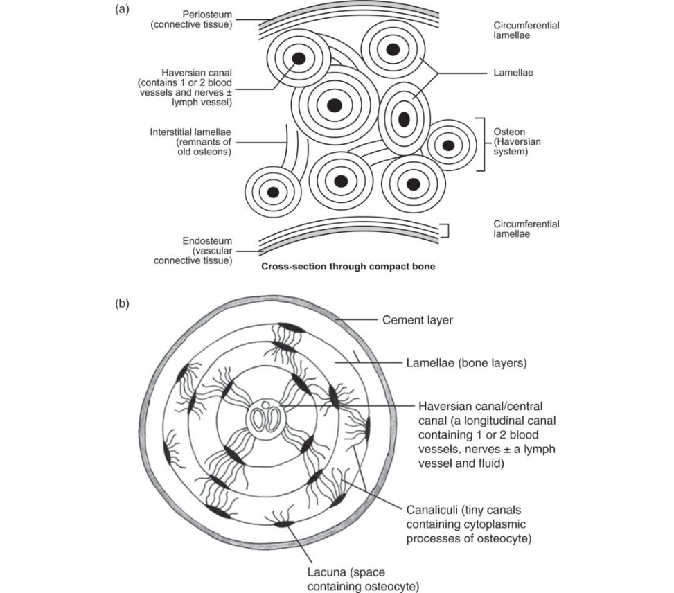
Figure 20.11 Candidate drawing. Cross-section of (a) long bone and (bos teon.
What do they all do?
Osteoblasts are large cells responsible for the synthesis and mineralization of bone during both initial bone formation and la ter bone remodelling. Osteoblasts form a closely packed sheet on the surface of the bone, from which cellular processes extend through the developing bone. Osteocytes lie within the substance of fully formed bone. They lie within a small space called a lacuna, which is contained in the calcified matrix of bone. Osteocytes are derived from osteoblasts, or bone-forming cells, and are essentially osteoblasts surrounded by the products they secreted. Cytoplasmic processes of the osteocyte extend away from the cell toward other osteocytes in canaliculi. These canaliculi allow nutrients and waste products to be exchanged to maintain the viability of the osteocyte. [Examiner cut me off, as he realized I would sit and regurgitate the whole book!]
Where are osteoclasts derived from?
Osteoclast precursors are a member of the monocyte/macrophage family, and, although the resorptive cell can be generated from mononuclear phagocytes of various tissue sources, the principal precursor resides in the marrow. RANK Lis produced by osteoblasts, binds to immature osteoclasts and stimulates differentiation in to active mature osteoclasts and macrophage colony stimulating factor (M-CSF). Osteoprotegerin inhibits bone resorption by binding and inactivating RANKL. A lot of candidates get confused in this section.
Tell me about mesenchymal stem cells.
MSCs are multipotent stem cells that can differentiate into a variety of cell types. Cell types that MSCs have been shown to differentiate in vitro or in vivo include osteoblasts, chondrocytes myocytes and adipocytes.
What types of cartilage do you know about?
There are three types of cartilage hyaline cartilage, fibrocartilage and elastic cartilage. Elastic cartilage exists in the epiglotis and the eustachian tube.
Draw a cross-section of articular cartilage (Figure 20.12).
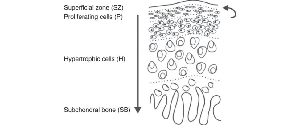
Figure 20.12 Articular cartilage layers.
Draw and talk about the different layers. This is one of the commonest questions to get asked in the basic science viva, know it well! Fail to draw this diagram at your peril! Practise the drawing and discussion of it at least 10 times.
What are the differences between articular cartilage and meniscus?
Articular cartilage is 68–85% water, 10–20% (type II) collagen and 5–10% proteoglycans. Meniscus is 60–70% water, 15–25% (type II) collagen and 1–2% proteoglycans. It’s easy to get confused with these facts.
Can you draw a picture of collagen and proteoglycans?
Draw the standard picture seen in many textbooks (Figure 20.13).
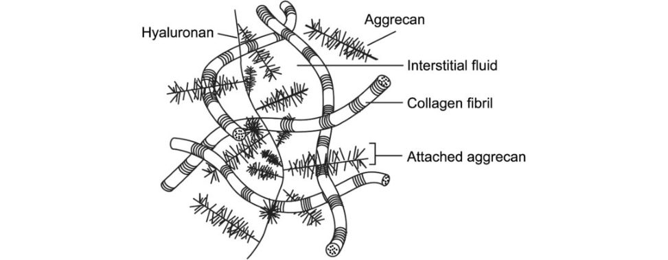
Figure 20.13 Collagen and proteoglycan arrangement in articular cartilage.
What’s the importance of water?
30% of the total water exists between the collagen fibres and this is determined by the negative charge of the proteoglycans which lie within the collagen matrix. Because the proteoglycans are bound closely, the closeness of the negative charges creates a repulsion force that must be neutralized by positive ions in the surrounding fluid. The amount of water present in cartilage depends on the concentration of pr oteoglycans and the stiffness and strength of the collagen network. The proteoglycan aggregates are basically responsible for the turgid nature of the cartilage and in articular cartilage they provide the osmotic properties needed to resist compressive loads. If the collagen network is degraded, as in the case of OA, the amount of water in the cartilage increases because more negative ions are exposed to draw in fluid. The increase in fluid can significantly alter the mechanical behaviour of the cartilage. That is a very specific question, but a common theme.
Bearing in mind what we have discussed, what do you want to talk about next?
Growth plates.
Correct answer! Tell me about any classifications you know of specific to the growth plate.
Salter–Harris fractures are classified from types I toV in the order of prognosis, with Salter– Harris Type V having the poorest prognosis and the greatest impact on potential deformity. SH I is a slipped growth plate, II the fracture lies metaphyseal, III is epiphyseal, IV both metaphyseal and epiphyseal and inV the physis suffers a compression injury. Salter–Harris Type II fractures are the most common. When all types of Salter–Harris fractures are considered, the rate of growth disturbance is approximately 30%. However, only 2% of Salter–Harris fractures result in a significant functional disturbance.
The fracture types described later, also less common, include:
Type VI (injury to the perichondral structures – rare).
Type VII (isolated injury to the epiphysis only).
Type VIII (isolated injury to the metaphysis).
Type IX (an injury to the periosteum which could interfere with membranous growth).
I would not mention an y of this unless you get asked. You may end up speaking to an orthopaedic paediatric professor!
Draw me a growth plate.
[I drew a simple schematic like Figure 20.14] Zone I is the reserve or resting z one with low rates of proliferation, pr oteoglycan synthesis and typeIIB collagen. These cells have a high lipid bodyand vacuole content which is involved withstorage for later nutritional requirements. This is nota germinal layer of ‘mother cartilage cells’. Zone II is the upper proliferative or columnarregion. The function of the proliferative zones ismatrix production and cell division that results in longitudinal growth. Chondrocytes are flat anda rearranged longitudinally. The zone is the truegerminal layer of the growth plate and Type IIcollagen synthesis is increased. Zone III is the mature proliferating z one,morphologically similar to zone II, but with less DNA synthesis. Zone IV is the hypertrophic zone, where cell size increases, and the columnar arrangement isless regular. Metabolic activity is high, with matrix synthesis approximately threefold compared to the proliferative zone; the main matrix componentssynthesized are types II and X collagenand aggrecan. [I got stopped at this point but will continue for completeness] Zone V is the zone of the matrix calcification as this calcified matrix becomes the scaffolding forbone deposition in the metaphysis. High levels of alkaline phosphatase synthesis of type X and type II collagen cell death by hypoxia. Zone VI is the junction of the growth plate with the metaphysis, the region where the transition from cartilage to bone occurs. Type I collagen, amarker of the osteoblast phenotype, isimmunolocalized to this area.
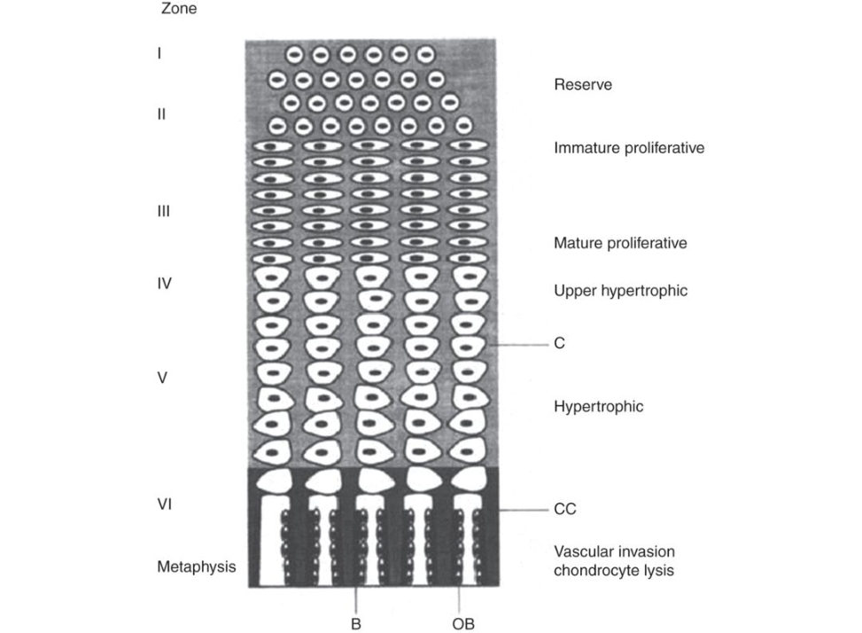
Figure 20.14 Diagram of a growth plate. B, bone; OB, osteoblast; CC, calcified cartilage; C, cartilage matrix.
There are a lot more facts in many textbooks, but if you know roughly about each zone that will be fine for a pass, more detailed knowledge will equal a good pass. The zones of the growth plate can be confusing as many textbooks use a variety of differing names, some referring to cell function, others to cell morphology. A non-controversial standard textbook zone description would be: (1) reserve zone, (2) proliferating z one, (3) hypertrophic zone which can be subdivided into (a) maturation z one, (b) degenerative zone, (c) zone of provisional calcification, (4) metaphysis subdivided into (a) primary spongiosa and (b) secondary spongiosa.
How is the growth plate regulated?
The growth plate is regulated by growth factors, hormones and vitamins. Growth factors include insulin-like growth factor, which is one of the most potent GF for skeletal tissue previously known as somatomedins. They have significant effects on the growth plate chondrocytes, and IGFs retained in bone matrix are important in the regulation of bone remodelling. IGFs stimulate osteoblast and chondrocyte proliferation, induce differentiation in osteoblasts and maintain the chondrocyte phenotype.
Transforming growth factors (TGFs) have an important role in skeletal tissue, particularly certain members of the TGF-b gene family which includes the bone morphogenetic proteins involved in morphogenesis and regulation of endochondral ossification and in bone remodelling. BMPs are the only molecules so far discovered capable of independently inducing endochondral ossification in vivo.
Urist 1965 is the paper to quote.4 Be able to mention something about BMP2 and BMP7. Key message5 Over 50 years ago, Urist made the key discovery that demineralized bone fragments implanted either subcutaneously or intramuscularly in animals induced bone formation. The extracellular matrix of bone contains substances that can stimulate new bone formation when implanted into extraskeletal sites in a host. These substances were later identified as bone morphogenetic proteins (BMPs). Both BMP2 and BMP7 are approved for use in acute tibial fractures and complex non-unions. They increase the local signals needed to initiate the cascade of bone healing. Both BMP2 and BMP7 have been shown to induce ectopic bone formation. At present, the use of BMP2 is preferred as studies suggest it may be more effective than BMP7 at promoting healing and it is also less costly. Study outcomes can be highly variable according to indication, implying that more rigorous prospective studies are needed to precisely identify the fracture population, timing and delivery mechanism via which BMP can be used to optimize bony healing. Fibroblast growth factor stimulates proliferation of mesenchymal cells in the developing limb that leads to limb outgrowth. This group is also involved in later stages of bone growth. Platelet-derived growth factor plays a role in bone development and growth, being important in the regulation of bone and cartilage cells, although liti leis currently known of their role in normal endochondral ossification. Tumour necrosis factors stimulate bone and cartilage resorption and division, and reversibly inhibit ectopic bone formation in animal models. If you get to this stage it’s a good pass. Other growth factors to think about are: interleukin I,6,8, interferons, colony-stimula ting factors, parathyroid hormone-related peptide and calcitonin gene-related peptide.
2. Structure and function of cartilage: (a) articular#
Introduction#
This is an A-list basic science viva topic that almost always appears with each diet of exams. In practical terms this means a candidate has a 1 in 4 or 25% chance of being asked this topic.
In the past, there was a predictable line of questions the examiners would ask. However, most candidates are now more aware of A-list topics than ever before and have pre-learnt their answers. As such the challenge for the Intercollegiate Board is to avoid asking the same questions each and every exam siting.
To make the subjectless predictable the topic focus can be changed mid viva onto different more detailed areas within this large topic such as proteoglycan structure and function.
The flip side is that regular examiners become more familiar with the topic and on occasion may ask esoteric questions to stretch you out to see if you are a possible score 7/8 candidate. What is the function of the tidemark and what insets into it, etc.
One word of caution with A -list topics is that candidates can continue to read further and further into a subject and end up concentrating on unfocused minutia details that have no relevance to any possible viva question likely to be asked.6 This is very different to reading extra details, but being able to apply these details into higher-order thinking to better answer a question. However, it is sometimes a thin line between the two.
Props#
Candidates may immediately be handed a laminated photograph of articular cartilage at the start of the viva or be asked a couple of warm-up questions before being asked to drawout the structure of articular cartilage Articular cartilage is definitely on the top 10 list for ‘must-know’ how-to-draw diagrams.
Structured oral examination question 1#
What are the functions of articular cartilage?
The two main functions are: To provide a smooth, low-friction, lubricated surface for joint motion lubrication). The surfaces roll or glide during motion. Hyaline cartilage should bethought of as a fluid-filled wear-resistant surface. It reduces the coefficient of friction down to 0.0020, which is 30 times superior to the best performing artificial joint. To resist the compressive forces encountered across the joint under loading (shock absorber). These are the two main functions of articular cartilage, although some textbooks mention other minor roles.7 There is some controversy as to the relative importance of articular cartilage as a shock absorber. Some authors have suggested that because it is a very thin structure it has negligible shock-absorbing capacity compared to the surrounding muscle and bone. Again, we suggest stay simple and don’t mention this standpoint unless it comes up in discussion (very unlikely).8
Can you draw the histological appearance of articular cartilage? [Draw the structure of articular c artilag e.9] What are the articular cartilage layers?
This is one of the commonest diagrams candidates will be asked to draw in the basic science viva. Fail to master this diagram at your peril. Candidates should be able to draw the various layers of articular cartilage without hesitation Candidates may then be asked to explain why the layers appear like this, with reference to the three-dimensional ultrastructure.10 The histological appearance of articular cartilage is structured into zones ...: Take the examiner sequentially through the layers. Focus your discussion on (1) the differing orientation of type II collagen fibrils, (2) orientation and cellular features of the chondrocytes. Why is there a different pattern of collagen orientation through the layers of cartilage? How does the differing pattern of collagen affect its properties? You ‘the candidate’ should explain this as you drawout articular cartilage and not allow the examiner to get in and ask you these questions. 11,12 Structure. The histological structure can be divided into four zones. 1. Superficial (tangential/ gliding) zone: 10–20% of thickness.
Collagen fibres are arranged parallel to the joint surface, forming a dense mat.
The most superficial partis called the lamina splendens,13 providing a very low-friction lubrication surface. It contains no cells, a clear film of collagen fibrils with litile pr oteoglycan.
This dense collagen arrangement reduces leakage of proteoglycans from the articular surface and protects it from the effects of harmful enzymes.
Below this is a cellular layer with chondrocytes parallel to surface, flat-shaped, high density, many cells 1–3 thick.
This layer provides good resistance to shear forces due to tangential arrangement of collagen and provides the greatest tensile strength.
Low metabolic activity , hence low healing potential.
Thinnest layer with the highest concentration of collagen and water and the lowest concentration of pr oteoglycan.
Water can be squeezed out of the layer to help create lubrication.
May function as a barrier to the passage of large molecules from the synovial fluid.
2. Middle (transitional) z one: 40–60% of thickness.
Collagen fibres arranged obliquely at right angles to each other.
Plentiful concentration of pr oteoglycan.
Chondrocytes arranged in random orientation round shape, progressively lower density and fewer cells.
Transitional z one between the shearing forces of the surface layer and resistance to compression in the deep layer.
3. Deep (radial) zone: 30% of thickness.
Provides resistance to compression.
Collagen fibres vertically arranged (perpendicular to articular cartilage) cross the tidemark and are anchored to subchondral bone.
Highest concentration of pr oteoglycans.
Chondrocytes spherically arranged in vertical columns.
Collagen fibres largest diameter.
Lowest water content.
4. Calcified zone.
This separates the cartilage tissue from the underlying subchondral bone.
Anchor for the various layers.
Collagen type X and hydroxyapatite crystals anchor articular cartilage to subchondral bone.
Forms a barrier to blood vessels supplying subchondral bone.
Matrix mineralization in the calcified zone allows gradual transition of mechanical properties between cartilage and bone.
Tidemark
The junction between the deep and calcified zone is called the tidemark.
As a candidate is describing the histological structure of articular cartilage an examiner may start to probe/interrupt/take over14 and ask esoteric15 questions.
What is the tidemark? What attaches to the tidemark?
The tidemark provides resistance to shear. It is the boundary between the calcified and uncalcified cartilage. It is cell-free and represents a calcification fr ont. The collagen fibres in the deep zone penetrate through the tidemark in to the calcified cartilage to provide structural stability for articular cartilage on the subchondral bone. It is a smooth, basophilic undulating line that is critical for the transmission of load from cartilage to bone.
What is the composition of articular cartilage (Table 20.3)?
The wet weight proportions of articular cartilage are water (65–80%), collagen (10–20%), proteoglycans (10–15%), chondrocytes (5%) and other matrix components such as adhesives and lipids. This isn’t a wrong answer, but it’s a bit unstructured and cumbersome. A better answer would be to say articular cartilage is composed of cells (chondrocytes) accounting for 5% of the wet weight and extracellular matrix the remaining 95%. The extracellular matrix is made up of fibres and ground substance. The fibres account for 10–20% and are almost exclusively Type II collagen. Water accounts for between 65% and 80% of the extracellular matrix, proteoglycans and glycoaminoglycans 10–15% with small amounts of glycoproteins and degradative enzymes. Or more simply, articular cartilage is mainly composed of chondrocytes, water, Type II collagen, proteoglycans and a variety of matrix proteins. Do not mix around the wet and dry weight percentages of articular cartilage components; preferably stick to wet weight. If you have to talk about dry weight mention that collagen accounts for 40–70% of the dry weight and that approximately 90% of the dry mass of articular cartilage is made up of proteoglycan aggrecan, type II collagen and hyaluronan.
What are the contents of articular cartilage?
There is an overlap between describing the layers and discussing the contents of articular cartilage. Chondrocytes16 Derived from mesenchymal stem cells, chondrocytes produce and maintain EC Mand are the main cell type of articular cartilage. Deeper cartilage zones contain no chondrocytes. Low metabolic rate. Chondrocytes have no contact with neighbouring cells and are spheroidal. There are distinct subpopulations of chondrocytes in the different zones of cartilage whose properties differ in terms of their morphology, metabolism, and their response to cytokines. Cell morphology varies according to zone. The superficial zone contains a high density of flat cells with approximately three times as many cells in the region compared to the larger cells of the deeper zone. In the deeper zones, they are arranged in columns, reflecting the articular cartilage role ingrowth of the epiphysis. Water Up to 80% of the extracellular matrix. Permits load-dependent deformation of articular cartilage by its movement both in and out and within cartilage. Increased water content leads to increased permeability, decreased strength and decreased elasticity . Provides a medium for lubrication. Collagen About 10–20% wet weight, 60% dry weight. Gives the articular cartilage its tensile stiffness. The main collagen in articular cartilage is type II accounting for 90–95% of the collagen. Types II, IX and XI form a mesh that serves to trap proteoglycans, providing for stiffness and strength. Type VI helps chondrocytes adhere to the matrix. Increases in early OA. Type XI constrains the proteoglycan matrix.
Type X is only found near the calcified zone.
Proteoglycans
Proteoglycans are complex macromolecules composed of a protein core to which many glycosaminoglycan side chains are attached.
Proteoglycans trap and hold water, providing the tissue with its turgid nature that resists compression.
They are secreted by chondrocytes.
The most common glycosaminoglycan in articular cartilage is chondroitin-sulpha te (two subtypes, chondroitin-4-sulpha te and chondroitin-6-sulpha te), then keratin sulphate and dermatan sulphate.
Chondroitin-4-sulpha te is the most abundant and decreases over the years; chondroitin-6-sulphate remains constant; and keratin sulphate increases with age.
Glycosaminoglycan can link to a protein core by sugar bonds to form a proteoglycan aggrecan (see
Figure 20.14).
A proteoglycan aggrecan has three globular domains, G1, G2 and G3. G1 and G2 are near the N terminus and separated by a short interglobular domain. The third globular domain, G3, is near the C terminal of the core protein.
Aggrecan molecules do not exist in isolation within the extracellular matrix, but as proteoglycan aggregates through the interaction with the polysaccharide, hyaluronan.
Each aggregate is composed of a central filament of hyaluronic acid with up to 100 aggrecan molecules radiating from it, with each interaction stabilized by the presence of a link protein.
PG aggregation promotes immobilization of the PG swithin the fine collagen meshwork.
PG shave an average lifespan of 3 months and have a great capacity for retaining water, which gives compressive strength and elasticity to the tissue.
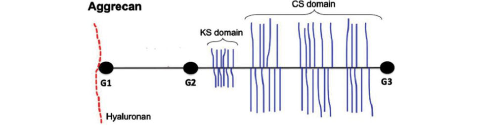
Figure 20.15 Proteoglycan aggrecan. A proteoglycan aggrecan has three globular domains, G1, G2 and G3.
What is the structure of a proteoglycan molecule?17,18
This involves differentiating between a proteoglycan aggregate and proteoglycan aggrecan molecule (Figure 20.14). It is not uncommon for candidates to be asked to drawout the structure of either a proteoglycan aggregate or aggrecan molecule (Figure 20.16) or sometimes both. Volunteer to draw both out for the examiners. As always, rehearse your drawing with dialogue as many times as needed to obtain a smooth, polished flow.
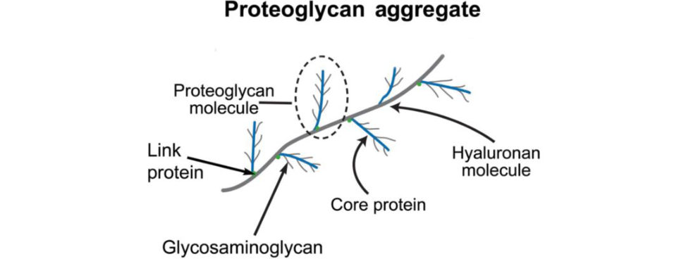
Figure 20.16 Candidate drawing of proteoglycan aggregate.
How does collagen synthesis take place? (Figure 20.17.)
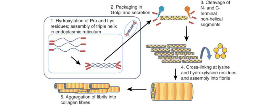
Figure 20.17 Collagen synthesis.
This question 19 may be asked as part of the main viva theme or as an add-on if a candidate has trail-blazed through previous viva questions and is on for a score 8. The secret is to try and simplify a complex process often poorly described in books. Split the process into intracellular and extracellular events. Procollagen is synthesized by a series of steps within the endoplasmic reticulum of cells such as fibroblasts. This molecule is then released into the extracellular space by transport through the Golgi apparatus and converted into collagen by procollagen peptidases that cleave the water-soluble, non-helical N- and C-terminal portions of the pro collagen molecule to form tropocollagen.
Collagen monomers are then covalently cross-linked with each other after certain residues are oxidized by lysyl oxidase.
Table 20.3 Constituents of articular cartilage.
Cells (chondrocytes) (5%)
Extracellular matrix (95%) Fibres Collagen (10–20%) Type II, IX, XI
Almost exclusively Type II Type VI, X
Elastin
Ground substance Water (65–80%)
Proteoglycans and glycosaminoglycans (10–15%)
Glycoproteins
Degradative enzymes (matrix metalloproteinases)
Steps that occur INSIDE the cell
Synthesis of pro-alpha chain.
Hydroxylation of selected proline and lysine residues.
Glycosylation of selected hydroxylysine residues.
Self-assembly of three pro-alpha-chains into triple helix.
Procollagen triple helix formation.
Then extrusion of procollagen from the endoplasmic reticulum/Golgi compartment into secretory vesicles and then secretion in to extracellular matrix.
Steps that occur OUTSIDE the cell
Cleavage of propeptides. Once secreted, procollagen peptidases remove the N-terminal and C-terminal propeptides.
Self-assembly into fibril.
Aggregation of collagen fibrils to form a collagen fibre. Formation of covalent cross-links by the enzyme lysyl oxidase. This enzyme allows hydroxyl groups on lysines and hydroxyl lysines to be converted into aldehyde groups that covalently bond between tropocollagen molecules to form a collagen fibril.
Closely related to synthesis is the structure of collagen and the line of questions may continue on with this subtopic.20
What is the structure of collagen (Figure 20.18)?
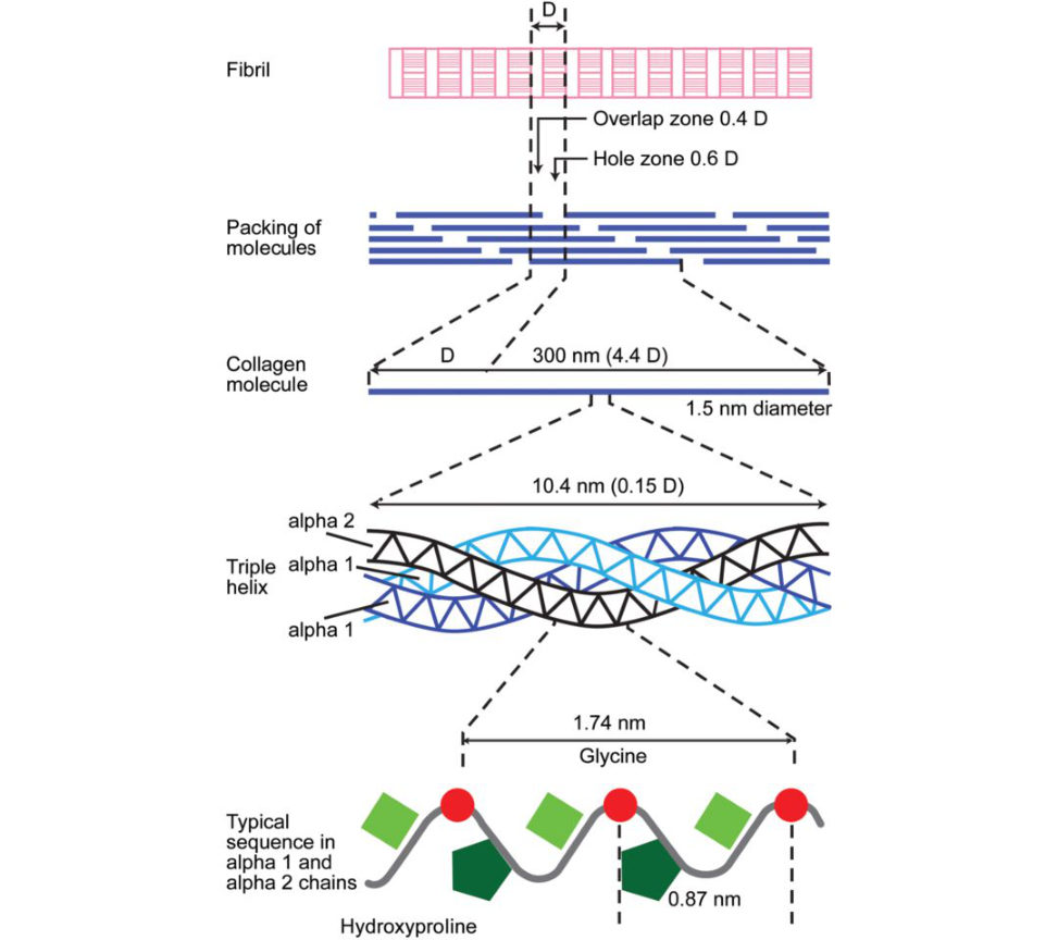
Figure 20.18 Hierarchical structure of collagen ranges from the amino acid sequence, tropocollagen molecules, collagen fibrils to collagen fibres.
Collagen is a triple helix of three polypeptide chains. The amino acids are glycine, proline and lysine. In articular cartilage collagen is mainly type 2.
What is the function of matrix glycoproteins?
These interact with collagen fibrils and stabilize the matrix framework.
They help chondrocytes bind to the macromolecules of the matrix.
Matrix glycoproteins are much smaller than aggrecans.
More simply, they act as a tissue glue binding to various matrix components.
What about matrix metalloproteinases?
These degrade collagen and proteoglycan aggregates as part of the normal turnover of the matrix.
What do we mean by tensegrity architecture?
Water is atir acted and retained in articular cartilage by the ionic pressure created by the high level of negative charges on glycosaminoglycan (GAG) chains on proteoglycan molecules. The ionic pressure creates a swelling pressure, which will keep imbibing water until it is resisted by the tension in the fibrous component of cartilage the collagen fibres. As proteogylcans are bound closely, the closeness of the negative charges creates a repulsion force that must be neutralized by positive ions in the surrounding fluid. The amount of water present in cartilage depends on the concentration of pr oteoglycans and the stiffness and strength of the collagen network. The proteoglycan aggregates are basically responsible for the turgid nature of the cartilage and in articular cartilage they provide the osmotic properties needed to resist compressive loads. If the collagen network is degraded, as in the case of OA, the amount of water in the cartilage increases because more negative ions are exposed to draw in fluid. The increase in fluid can significantly alter the mechanical behaviour of the cartilage. Candidates that do not understand this concept particularly well may become trapped quite easily by the examiners (just like water gets trapped by proteoglycans).
Tell me about matrix metabolism.
The production of extracellular matrix, its organization maintenance and breakdown are controlled by the chondrocyte. Chondrocyte regulators include hormones, cytokines and growth factors. Turnover of the matrix is needed for remodelling and maintenance. Enzymes include aggracanase and metalloproteinases degrade the matrix. The chondrocytes also make inhibitors of these enzymes called tissue inhibitor of metalloproteinases (TIMP).
What do we mean by the term matrix region?
An alternative zonal classification is by the matrix regions. The matrix is divided into three distinct regions with respect to the distance from the chondrocyte cell membrane.
Matrix regions differ in their collagen content, collagen fibril diameter, collagen fibril orientation and proteoglycan and non-collagenous protein content and organization.
There are three regions.
1. The pericellular matrix is a thin layer adjacent to the cell membrane, completely surrounding the chondrocyte. It contains mainly proteoglycans, as well as glycoproteins and other non-collagenous proteins.
The pericellular matrix region may play a functional r ole to initiate signal transduction within cartilage with load bearing.
2. The territorial matrix surrounds the pericellular matrix; it is composed mostly of fine collagen fibrils, forming a basket-like network around the cells.
The territorial matrix may protect the cartilage cells against mechanical stresses and may contribute to the resiliency of the articular cartilage structure and its ability to withstand substantial loads.
3. The interterritorial region is the largest of the three matrix regions; it contributes most to the biomechanical properties of articular cartilage.
This region is characterized by randomly oriented bundles of large collagen fibrils and large amounts of proteoglycans.
Structured oral examination question 2#
Articular cartilage changes with ageing versus osteoarthritis is more of a section 3 (pathology) topic but there will be a large overlap with section 2 material (structure and function of articular cartilage Throw in management options for a cartilage defect and this topic is equally at home in an adult and pathology viva.
Describe the changes in articular cartilage with ageing.
Articular cartilage undergoes significant structural, matrix composition and mechanical changes with age. With increasing age there is an age-related decline in the ability of chondrocytes to maintain the tissue Chondrocytes become less responsive to the proliferative and anabolic effects of growth factors. There is a marked increase in the formation of advanced glycation end-pr oducts (AGEs). This results in increased cross-linking of collagen molecules altering the biomechanical properties of cartilage resulting in increased stiffness and an increased susceptibility to fatigue failure. Candidates may be asked to directly compare the biomechanical changes of ageing with osteoarthritis inc artilag e. This can be rote-learned from various tables, but a more detailed understanding is a safer bet in case follow-up questions focus in on more comprehensive detail.
Describe the changes in articular cartilage with osteoarthritis. What pathological processes are involved in the development of osteoarthritis?
The process can be divided into three overlapping stages: (1) cartilage matrix damage, (2) chondrocyte response to tissue damage (3) decline of the chondrocyte synthetic response with progressive loss of tissue. In the early stages of disease, loss of proteoglycan is reversible, whereas at later stages there is irreversible loss. The earliest visible change is loss of collagen integrity resulting in tissue fibrillation and increased water content. Decreased aggrecan concentration and aggregation and decreased glycosaminoglycan chain length all increase the permeability and stiffness of the matrix and make it vulnerable to further mechanical damage. Despite chondrocyte proliferation and increased collagen and proteoglycan synthesis this response fails to halt disease progression. There is progressive loss of articular cartilage and a reduced chondrocyte anabolic and proliferative response. This is related to adown regulation of chondrocyte function. With osteoarthritis, the cartilage may show areas of softening, fibrillation, fissures or gross erosion. There may be areas of full-thickness cartilage loss with the subchondral bone exposed and often sclerotic.
Microscopic appearances include surface irregularities and erosions, deterioration of the tidemark, fissuring and damage to the cartilage structure. Water content is increased in OA, with a decrease in the proteoglycan content. The proteoglycan chain is shorter, and the chondroitin/k eratin sulphate ratio is increased.
Overall, the collagen content is maintained but the presence of collagenase disrupts its organization and orientation.
Have an answer rehearsed.
What are the management options for osteoarthritis?
Conservative measures include targeted physiotherapy, regular oral or topical analgesia/anft-in flammatories, intra-articular injections (corticosteroids or hyaluronic acid) and activity modification. Surgical management may include joint debridement, osteotomy, arthroplasty and arthrodesis.
What are the options for treating an articular cartilage defect?
There are three main types of cartilage injury: (1) superficial matrix disruption, (2) partial thickness defects and (3) full-thickness defects. Superficial matrix disruption arises from blunt trauma whereby the EC Mis damaged but viable chondrocytes aggregate into clusters and are capable of synthesizing new matrix. Partial thickness defects disrupt the cartilage surface but do not extend into the subchondral bone. These defects are unable to self-repair. Full-thickness defects arise from damage that penetrates deep into the subchondral bone. These defects can elicit a repair response due to access to marrow cells; however, they are typically filled with fibrocartilage This type of repair tissue is much weaker than hyaline cartilage and displays poor long-term performance due to poor compressive strength and durability.
What are the options for treating a symptomatic focal articular cartilage defect in the medial femoral condyle of the knee of a young active patien t?21
Appropriate management of an articular cartilage defect in a younger patient is often very challenging. Although the natural history of an isolated articular cartilage lesion is not completely understood, these defects may to lead to significant morbidity and progress to diffuse osteoarthritis in time. Current treatment options f all into three broad categories: 1. Mesenchymal stem cell (MSC) stimulation (micro fracture, drilling, abrasion chondroplasty). 2. Substitution options (osteochondral autograft transfer system [OATS], osteochondral allograft).
3. Cell-based, biological replacement options autologous chondrocyte implantation [A CIs tem cell therapy, tissue engineering).
Marrow stimulation techniques such as abrasion arthroplasty and microfracture penetrate the subchondral bone, causing bleeding within the cartilage defect that leads to fibrin clot formation.
Undifferentiated MSCs from the bone marrow migrate into the defect, proliferate and differentiate into fibrochondrocytes. These induce the formation of fibrocartilage repair tissue.
Although excellent short-term clinical outcomes have been reported, the clinical durability of marrow-stimula ted repair tissue declines with longer follow-up. The repaired articular cartilage generally fails to replicate the structure, composition and function of normal articular cartilage.
OATS is recommended for smaller lesions, lesions in high-demand athletes, and lesions with associated bone loss.
Microfracture is suited for medium-size defects with litile or no bone loss in lo wer-demand older patients.
Candidates may be asked more specific details about each option such as indications, complications results, especially if in the adult pathology viva and aiming for score 8.
Structured oral examination question 3#
As viva question 1 initially: what are the functions of AC, drawout and describe the layers of AC, etc. and then a focus midway through on the biomechanical properties of articular cartilage.
What are the biomechanical properties of cartilage?
Cartilage is a biphasic, viscoelastic and anisotropic material demonstrating both creep and stress relaxation. There is a debate whether cartilage is a biphasic (fluid and solid phase) or triphasic material (see below).
What do you mean by viscoelastic?
A viscoelastic material will exhibit a time-dependen t behaviour when subjected to a constant load or constant deformation.
What are the properties of a viscoelastic material?
A viscoelastic material demonstrates creep and stress relaxation. Creep is time-dependen t deformation of amate rial under constant load that is below its yield strength. Stress relaxation is the decrease in stress required to maintain constant strain over time. When a constant compressive stress (load/area) is applied to the tissue, its deformation will increase with time; it will creep until an equilibrium value is reached. Creep produces plastic deformation of amate rial. When the tissue is deformed and held at a constant strain, the stress will rise to a peak, followed by a slow stress–relaxation process until an equilibrium value is reached. Viscoelastic materials display four characteristics: 1. Creep. 2. Stress relaxation. 3. Hysteresis. 4. Strain rate-dependent mechanical properties. Hysteresis occurs when a viscoelastic material is cyclically loaded and unloaded. It is the ability of the material to dissipate energy between loading and unloading cycles. It is due to the fact that materials do not perfectly obey Hooke’s law. A viscoelastic material is harder to deform when loading than unloading.
Viscoelastic materials are stiffer, tougher and stronger when loaded at a faster rate (higher strain rate) because there isless time for them to strain. A given load produces less deformation over a shorter period of time than a longer period of time.
Can you drawout the graphs of creep and stress relaxation ( Figures 20.19 and 20.20)?
Yes, but my mind has gone blank.
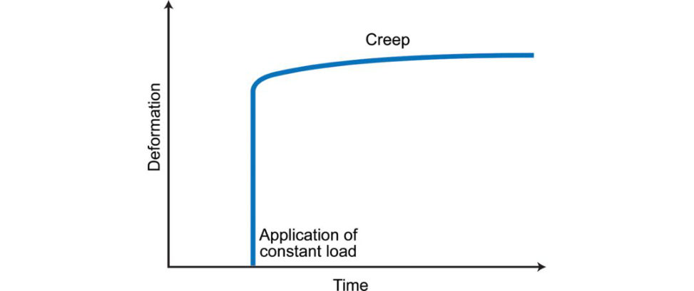
Figure 20.19 Creep. Continuous deformation overtime in response to constant load.
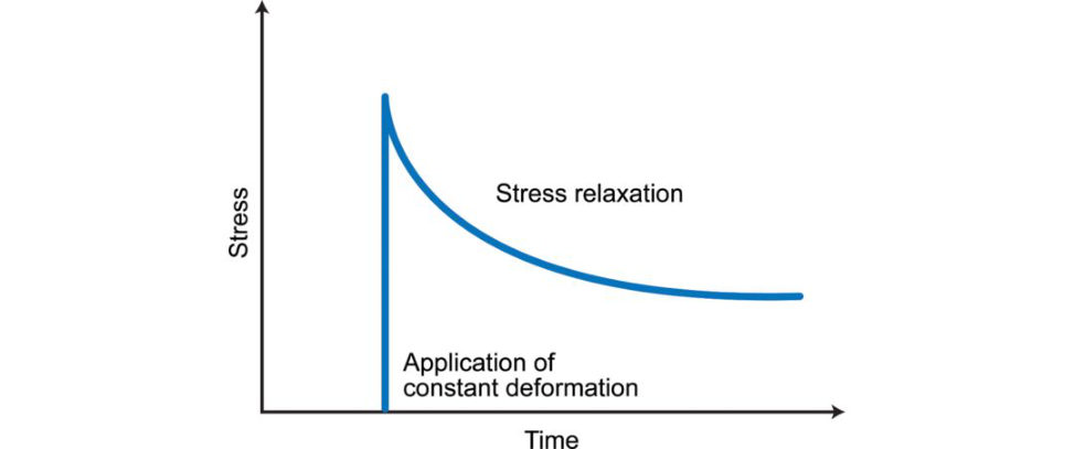
Figure 20.20 Stress relaxation. Time-dependen t decrease in stress required to maintain strain.
What about hysteresis and strain-dependent mechanical properties ( Figures 20.21 and 20.22)?
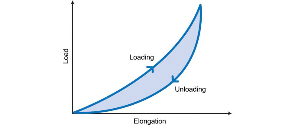
Figure 20.21 Hysteresis. Strain energy loss as heat due to internal friction between loading and unloading.
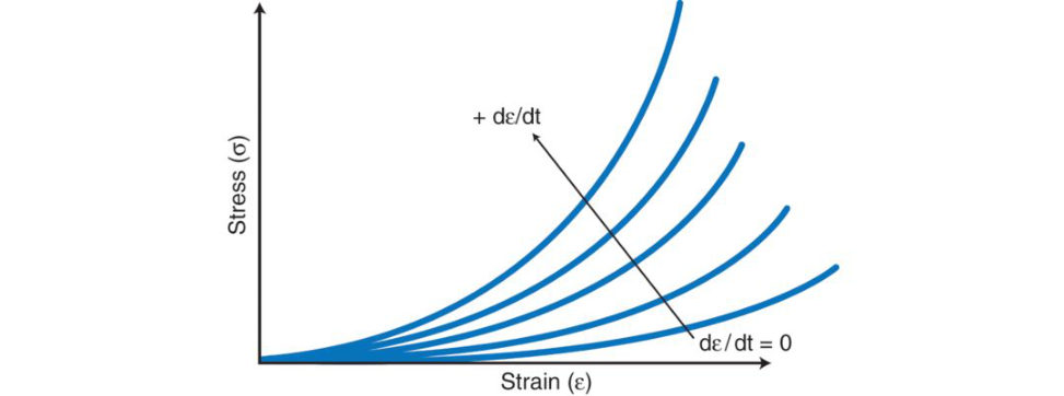
Figure 20.22 Time-dependent strain behaviour. Stress is proportional to strain rate.
No, sorry. These are predictable questions. The vi vais heading for a score 4 or at best 5.
What about articular cartilage permeability with compression?
Articular cartilage permeability decreases non-linearly with compression. This serves to regulate the response of cartilage to compression by preventing rapid and excessive fluid exudation from the tissue with compression loading and by promoting in terstial fluid pressurization for load support. It also regulates the ability of cartilage to dissipate energy during cyclic loading. There are two causes for this nonlinear effect. As the tissue is compressed: (1) The water content or porosity is reduced. (2) The density of the negative charges on the proteoglycans is increased.
There is a direct relationship between permeability and water content and an inverse relationship between permeability and proteoglycan content.
This is an esoteric question that tests knowledge of the interaction between proteoglycan and water. This is in the score 8 zone for candidates.
How does the internal architecture of articular cartilage relate to its biomechanical properties?
The presence of water within the tissue allows the support of most of the load through pressurized fluid. This fluid support is not uniform between the different zones of the tissue, with the superficial zone demonstrating the highest support (95%) compared to the deep zone (70% of applied load). Over timet here is a decreased interstial fluid support that causes an increased loading of the solid phase including chondrocytes. As such, articular cartilage can bethought of as a biphasic, porous model. However, dissolved electrolytes together with fixed charges of the solid matrix bring about mechanoelectrochemical phenomena adding to the load bearing of the tissue and described as a third phase. The triphasic nature of cartilage explains how fluid is drawn back into the cartilage matrix after being excreted during compression. While the triphasic nature of cartilage is a physically intuitive way to model cartilage, it is also very complex. The biomechanical properties of articular cartilage can be complicated to understand and tests higher-order thinking relating to structure. Biphasic creep behaviour of articular cartilage during compression. Rate of creep is governed by the rate at which fluid is forced out from the tissue, which, inturn, is governed by the permeability and stiffness of the por ous-permeable, collagen–proteoglycan solid matrix.
Structured oral examination question 4#
What are the properties of articular cartilage?
Cartilage is avascular, aneural anda lymphatic.
What are the articular cartilage changes that occur with ageing and osteoarthritis ( Table 20.4)?
This is an A-list basic science question. While tables provide a succinct summary they do not give a candidate the opportunity to practise out loud and rehearse their answer. Perhaps best remembered as changes occurring in the composition of articular cartilage, i.e. water, chondrocytes, collagen and proteoglycans.
What are the changes that occur with osteoarthritis?
With osteoarthritis, the cartilage may show areas of softening, fibrillation, fissures or gross erosion. There may be areas of full-thickness cartilage loss with the subchondral bone exposed and often sclerotic. Microscopic appearances include surface irregularities and erosions, deterioration of the tidemark, fissuring and damage to the cartilage structure. Water content is increased in OA, with a decrease in the proteoglycan content. The proteoglycan chain is shorter, and the chondroitin/k eratin sulphate ratio is increased. Overall, the collagen content is maintained, but the presence of collagenase disrupts its organization and orientation. The three main areas to consider with OA are (1) macroscopic changes, (2) microscopic changes and (3) synovial joint changes. Synovial joint changes include changes in periarticular musculature, and in articular and periarticular tendons and ligaments. There is synovial inflammation, joint capsule hypertrophy, meniscal degeneration thickening of subchondral bone and formation of osteophytes.
What is the relationship between ageing and osteoarthritis?
Ageing does not necessarily cause OA, but age related changes may provide a basis upon which OA can be initiated. During ageing, an imbalance between the catabolic and anabolic processes occurs. Age-related loss of the ability of chondrocytes and tissues within the E CM to maintain a homeostasis between these pathways leads to a procatabolic state favouring matrix degradation. This loss of homeostasis and inability to adapt to external mechanical stressors can in time become a precursor for the development of OA.
Despite ageing being a significant risk factor for OA, not all aged joints develop the disease.
What are advanced glycation end products (AGEs)?
Advanced glycation end products (AGEs) are the products of uncontrolled, non-enzymatic glycation, and oxidation reaction between proteins and sugars, and accumulate in the AC as a part of ageing, making the tissue britile. Due to its low metabolic activity articular cartilage is particularly susceptible to AGEs accumulation. The effects of AGEs formation include: Modification of type II collagen by cross-linking of collagen molecules: increasing stiffness and britileness, increasing susceptibility to fatigue failure. Fragments of collagen and fibronectin are formed because of ageing. These fragments can induce production of inflammatory cytokines and MMPs to continue E CM destruction and also activate innate immune responses or the classic complement pathway. The combination of changes in the mechanical properties of the cartilage tissue, the pro catabolic environment, and the innate low capacity for self-repair leads to a tissue that is unable to withstand normal joint loading, which gradually leads to total joint failure.
What about the use of hyaluronic acid? What is the evidence for its use in osteoarthritis?
HA is a macromolecule found naturally within cartilage with reduced levels found in joints where osteoarthritis is present. HA injection in to degenerative joints has been shown to improve function and to provide good pain relief in knees. I am not sure about the specifics of the literature, but I believe there has been some recent evidence suggesting beneficial responses inpatients with early disease.
What about PRP. Does this work?
PRP can be defined as the volume of the plasma fraction from autologous blood with platelet concentration above baseline. Platelets contain many important bio active proteins and growth factors that regulate key processes involved in tissue repair, including cell proliferation, chemotaxis migration, cellular differentiations tem cell recruitment, extracellular matrix synthesis and local increased vascularity. There have been early encouraging clinical results shown inactive patients with early knee OA. NICE guidelines have suggested although there are no concerns regarding safety of PRP in knee osteoarthritis, the evidence for efficacy is weak. It should only be used as a second-line treatment with special arrangements for clinical governance, consent and audit/research.
What would you say to a colleague who has listed 10 patients with early knee osteoarthritis for PRP injections?
This is a delicate situation. I would discreetly suggest PRP injections are quite expensive to perform and can be problematic in blocking upo per ating list capacity. They perhaps should be used more selectively rather than as a first-line standard treatment for early OA as the evidence for efficacy is fairly weak. If my colleague was still k een to undertake large amounts of PRP injections to patients with early knee OA it could bepart of a multic entre RCT with a view to publishing results and giving national guidelines and recommendations for use. Table 20.4 Osteoarthritis versus ageing.
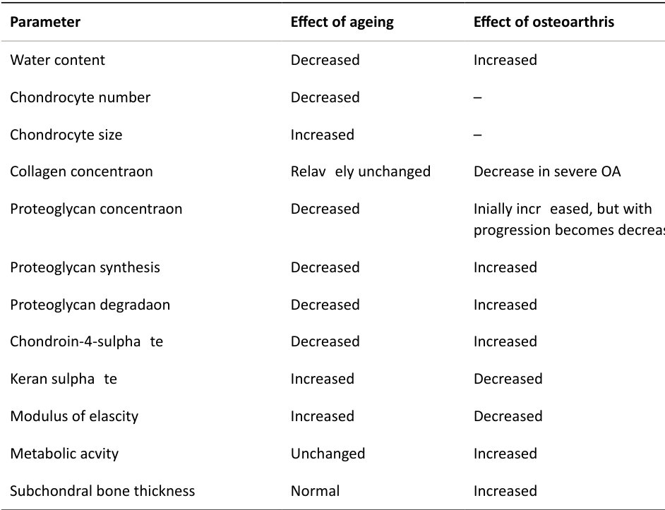
2. Structure and function of cartilage: (b) meniscus#
Introduction#
This is an A-list topic that candidates should have viva practised beforehand. For a basic 6 the candidate should learn the usual core questions on meniscal structure, function, biomechanics and hoops tresses.
Candidates aiming to score a 7 or 8 will need to put in some detective work to uncover the higher-order thinking and judgement questions that follow on from this. As a general rule the application of basic science in a clinical content is always an excellent starting position.
There are several possible routes into the topic.
Structured oral examination question 1#
1 . EXAMINER : Here is a picture of the menisci of the knee, which I am sure you recognize. Can you talk me through this picture (Figure 20.23)?
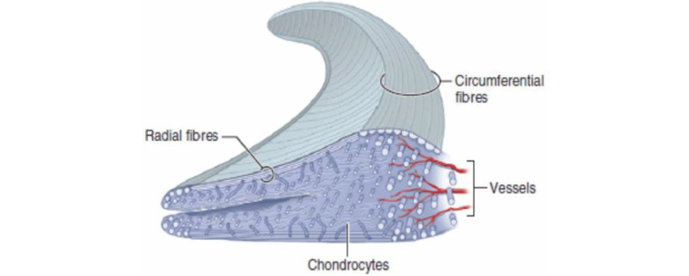
Figure 20.23 A cross-section of the meniscus showing the radial and circumferential collagen fibre orientation. Also shown are blood vessels penetrating the peripheral one-third of the tissue and location of chondrocytes.
No marks for recognizing the menisci as the examiners have already told you this. Being handed a meniscal diagram is a lucky escape into the topic for those candidates with poor drawing skills. The diagram can be tricky to draw and this side-steps the potential for a stumbling start into the topic.22 2 . EXAMINER : Can you drawout a meniscus for me concentrating on the collagen arrangement within the menisci (Figure 20.24)?
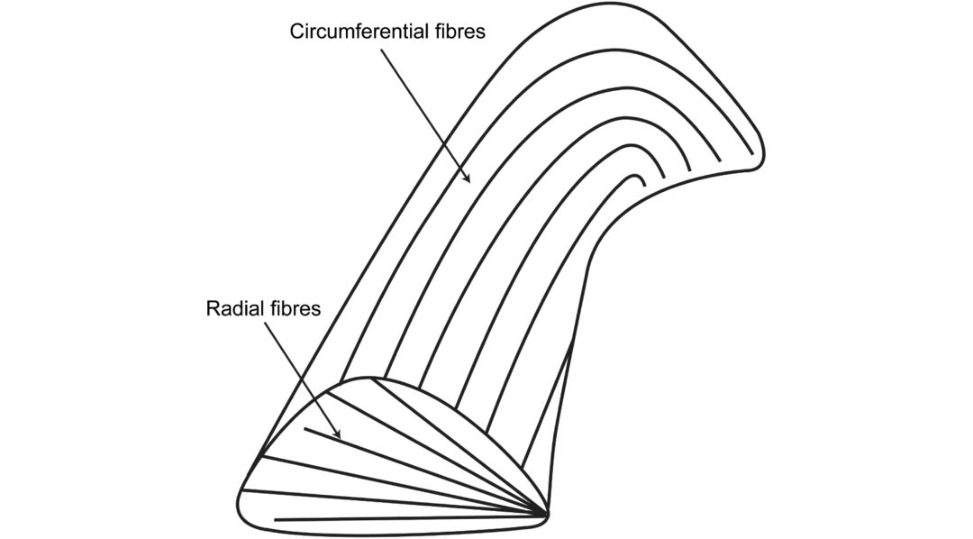
Figure 20.24 Candidate 20-second diagram of meniscal structure.
Well-prepared candidates would have anticipated this well-known route into the topic and worked out how to drawout and explain the structure within a time period of around 30 seconds. The large collagen fibres within the meniscus are mainly arranged in a circumferential pattern, with a smaller number of fibres in a radial orientation acting as ties. 3 . EXAMINER : [Birdeye nest picture of the meniscus shown] Can you identify the unlabelled structures on the diagram (Figure 20.25)?
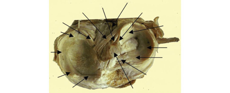
Figure 20.25 Unlabelled axial view of a right tibial plateau showing sections of the meniscus and their relationship to the cruciate ligaments.
This diagram could be shown either unlabelled (Figure 20.25) or labelled (Figure 20.26). The unlabelled version is a more precarious way into the topic.
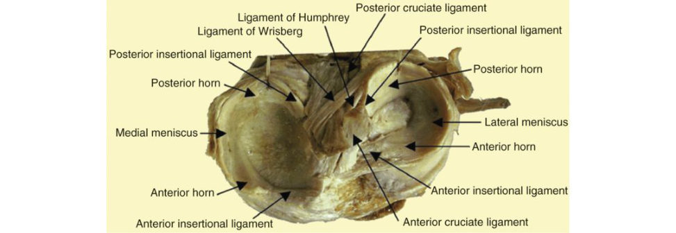
Figure 20.26 Labelled axial view of a knee showing sections of the meniscus and their relationship to the cruciate ligaments.
4 . EXAMINER : [Birdeye nest picture of the meniscus shown (Figure 20.26)] Describe what you see.
A birdeye nest picture of the meniscus generally leads to a more focused initial testing of meniscal anatomy.23 Candidates need to avoid stumbling around describing the diagram. A well- rehearsed answer would allow a candidate to progress on to the next part of the topic in a timely fashion.
Describe the anatomy of the medial and lateral meniscus (Figure 20.27).
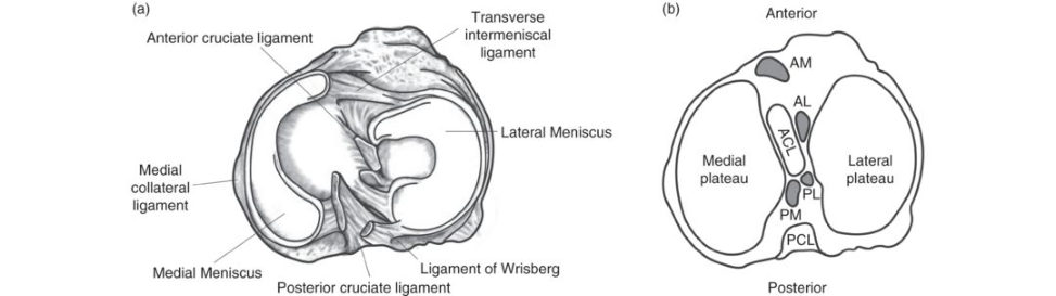
Figure 20.27 (a) Anatomy of the meniscus viewed from above. (b) Axial view of a right tibial plateau showing sections of the meniscus and their relationship to the cruciate ligamentAL, anterior horn lateral meniscus; AM, anterior horn medial meniscus; PCL, posterior cruciate ligament; PL, posterior horn lateral meniscus; PM, posterior horn medial meniscus.
The menisci are specialized intra-articular fibrocartilaginous structures of the knee. They are triangular in cross-section, with an average thickness of 3–5 mm. They are divided into three regions: anterior horn, body and posterior horn. The peripheral, vascular border of each meniscus is thick, convex and attached to the joint capsule. The innermost border tapers to a thin free edge. The superior surfaces of menisci are concave, enabling effective articulation with their respective convex femoral condyles.
Lateral meniscus
The lateral meniscus is O-shaped and covers a larger area than the medial meniscus (80–85% of the lateral tibial plateau).
It is more constant in size between the anterior and posterior horns.
The anterior horn is attached to the tibia anterior to the intercondylar eminence and posterior to the attachment of the ACL, with which it partially blends.
The posterior horn is attached posteriorly to the intercondylar eminence of the tibia anterior to the posterior horn of the medial meniscus.
No attachment of the lateral meniscus to the LCL, only a loose attachment to the joint capsule that is interrupted posteriorly by the popliteus tendon.
Occasionally a few fibres of the popliteus are attached to the posterior convexity of the LM.
Medial meniscus
The medial meniscus is C-shaped and covers 60–65% (~two-thirds) of the medial plateau.
The posterior horn is significantly wider than the anterior horn.
It has a larger anterior–posterior dimension than width.
The anterior attachment is approximately 7 mm anterior to the ACL attachment, inline with the medial tibial tubercle.
The posterior attachment to the posterior intercondylar fossa of the tibia anterior to the PCL attachment.
The MM attaches to the deep part of the MC Land to the capsule around its periphery via the coronary (meniscotibial) ligament. This results in the MM beingless mobile than the LM.
What are the ligaments associated with the meniscus?
A number of important ligaments are associated with the menisci. Both menisci have firm attachments to the tibial surface at their anterior and posterior horns via the insertional ligaments, which are very strong and stiff. Anteriorly, the transverse meniscal ligament connects the anterior convexity of the lateral meniscus to the anterior horn of the medial meniscus. Posteriorly, the lateral meniscus may be connected to the lateral side of the medial femoral condyle by one or two meniscofemoral ligaments. The anterior meniscofemoral ligament (ligament of Humphrey) passes in front of the PCL, the posterior meniscofemoral ligament (ligament of Wrisberg) passes behind the PCL.
What are the functions of the meniscus?
The function of the meniscus includes: Load transmission (bearing). Shock absorption. Lubrication. Distributes synovial fluid throughout the joint. Contributes to joint stability. Prevents hyperextension. Assists in gliding motion. Prevents synovial impingement. Proprioception. Nutrition. This is a predictable question and candidates wanting to score higher marks than a basic pass should be able to discuss meniscal function in a more detailed way than the usual bulleted list.
What are the biomechanical functions of the meniscus?
This is a slightly more probing question than the preceding one and needs a bit more thought. Typical leading-on questions may focus on the development of osteoarthritis. The meniscus serves several important biomechanical functions. The y contribute to load transmission, stability, nutrition, joint lubrication and proprioception. The y also function to decrease contact stresses and increase contact area and congruity of the knee. In most textbooks menisci are described as functioning as shock absorbers in the knee. However, recent evidence has suggested this might not actually be the case.24
What about the load transmission functions of the meniscus?
Inextension, the posterior menisci bear 50% of the compressive load compared to 85% at 90° flexion. Following meniscectomy, contact areas can be reduced by over 75% resulting in an increase in peak contact pressures as high as 235%.
How does this lead to the development of osteoarthritis?
Medial meniscectomy results in a 50–70% reduction in femoral condyle contact area and a 100% increase in contact stress. Total lateral meniscectomy results in a 40–50% decrease in contact area and increased contact stress in the lateral component to 200–300% of normal. Increased contact pressures lead to overload of the articular cartilage and the development of osteoarthritis.
So why is lateral meniscectomy worse than medial meniscectomy?
The medial tibial plateau is slightly concave, giving some degree of congruency with the curved femoral condyle. However, with the lateral tibial plateau this is convex, causing a natural tendency to point loading. The lateral meniscus is therefore more important as a load-bearing structure than the medial meniscus and partial or total lateral meniscectomy much more significant than the equivalent medial meniscectomy.
How are menisci viscoelastic? What do you mean?
Viscoelastic materials display properties of both a solid and liquid. The elas tic quality or solid phase of the meniscus is due to its collagen–proteoglycan structure, whereas the viscous or fluid phase is due to its permeability and water content. Under compression, meniscal permeability determines the rate at which fluid is extruded. Meniscal permeability is much lower compared to articular cartilage, giving menisci the ability to maintain their shape during axial loading. The viscoelastic nature of the meniscus functions to dampen the intermift ent shockwaves generated by impulse loading of the knee during gait. This shock-absorbing mechanism reduces the risk of osteoarthritic development. Articular cartilage, tendons and ligaments, intervertebral discs and menisci display viscoelastic properties.
How does the meniscus assist in lubrication?
The menisci serve to increase the congruity between the condyles of the femur and tibia; they contribute significantly to overall joint conformity. This assists in the overall lubrication of the articular surfaces of the knee joint. Like shock absorption, the reis no firm evidence of menisci involvement in knee joint lubrication. Again, similar to shock absorption, it is mentioned as a meniscal function in various textbooks.
What else?
There is fluid exudation across meniscal surfaces. Compression squeezes fluid out into the joint space to allow smoother gliding of the joint surfaces.
How does the meniscus function in proprioception?
The menisci provide a feedback mechanism for joint position sense. Neural elements have been identified within the meniscal tissue. It is thought that mechanoreceptors located in the meniscal horns sense a taut meniscus during extremes of flexion and extension and feed this back to the central nervous system, which contributes to a reflex arc that stimulates protective or postural muscular reflexes.
What are hoop stresses in the meniscus?
This is very much pass/fail material. Candidates would be expected to know this to score a 6. The arrangement of collagen fibres in the meniscus convert compressive forces into a radially directed force, which is distributed and resisted as hoop stresses within the meniscus. The radial fibres act as intrasubstance tier ods to provide structural rigidity and resist against longitudinal spliting of the circumferential collagen bundles. The development of hoop stresses within the meniscus depends on intact anterior and posterior attachments. Hoop stress also relies on the conversion of axial load into tensile strain through intact longitudinal-orientated collagen fibres. No matter how many times I described hoops tresses in the meniscus, for some reason I never appeared very convinced that I knew what I was talking about. The answer is textbook reading, but something in my delivery either a lack of confidence, poor body language or just not being persuasive enough meant I always failed this question. Equally frustrating was observing other candidates giving either exactly the same answer or even a slightly incorrect answer but still being passed Very frustrating! The candidate has probably rote-learnt the topic rather than fully understood it and the examiners may have sensed this. There may be other complicated reasons that are difficult to analyze in cold print on paper without observing the scenario. Try more to understand basic science principles rather than memorize the subject and sound convincing in your answer, even if you don’t feel very confident.
What is the blood supply of the meniscus?
The blood supply to the meniscus is mainly from the medial and lateral genicular arteries (superior and inferior branches). Branches from these vessels give rise to a perimeniscal capillary plexus within the synovial and capsular tissues of the knee joint. These perimeniscal vessels are orientated predominantly in a circumferential pattern with radial branches directed towards the centre of the joint. The middle genicular artery also supplies the menisci through the vascular synovial covering of the anterior and posterior horn attachment. Approximately 10–30% of the periphery of the MM and 10–25% of the LM are relatively well vascularized. The remaining portion of each meniscus (65 –75%) receives nourishment from synovial fluid via diffusion or mechanical pumping (i.e. joint motion).
What factors influence your decision whether to repair a meniscal tear or resect?
Location oft ear
The vascular supply only reaches the peripheral 25–30% of each meniscus. Repair of tears in the well-vascularized red zone (0–5 mm from the periphery) have a good chance of healing. Red–white zonal tears have a reasonable chance of healing, whereas tears in the white zone (3–5 mm from the periphery) are unlikely to heal.
As a general rule, red–red should, white–white won’t and red–white might.
Age of tear
Fresh tears are more likely to heal than older tears.
Age of the patient
Meniscal repairs in older patients (> 30 years) have a significantly worse outcome than in younger patients There isless vascularity in the older tear.
Tear pattern
Displaced bucket-handle tears should be repaired on an urgent basis whenever possible. Delay of more than a few weeks will lead to scarring and retraction of the buck et-handle fragment.
Repetiv e compression and abrasion in the displaced position will lead to macerated and damaged tissue and preclude repair.
Peripheral, vertical longitudinal tears are ideal for repair in the red–red or red–white zone.
Complex bucket-handle tears, flap tears, degenerative and radial tears often perform poorly with repair and are more often amenable to excision.
Horizontal cleavage tears are not repairable, and the unstable leaf should be excised, leaving up to
3 mm of the leaf.
Ligament stability
A meniscal tear should not be repaired in an unstable ACL-deficient knee. Due to the abnormal kinematics of the ACL-deficient knee, the failure rate in the unstable knee is much higher than in a stable or reconstructed knee.
What percentages of tears are amenable to repair?
This depends to a certain extent on the expertise of the surgeon, but a figure around 15% is generally accepted. There is very litile guidance in the literature. If acute and/or a relatively well-preserved joint, consider repair. If the patient is older (> 50 years), degenerative changes are presentor the patient is obese or has inflammatory arthritis resect.
How do radial and longitudinal tears differ?
A radial tear disrupts the continuity of the circumferential fibres interfering with the distribution of hoops tresses within the meniscus. If the tear reaches the periphery it transects the meniscus and renders the hoop stress distributing capacities of the meniscu sine ffectiv e. This is the equivalent of a total meniscectomy. In this situation consider repair. In contrast, longitudinal tears do not disrupt the continuity of the circumferentially orientated fibres that bear load. They occur due to fracture of the weak radial tie fibres.
If radial tears interfere so much biomechanically with hoop stress distribution, why do we not repair more of them?
These tears are difficult to repair. There may be a case for attempting repair in a young active person, especially if the lateral meniscus is involved or if the tear extends to the periphery, but success rates can be unpredictable, and the patient needs to be partial w eight-bearing for at least 6 weeks following the repair.
What is a meniscal root tear?
A meniscal route tear is where the tear extends to either the anterior or posterior meniscal root attachment to the central tibial plateau. They often tend to be radial tears extending into the root. The tear may lead to meniscal extrusion, secondary osteoarthritis, and subchondral insufficiency fracture.
Biomechanically?
Loss of the root attachment impairs the ability of the meniscus to resist hoop stress when the tibiofemoral joint is loaded. This produces increased joint contact pressure and leads to rapid articular cartilage damage, subchondral bone oedema and sometimes collapse.
What else?
There are two main types of meniscal root tears. The first type is low-energy tear occurring in older patients with pre-existing osteoarthritis. These cond type occurs in young athletic patients from a sporting knee injury and are generally repaired. Repair techniques can be difficult but include pull-out (transosseous) techniques, suture anchor repair, and side-to-side suture repair.
What is the role of meniscal replacement?
Meniscal replacement may be indicated in a young patient who develops significant pain and swelling in a compartment specific to a major or total meniscectomy. There should be only early or minimal chondral changes, normal limb alignment and a stable knee. Candidates should be prepared to modify sporting activities after surgery to improve the chances of success. Cadaveric menisci are matched by size and site and are implanted by various techniques that include a free soft -tissue allograft implantation, separate anterior and posterior bone plugs and bone bridges.
A meniscal allograft may partially replicate the normal meniscus function and significantly reduce pain and improve knee function.
Structured oral examination question 2#
Meniscus – draw, structure, function contents, hoop stresses.
Similar to question 1, but meniscal contents need to be described.
What are the contents of the meniscus?
The meniscus is a dense extracellular matrix (ECM) composed mainly of water (72%) and collagen (22%), interposed with cells. The remaining dry weight is composed of proteoglycans, non- collagenous proteins and glycoproteins (Figure 20.28). The cells of the meniscus are called fibrochondrocytes because they appear as a mixture of fibroblasts and chondrocytes. Cells in the more superficial layer of the meniscus are more fibroblastic spindle-shaped while those cells located deeper in the meniscus are more chondrocytic o void- shaped (Figure 20.29). The proteoglycans retain water within the meniscus, thus permiting its specific viscoelastic properties. The outer region of the meniscus is composed of type I collagen while the inner region is 60% type II and 40% type I. As such the outer portion is more fibrous while the inner region displays more hyaline cartilag e-like properties. The collagen fibre arrangement is either circumferential radial or random. The meniscus is divided into five layers. Superior superficial layer: random. Superior lamellar: random but with some short radial-orientated fibres at the posterior and anterior horns. Deep layer: circumferential fibres interspersed with a few radial fibres. Inferior lamellar: radial and random. Inferior superficial layer: radial.
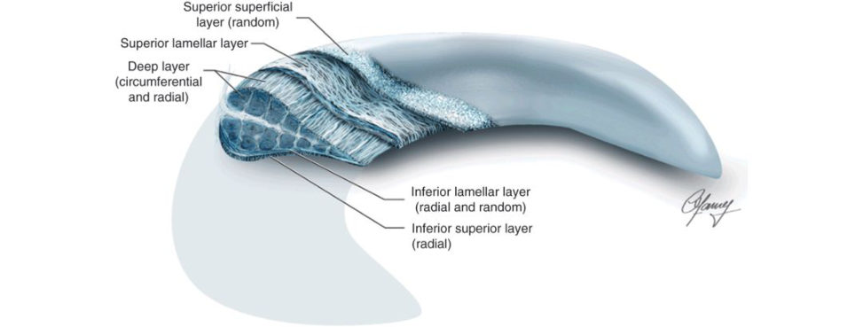
Figure 20.28 Complex collagen arrangement in meniscus. The outer region is the outer third of the meniscus; the inner region is the inner two-thirds of the meniscus; the superficial region is the surface of the meniscus.
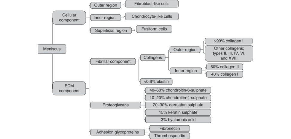
Figure 20.29 The complex composition of the meniscal cellular and meniscal extracellular matrix (ECM) components.
Structured oral examination question 3 (Figure 20.30)#
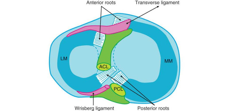
Figure 20.30 Birdeye picture of the tibia.
Draw me the top of the tibia! Label the structures. How are the menisci attached?
What is the function of menisci – load transfer, shock absorption, hoops tresses, etc.?
How do menisci get injured?
Birdeye picture of the meniscus (discussed in earlier questions). Ideally , the anatomical description of each meniscal attachment should be explained while the diagram is being drawn out. Candidates wanting to score well need to get through this opening test material in a timely fashion to set themselves up for the later, more difficult score 7/8 questions.
What do we mean by hoop stresses?
As a result of circumferential collagen fibres and the insertional features of the anterior and posterior horns, vertical compressive forces are converted into a radially directed force that is taken up as circumferential hoops tresses within the meniscus (Figure 20.31).
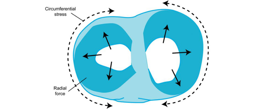
Can you draw this for me? Can you explain this free body diagram of forces acting on the meniscus ( Figure 20.32)
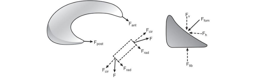
As the femur presses down on the meniscus during normal loading, the meniscus deforms radially but is anchored by its anterior and posterior horns (Fant and Fpost). During loading, tensile, compressive and shear forces are generated. A tensile hoop stress (Fcir) results from the radial deformation, while vertical and horizontal forces (Fv and Fh) result from the femur pressing on the curved superior surface of the tissue. Ara dial reaction force (Frad) balances the femoral horizontal force (Fh).
How do menisci get injured?
There are two types of meniscal tears, traumatic and degenerate. Traumatic t ears usually occur with rotation as the flexed knee moves towards an extended position. The most common location for injury is the posterior horn and the most common tear pattern is longitudinal. Degenerative tears occur as the meniscus becomes less compliant and elastic with age.
Figure 20.31 Menisci convert a compressive stress into a radial stress that is taken up by a circumferential (hoops tress within the meniscus.
Figure 20.32 Free body diagram of forces acting on the meniscus during loading.
Structured oral examination question 4#
Meniscus anatomy and function (see above).
What is the histology of the meniscus?
The meniscus is primarily constituted of interlacing networks of collagen fibres (predominantly type 1 interposed between cells and an extracellular matrix (ECM) of proteoglycans and glycoproteins.
What about the layers of the meniscus?
There are three collagen layers: superficial, lamellar and deep. The surface layer has a random organization of collagen fibres. There are circumferential collagen fibre bundles and radial ‘tie fibres’ or tie sheaths. The radial fibres stabilize the meniscus, preventing circumferential splits and also resisting excessive compressive loads. The circumferential fibres function inhoops to accept stress without gross deformation or extrusion from the joint surface. The radial displacement is opposed by posterior and anterior attachments on the tibial plateau.
When does a meniscus stop growing in a child?
The meniscus stops growing in a child when the growth plate closes. Anatomically, the meniscus is fully vascularized at birth, but the area of vascularity recedes toward the periphery with age, such that, by the age of 10, only the peripheral 10–30% of the meniscus is vascularized, as is seen in the adult meniscus. The menisci growth closely matches the growth of the femur and tibia.
What meniscus is usually damaged with ACL injury?
Medial meniscus tears are more common inpatients with chronic ACL insufficiency, lateral meniscus tears are predominately found in acute ACL injuries. The lateral meniscus is more mobile than the medial meniscus and can become trapped between the femur and tibia during ana cute pivoting episode. Most commonly, the tear occurs in the posterior third of the lateral meniscus, which does not have a strong attachment to the surrounding capsule. The medial meniscus is a secondary stabilizer of the knee against anterior instability and can be commonly torn in chronic ACL injuries associated with significant knee instability.
3. Invertebral disc structure and function#
Introduction#
This subject just about makes it as an A-list basic science viva topic. Tipping the balance is that the basic science of degenerative disc disease may additionally find its way into a general adult and pathology viva on PID.
A good starting point is a basic understanding of the anatomy and function of the spine. Although candidates may not be directly asked about spine anatomy, they still need to be familiar with it. If a candidate appears unsure, the examiners may switch focus and start asking them pass/fail questions on spinal anatomy.
Structured oral examination question 1#
Describe the anatomy of the vertebral column
The normal adult vertebral column typically consists of 33 vertebrae (seven cervical; 12 thoracic; five lumbar; five fused sacral and four fused coccygeal). It extends from the occipital condyles of the skull at the atlanto-occipital joint to the apex of the coccyx. Movement of the vertebral column occurs through the 23 discs in the human spine. Apart from the fused vertebrae of the sacrum and coccyx, the only vertebrae not connected by discs are the atlas and axis, which pivot at the specialized atlanto-axial joint and the articulation between the atlas and the base of the skull at the occipitoatlantal joint.
What are the individual anatomical features of each vertebral region?
In the cervical region there are C1 (atlas) and C2 (axis) which are considered specialized vertebrae. The C3 to C7 section is referred to as the subaxial region. The spinous processes are usually bifid. The vertebral bodies of the subaxial cervical spine have upward projections on the lateral margins called uncinate processes. In the thoracic region there is a progressive increase invertebral body mass from T1 to T12, pedicles are small in diameter, laminae are vertical with a ‘roof tile ’ arrangement, the spinous processes are long, overlapping and projected downwards and the intervertebral foramen is larger with less incidence of nerve compression. In the lumbar region there is a progressive increase invertebral body mass, pedicles are long and wider than the thoracic area and oval-shaped, the spinous processes are horizontal, square-shaped, transverse processes smaller than the thoracic region and the intervertebral foramen, although large, has an increased incidence of nerveroot compression.
What are the functions of the spine?
Functions of the spine include: Protection of the spinal c ord. Providing for balance and stability of the body. It offers attachment points for the ribs and muscles of the back and trunk. Allows for flexibility and mobility. Supports the structure and weight of the body in various activities.
What are the functions of the intervertebral disc?
The intervertebral disc: Allows the spine to twist and bend throughout a wide range of positions. Functions to absorb energy and distribute loads applied to the spine. Redistributes compressive loads, resists tensile, rotational and shear forces. Restricts excessive motion. If all else fails, reply that the intervertebral discis a load-bearing structure that gives mobility to the vertebral columnA misconception in the literature is that they act as shock absorbers, but there is no good evidence to support this function. 25 No need to complicate matters for yourself and mention this in the exam lest it alerts the examiners, who may then start asking you difficult questions.
What is a motion segment?
The motion seg mentis the functional unit of the spine. It consists of the lower half of the upper vertebra, the upper half of the adjacent lower vertebra and the facet joints, intervertebral disc and the ligaments that lie between the adjacent vertebrae.
What is the structure of the intervertebral disc?
There are three main regions to the intervertebral disc each with differing structural compositions: (1) outer annulus fibrosis, (2) inner nucleus pulposus and (3) endplates. Each has different functions.
What is the structure of the annulus fibrosus?
The annulus fibrosis is the outer structure that encases the nucleus pulposus. It is attached to both the anterior and posterior longitudinal ligaments of the spine and the vertebrae on either side. It is mainly composed of type I collagen, water and proteoglycans. It has a high collagen/low proteoglycan ratio. Fibroblast-like cells are responsible for producing type I collagen and proteoglycans. Characterized by high tensile strength and its ability to prevent intervertebral distraction. It remains flexible enough to allow for motion. Compared to the NP it is a much more fibrous structure with a much higher collagen but lower water content. The AF can be divided into two regions, the outer third and the inner two-thirds. The outer annulus fibrosus consists of a number of densely packed layers composed of predominantly type 1 collagen called lamellae. Fibres of each lamella run obliquely between vertebrae at about 30° to the horizon, with adjacent lamella typically running at right angles.
Why is this?
This arrangement allows the disc to resist both torsional, axial and tensile loads (distraction and shear). This is sometimes referred to as a hands-in-the-pocket configuration or plies in a tyre tread. The lamellae are more abundant and stronger in the anterior and lateral aspects of the disc. The larger fibrocartilaginous inner annulus fibro sus layer is found more centrally, containing chondrocytes and a less-dense, predominantly type II collagenous matrix lacking lamellar organization. Therefore, on progressing from the outer to the inner annulus, the type I collagen level declines and that of the type II increases.
Describe the structure of the nucleus pulposus.
The nucleus pulposus is the water-rich, gelatinous central portion of the intervertebral disc that is surrounded by the annulus fibrosis. It is mainly composed of type II collagen, water and proteoglycans and polysaccharides. It has a greater water content than the AF. Chondrocyte-like cells are responsible for producing type II collagen and proteoglycans. These cells continually maintain the matrix and rely on diffusion of nutrients from the endplates surviving in relatively hypoxic conditions. The high density of negatively charged sulphate and carboxyl groups on the glycosaminoglycan chains of the major proteoglycan in the disc, aggrecan, are responsible for atir acting the high water content. Aggregates are held together by type II collagen that is cross-linked by type IX collagen. Equilibrium between the aggrecan and type II collagen helps in creating a load-bearing and c ompression-resisting tissue that gives stability to the disc. A pressure is generated by swelling from the atir action of water into the IVD from surrounding tissues resulting in the vertebral bodies being pushed apart. This pressure is resisted by tension in the collagen fibres of the AF. The balance between expansion of the NP and tension in the AF leads the IVD to resist compression. The candidate is relating NP structure to function. This should not draw any examiner criticism ast o not answering the question, 26 as linking structure to function brings higher -order thinking into the topic.
What are the functions of the NP?
The NP resists compressive loads, dampens mechanical loads and evenly distributes forces onto the endplates.
What is the function of the matrix?
The disc matrix is an elaborate framework of macromolecules that atir act and hold water. This viscoelastic matrix distributes forces smoothly to the annulus and the endplates. The NP contains proteoglycan aggregates entrapped in a collagen fibre network. The hydration properties of the gly cosaminoglycan chains of aggrecan cause the tissue to swell until an equilibrium is reached, in which the swelling potential is balanced by tensile forces in the collagen network.
What about the endplates? What is the function of the endplates?
The endplates are positioned above and below the nucleus and most of the annulus and are thin layers of hyaline cartilage that are considered part of the disc, not part of the vertebral body. The annulus and nucleus are firmly attached to the endplates and separation is difficult. The major function of the endplates is to act as a semipermeable membrane allowing nutrients and metabolites to diffuse into the disc from the capillary blood of the vertebral body and to allow waste products to diffuse out.
How can oxygen and glucose and other nutrients diffuse through the usually impermeable tough periosteum of the vertebral body?
Under normal circumstances, no diffusion would occur; however, the subchondral bone of the vertebral bodies has special channels called marrow cavities that allow for diffusion to occur (Figure 20.33). Therefore, as longas these channels are open, the cells of the disc can receive oxygen, glucose and other amino acids, as well as have an avenue forwaste removal.
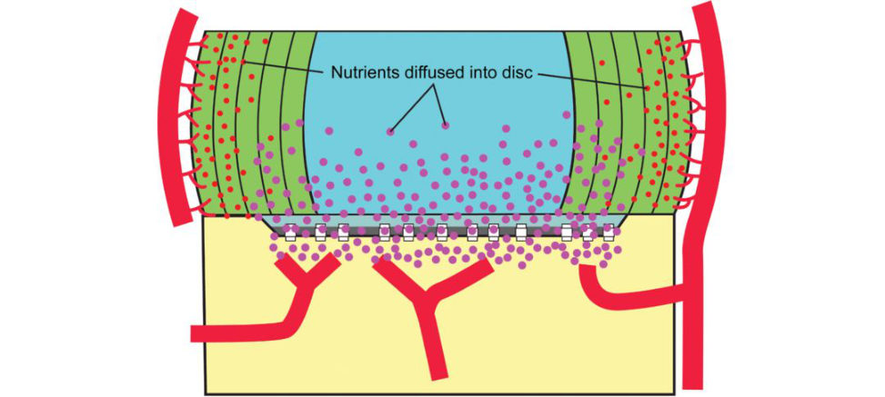
What about the blood supply of the intervertebral disc?
Intervertebral discs have no significant vascular supply. They receive their blood supply by diffusion through the vertebral body endplates. A network of vessels located centrally in the endplate allows nutrients to diffuse into the nucleus pulposus and annulus fibrosus (Figure 20.34). Nutrients are supplied to the disc primarily through diffusion. Because the disc has a very limited blood supply, invertebral disc cells, particularly those in the centre of the avascular NP, operate in an environment that would be unviable to most other cells. They appear to have adapted to this hypoxic, acidic environment.
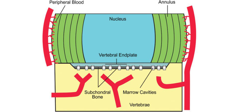
What happens to the disc with ageing?
With ageing there is decreased vascularity of the endplates. This reduces the nutrition supply to disc cells resulting in decreased synthesis and concentration of pr oteoglycans.
This leads to an overall loss of water content within the NP altering the biomechanical response of the disc to physiological loading stresses. There is an increase in the proportion of type I to type II collagen and an increased ratio of keratin sulphate to chondroitin sulphate. There is conversion to fibrocartilage which causes a generalized stiffening of the NP, with the net effect being a less biomechanically competent disc.
What is the nerve supply to the intervertebral disc?
The posterior and posterolateral disc are innervated by the sinuvertebral nerve, the lateral disc by the grey ramus communicans (a sympathetic nerve of the autonomic system), and the anterior disc by sympathetic branches from the sympathetic trunk or ganglion that courses anterolaterally over the vertebral bodies (Figure 20.35). Only the outer third (to a ~3 mm depth) of a healthy annulus is innervated and capable of transmiting pain. The inne rtw o-thirds of the annulus and the entire nucleus are completely avascular (no blood vessels) anda neural (no nerves).
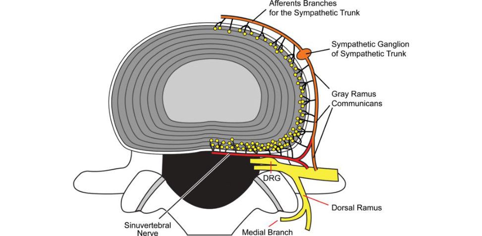
Describe the natural history of a lumbar disc prolapse.
Recurrent torsional strain leads to tears of the outer annulus that lead to herniation of nucleus pulposus. Size of herniation decreases over time duet o reabsorption via macrophage phagocytosis. Smaller disc fragments are less likely to be resorbed while large sequestered disc herniations show the greatest degree of spontaneous resorption. Approximately 90% of patients will have improvement in symptoms by 3 months.
What are the risk factors for developing a disc prolapse?
Disc prolapse is related to failure of the annulus fibrosus due to either increased load/pressure or decreased mechanical strength. Pressure within the discis maximized by forward flexion in a seated position Torsional movement increases this further. There is also a role of ageing causing degeneration of the annulus, increasing the predisposition to tear. This means the annulus isless well supported and more prone to tearing.
What are the suggested theories for the mechanism of production of pain in disc disease?
There are several possible mechanisms for development of pain in disc disease. The first is due to simple nerve compression and accounts for the radicular pain seen in disc prolapse. There is also a degree of compression in degenerative disc disease due to reduced disc height, causing foraminal stenosis. The second type of pain is mechanical in nature and is due to the degenerative disc reducing in height and therefore providing a reduced structural role, which causes increased load through the posterior elements (e.g. facet joints) and their subsequent degeneration. The annulus has nociceptors inits outer third and therefore the micro tears due to trauma may be directly painful.
Finally, there is an altered cytokine profile within the degenerative disc with increased IL-1 that may be implicated in discogenic pain.
Figure 20.33 Diffusion of nutrients into the intervertebral disc.
Figure 20.34 Blood supply of the intervertebral disc.
Figure 20.35 Nerve supply of the intervertebral disc.
Structured oral examination question 2#
Intervertebral discs, describe parts, contents, effect of ageing, disc function, nutrition, pathophysiology of prolapsed disc, hoop stresses, shock absorption, and what is lumbago vertebra.
What is this (Figure 20.36)?
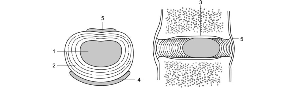
It is a picture of an intervertebral disc.
Figure 20.36 The intervertebral disc: 1, nucleus; 2, annulus; 3, cartilaginous endplate; 4, anterior longitudinal ligament; 5, posterior longitudinal ligament.
Can you name the various blank labels of the disc?
Be able to name the various structures correctly and if you feel confident and the overall viva table is proceeding well go on to talk about how IVD structure is related to its function.
Can you describe the various parts and contents of the disc?
Go through the three components of the disc (AF, NP and endplates) and try to link structure to function (see above question 1). 27
What are the functions of the disc?
The disc essentially functions as a shock absorber to redistribute compressive loads and resist tensile, rotational and shear forces. It allows spinal movement and provides stability.
What happens to the disc with ageing and how does this affect function?
With ageing, the inner AF expands, the size of the NP decreases while the outer AF remains the same. In the NP the concentration of viable cells decreases, proteoglycan and water concentrations decrease and there is a partial loss of structural integrity. The NP undergoes a transition from fluid-like to solid-like behaviour, severely limiting the ‘shock- absorbing’ properties of the disc. This is a solid score 6, but the answer could be better explained in terms of the changes.
The rate of synthesis of glycosaminoglycans, proteoglycans, link proteins and hyaluronan decreases progressively with age as they undergo continuous proteolytic degradation (MMP s and
ADAMs). At the same time, the production of collagen type I increases.
This results in a number of changes.
Aggrecan content in the nucleus pulposus drops significantly, and with it the ability of the ECM to atir act, bind and maintain water. The NP becomes progressively more fibrous and opaque, and with increased pigmentation. As the collagen content increases and changes from type II to type I, demarcation between the NP and AF becomes less distinct and separation of adjacent annular laminae occurs. This delamination leads to the development of concentric tears in the annular laminae.
Score 7/8.
How is ageing different to degeneration?
This is a difficult, controversial topic that is testing higher -order thinking. Although age-related disc changes and disc degeneration are considered separate enties, both share similar biomechanical alterations in the NP with the distinction between the two conditions often blurred. Intervertebral discs receive the vast majority of their nutrient supply from diffusion across their endplates. With ageing, the number of vascular channels perforating the osseous vertebral endplates diminishes. This leads to reduced porosity across the vertebral endplates and an accumulation of cell waste products and degraded matrix molecules that impair cell nutrition and function. This gradual loss of viable cells within the NP further compromises matrix synthesis. Similar to ageing, the major pathological process associated with disc degeneration is a declining nutritional support reaching the intervertebral disc cells. This leads to a decrease in the number and activity of disc cells. Factors thought to accelerate disc degeneration include mechanical disc overload, genetic factors, immobilization and obesity . Additional influences that may compromise disc blood supply leading to disc degeneration include smoking, diabetes and peripheral vascular disease. Each factor mediates its effect by either altering the balance of protein synthesis and degradation and/ or the rate of cell death or apoptosis.
What is the pathophysiology of the prolapsed disc?
Degenerative changes in the IVD alter the structural properties of disc components. The NP becomes stiffer with loss of water content and increase in tissue density . There is a more anisotropic stress state with a more non-uniform distribution of stresses. More stress is placed on the collagen fibres of the AF that they are not designed to tolerate. As such, the collagen structure of the AF deteriorates due to impaired formation, increased cross-linking and increased breakdown.
The endplates become thin, sclerotic and pr one to microfracture.
This reduces a disc’s ability to recover from deformation and predisposes to weakness of the AF, leaving it susceptible to annular fissuring and tearing.
What are the macroscopic changes that occur?
There is narrowing of the disc space osteophytes at the margins of endplates, increased stress at the facet joints, facet joint degeneration with osteophyte formation, bulging of the annulus and herniation of the NP throu ghan annular fissure Osteophytes develop to attempt to stabilize the motion segment but may encroach on neural structures.
How do hoop stresses occur in the spine (Figures 20.37 and 20.38)?
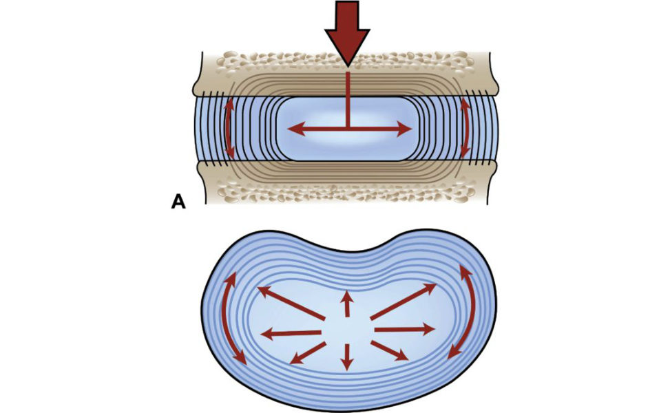
Figure 20.37 Compression force from bodyweight contraction straight arrows) raises the pressure in the NP. This, inturn, increases the tension in the AF (curved arrows) and muscle.29
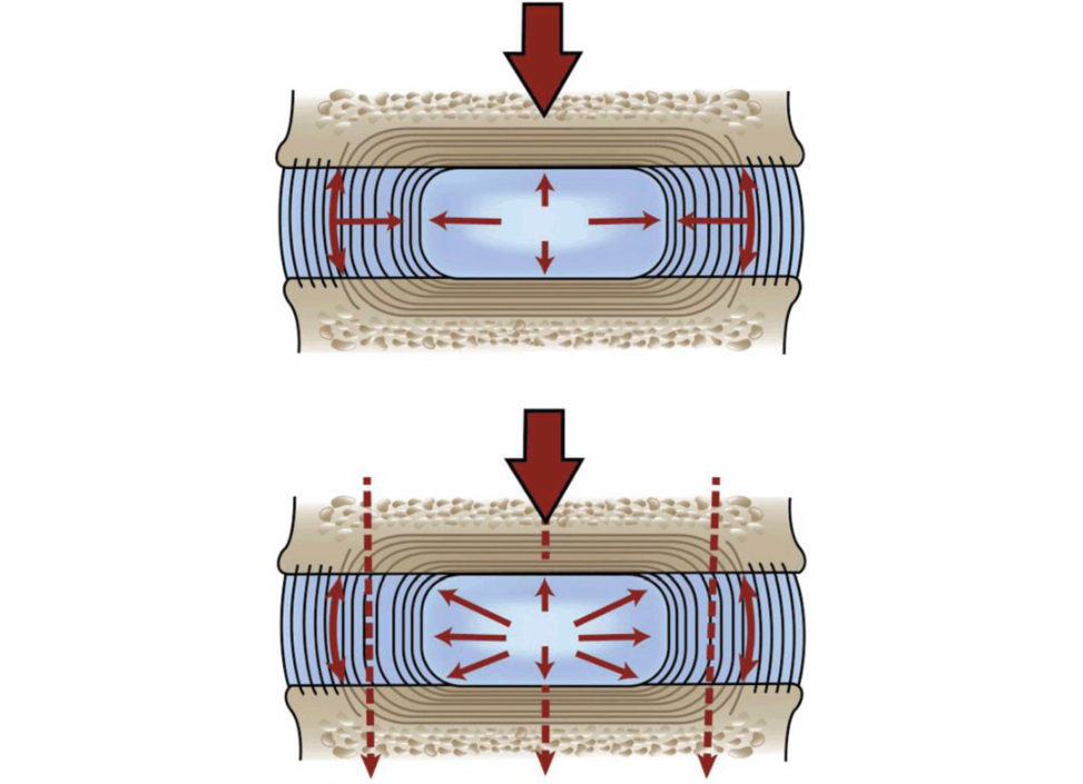
Figure 20.38 The increased tension in the AF inhibits radial expansion of the NP. The rising pressure in the NP is also exerted upward and downward against the vertebral endplates. The weight is partly borne by the AF and NP and is then transmift ed across the endplates to neighbouring vertebrae.30
When loaded from above, the height of the NP is reduced. It attempts to expand out against the annulus and the collagen rings (lamella) of the annulus are stretched. The radial pressure exerted by the NP is quickly balanced by the elastic tension that develops in the lengthening fibres of the annulus. At the same time, the nucleus is constrained in the up–down sense by the endplates and vertebral bodies. In this way, pressure applied to the NP is passed on to both the AF and the endplates. The fibres of the annulus are braced and prevented from buckling.
How does the disc function as a shock absorber?
The conventional view was that a disc works as a shock absorber when the spine experiences a rapidly applied force; the force could be momentarily diverted into the annulus, easing the speed with which it must be transmift ed down the chain of vertebrae. The idea that the intervertebral discs act as a shock absorber has been challenged in recent years, with the view that by far the greatest amount of energy absorbed is from the muscles and tendons surrounding the spine, rather than by the disc.
A controversial and misunderstood concept that perhaps should not have been asked.28 The fibre orientation of the AF resists hoop stresses generated by the hydrostatic pressure from the NP (Figure 20.39). The AF provides the ability to absorb significant hoop stresses and maintain stiffness in the presence of tensile forces induced by bending and twisting of the spine. Animal experiments have demonstrated that even a partial thickness laceration in the annulus rapidly produces advanced disc degeneration.
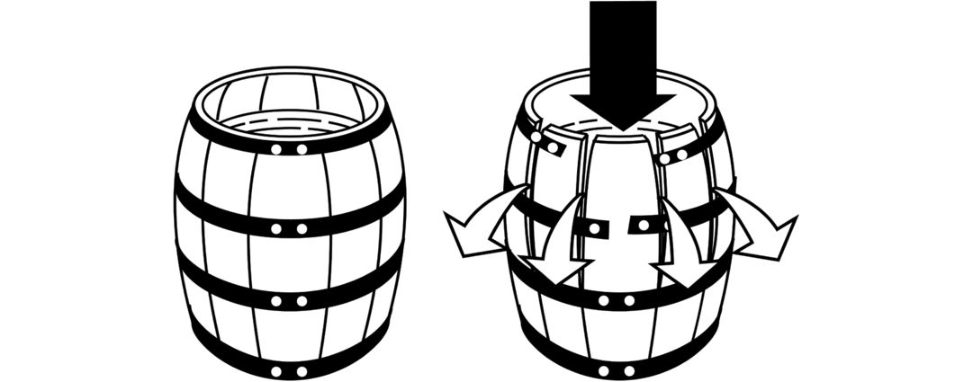
Figure 20.39 Hoop stress. A load of water in a barrel is resisted by the hoops around the barrel. When too great a load is applied, the hoop will break. The annulus functions in a similar manner to that of the hoops around a water barrel.
This is probing for a more detailed answer than just concentrating on the g el-like material features of the NP. Because the nucleus pulposus is gelatinous, the load of axial compression is distributed not only vertically but also radially throughout the nucleus. This radial distribution of the vertical load (tangential loading of the discis absorbed by the fibres of the annulus and can be compared with the hoops around a barrel.
What is lumbago vertebrae?
Sorry, I have no idea. Lumbago is a seldom-used term to mean mild to severe low-back pain. The pain can be acute or chronic and affect old or young patients Many years ago, lumbago was associated with ‘rheumatism ’, another loosely used term, seemingly brought on by exposure to cold damp winter weather. Low-back pain is a more precise term and should preferably be used instead.
What is the role of aggrecan and collagen in the ability of discs to resist compression?
The NP contains proteoglycan aggregates entrapped in a collagen fibre network.
The hydration properties of the gly cosaminoglycan chains of aggrecan cause the tissue to swell until an equilibrium is reached, in which the swelling potential is balanced by tensile forces in the collagen network.
Compressive loading of the spine forces some water from the disc effectively increasing the aggrecan concentration and itss welling potential and resisting further compression.
On removal of the compressive load, disc height is restored as water is drawn back into the tissue to restore the original equilibrium conditions. An y parameter that decreases proteoglycan concentration or weakens the collagen network will be detrimental to disc function .
Structured oral examination question 5#
What is the purpose of the spine?
The primary purpose of the spine is to provide protection for the spinal cord and axial support system to allow locomotion and function of limbs. It has three natural curves which provide an S-shape. [I genuinely didn’t know what to say at this stage, which can beseen by the rather pathetic statement above.]
OK, what is the primary site of movement in the spine?
[The penny drops!] Much of the ability to rotate and move within the spinal column is possible due to the intervertebral discs.
Tell me about the anatomy of the intravertebral disc?
Essentially the discis made up of two parts, the outer annulus fibrosus and the inner nucleus pulposus. In the cervical and lumbar spine, the discs are thicker anteriorly and in the thoracic spine the discis equal. The largest discis at the level of L5/S1. [The largest discis actually L4/5, but the examiner either didn’t know himself or didn’t want to push me at this stage.] The classic paper to quote and read is Coventry et al.31 There are three parts to this.
OK, can you tell me about the structure in more detail?
The annulus fibrosus makes up the peripheral portion of disk structure and is predominantly made up from fibrocartilage and type I collagen. The fibres run obliquely and are arranged primarily in concentric layers. The orientation of the fibres varies in successive layers and alternates at about 45°. The nucleus pulposus is predominantly type II collagen and has a high water content, which enables it to resist compressive loads. With age, the water content declines, which reduces its resilience.
What is the nerve supply?
The majority of the nerve supply lies in the outer rings of the annulus fibrosus, with supply from the sympathetic chain interiorly.
And posterior?
I can’t recall! [Sinus vertebral nerve dorsally]
Why is discitis common in children then?
Blood vessels occur in the annulus up to late teens, and into the cartilage endplates up to 8 years, which is why discitis occurs in this specific paediatrics group. A good paper to explain this question is by Rudert et al.32 The vascular pattern of human intervertebral discs and the surrounding tissue a t different ages were investigated using histochemical methods. The occurrence of blood and lymph vessels ingrowing intervertebral discs helps us to understand childhood discitis without simultaneous affection of the vertebral body.
4. Muscle structure and function#
An appreciation of muscle anatomy and physiology is important in the understanding of muscle injury.
However, a passive read through of this topic in a standard orthopaedic textbook is quite poor preparation for a viva question. Like most basic science subjects, it takes time and effort to fully appreciate the topic and develop the higher-order thinking needed to master the subject. It is worth knowing this subject well as it is definitely an A-list topic.
Structured oral examination question 1#
What is this picture (Figure 20.40)?
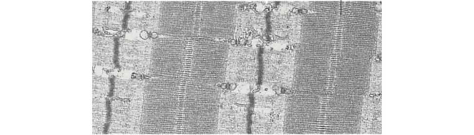
This is an electron micrograph picture of skeletal muscle.
Why do you say this?
We can see striations alternating light (I) and dark (A) bands.
Can you identify sarcomeres, A bands, I bands, Z lines, M lines and H zones (Figure 20.41)?
A sarcomere is the basic unit of striated muscle tissue. It is the contractile unit of a muscle cell. It spans from one Z line (disc) to the next. The Z line represents the attachment of adjacent sarcomeres. This is taken from the German word ‘Zwischen’, meaning between. Actin fibres are anchored on the Z line. The I band represents just actin filament sin adjacent sarcomeres where there is no overlap with myosin filaments. In between the I bands is the A band of the sarcomere. It is darker on the edges where there is a double, overlapping, hexagonal array of thick filaments (mostly myosin) and thin filaments (actin plus the regulating proteins: troponin and tropomyosin). It is anisotropic. The central H zone of the A band contains only thick myosin filaments. It is a less-dense, lighter region from the German ‘Heller’ for ‘brighter’. The M-line (or M-band) maintains (anchors) the myosin filaments in a hexagonal latice and the Z - line (or Z-band) maintains the actin filament sin a tetragonal latice. The relationship between the A band and H zone can be confusing. TheA bandis a dark overlap of thin and thick filaments that also contains the central H zone composed of thick myosin filaments only (Figure 20.42). Mentioning the German w ord ‘Zwischen’ (between) for some reason seemed to greatly impress the examiners. As the I bands are uniform (iso = same/uniform) in appearance throughout they are called isotropic bands. The dark A bands are not uniform as they have both thick and thin filaments and are therefore called anisotropic.
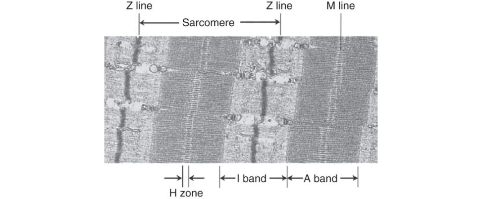
What do you mean by isotropic and anisotropic bands?
Candidates need to be careful (especially in the basic science viva) not to use terms they do not fully understand. Better to just stay quiet and not be caught out!
Each sarcomere contains an anisotropic (doubly refractive therefore dark inphase microscopy) band bounded by two isotropic (singly refractive therefore light) bands. The anisotropic bandis called the A band; the isotropic bandis called the I band. Actually, each sarcomere contains two half-I bands (one at each end) because a single I band straddles the Z line and therefore is part of two adjacent sarcomeres. In the centre of the A band, there is a lighter region known as the H zone or H band.
What is the structure of actin and myosin?
Each myofibril is made of parallel filaments, thick and thin filaments. The thick filaments are made of a protein called myosin. It is shaped like a golf club with two heads. The thin filament is made of a protein called actin. Actin is a complex globular molecule represented by two chains of beads in a double helix. Tropomyosin is situated between two actin strands in a double helix configuration. In the resting state, tropomyosin blocks the myosin binding sites on actin. Troponin is a complex of three separate proteins that is closely associated with tropomyosin. When troponin binds to calcium a conformational change in the troponin complex occurs. This results in a conformational change in tropomyosin, exposing the myosin binding sites on actin.
What happens to the I and A bands with muscle contracture? Can you identify which sarcomere is contracted in these pictures (Figures 20.43 and 20.44)?
In a relaxed muscle, actin and myosin myofilaments lie side by side and the H zones and I bands are at maximum width. During contracture the actins are pulled towards the centre of each myosin myofilament. As a result, the sarcomeres shorten. The Z lines move closer together. The I band becomes shorter. The A band stays at the same length. In a fully contracted muscle, the ends of the actin myofilaments overlap, the H zone disappears and the I band becomes very narrow. Observations that during muscle contraction, the I and H bands become narrower, while the A band did not, coupled with the observation that thick and thin filaments do not shorten led to the sliding theory model of contracture.
Can you draw what is happening to the actin and myosin within the muscle sarcomere when the muscle contracts?
(Figure 20.45)
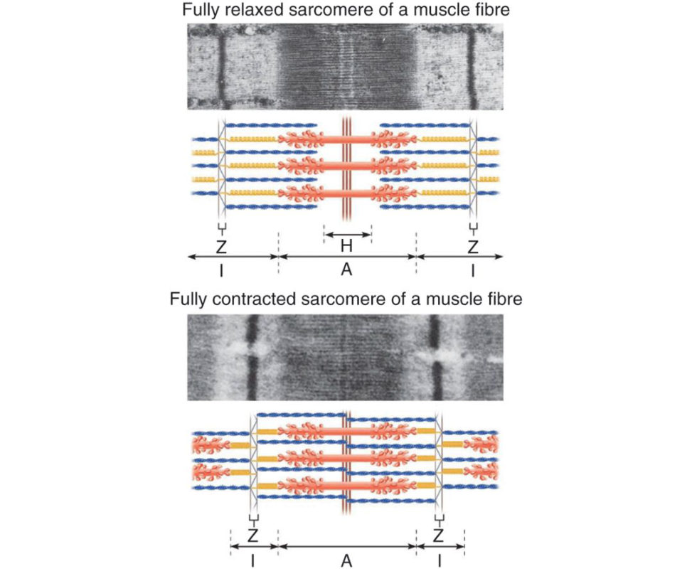
This question (we think) tests inequal measure a candidate’s drawing ability and muscle sarcomere knowledge.
How do muscles contract? Explain how skeletal muscles contract.
Having dealt with the microstructure of the sarcomere the second part of the topic deals with how the myosin heads interact with the actin filaments. An answer would be expected to cover the following points: The release of calcium ions from the sarcoplasmic reticulum. The formation of cr oss-bridges. The sliding of actin and myosin filaments. The use of ATP to break cross-bridges and reset myosin heads. When muscles contract, actin slides over the myosin and causes the sarcomere to shorten. In resting muscle fibres, Ca2+ is stored in the sarcoplasmic reticulum. Muscle cells have a unique membrane structure, called the transverse tubule or simply the T-tubule. The T-tubule is an invagination of the muscle membrane that plunges repeatedly into the interior of the fibre. The T-tubules terminate near the calcium-filled sacs of the sarcoplasmic reticulum. The arrival of the action potential a t the ends of the T-tubules triggers the release of Ca2+. The Ca2+ diffuses among the thick and thin filaments where it binds to troponin on the thin filaments. This turns on the interaction between actin and myosin and the sarcomere contracts. Due to the speed of the action potential (milliseconds), the action potential arrives virtually simultaneously at the ends of all the T-tubules, ensuring that all sarcomeres contract in unison. When the process is over, the calcium is pumped back into the sarcoplasmic reticulum using a Ca 2+ ATPase. The hydrolysis of ATP causes the myosin heads to change shape and swivel which moves them towards the next actin binding site. The movement of the myosin heads causes the actin filaments to slide over the myosin filaments, shortening the length of the sarcomere. Via the repeated hydrolysis of ATP, the skeletal muscle will contract.
What do we mean by the cross-bridge cycle (Figure 20.46)?
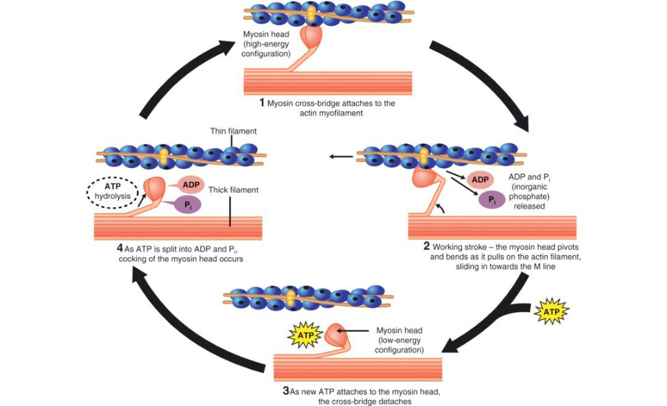
The cross-bridge cycle consists of four steps: 1. ATP hydrolysis. Hydrolysis of ATP reorients and energizes the myosin head. 2. Formation of cr oss-bridges.
Myosin head attaches to the myosin-binding site on actin.
3. Power stroke.
During the power stroke the cross-bridge rotates, sliding the filaments.
4. Detachment of myosin from actin.
As the next ATP binds to the myosin head, the myosin head detaches from actin. The contraction cycle repeats as longas AT Pis available and the Ca2+ level is sufficiently high.
Continuing cycles applies the force that shortens the sarcomere.
What is the role of AT Pin muscle contracture?
AT Pis the immediate source of energy for muscle contraction. Although a muscle fibre contains only enough ATP to power a few twitches, its ATP ‘poolis replenished as needed.
What are the actual events occurring in muscle contraction and relaxation?
Membrane excitation Arrival of motor neuron action potential from a nerve cell at the neuromuscular junction. Synaptic transmission at the neuromuscular junction with release of acetylcholine into the synaptic cleft and diffusion across to bind to sarcomere receptors. Local depolarization occurs with the action potential propagating along the sarcolemma leading to hypopolarization ofT -tubules. Excitation –contraction coupling At the end of the T-tubules the depolarization triggers Ca2+ release from the sarcoplasmic reticulum. Ca2+ binds to the troponin C molecule, resulting in a cooperative configurational change in the troponin–tropomyosin complex. Release of inhibition of m yosin-ATPase. Link between thick and thin filaments, swivel of myosin head. Tension exerted. Shortening by sliding filament. Muscle recovery Ca2+ removed from sarcoplasm and transported back into the sarcoplasmic reticulum.
Mg2+ ATP bound by actinom yosin.
Cross-bridges disconnected.
Actinom yosin-ATPase inhibited.
Active tension disappears.
Figure 20.40 Unlabelled electron micrograph picture of skeletal muscle.
Figure 20.41 Bare labelled electron micrograph picture of skeletal muscle.
Figure 20.42 Labelled diagram of skeletal muscle.

Figure 20.43 Relaxed muscle.

Figure 20.44 Contracted muscle.
Figure 20.45 Skeletal muscle actin/m yosin arrangement with relaxation and contracture.
Figure 20.46 Cross-bridge cycle.
Structured oral examination question 2#
Viva question 2 tests similar knowledge to viva question 1, but they are not identical and are probably different diets of the exam rather than a candidate 6 versus a candidate 8 situation.
How do you classify skeletal muscles?
Muscles can be classified according to: Shape and fascicular architecture. Myoglobin content (red, white). Type of contractile activity (isometric , isotonic) (concentric, eccentric and isokinetic). The relative magnitude of their stabilizing and rotatory components. Orientation of the line of pull to the joint surface (flexors, extensors, abductors and adductors). The number of joints over which the muscle crosses. Type of muscle action or function (their interaction injoint movement) (agonists, antagonists, synergists and fixators).
Tell me about the types of muscle contraction that can occur. What do we mean by isometric and isotonic muscle contracture?
1. Isometric contraction (iso = equal + metric = length): Force is generated but the muscle does not shorten (no movement). Muscle is held at a fixed length. The muscle contraction is activated , but instead of being allowed to lengthen or shorten, it is held at a constant length. Examples include when one pushes against an immoveable object such as a wall or the rotator cuff and its ability to compress the humerus into the glenoid surface. 2. Isotonic contraction (iso = equal + tonic = tone or tension): Constant force with change in muscle length (movement). a. Concentric contracture. b. Eccentric contracture. Concentric contraction (con = towards + centric = centre). It is a contraction in which the origin and insertion of the contracting muscle are brought closer together due to the action of the muscle Generation of a force leads to muscle contracture and shortening if the load on the muscle isless than the force the muscle creates.
Eccentric contraction : (Ecc = away from + centric = centre)
It is a contraction in which the origin and insertion of the contracting muscle are moved away from each other by an external force. Despite the muscle contracting the muscle lengthens, as the force generated isless than the external force.
Biceps curls exhibit both concentric and eccentric contraction. During flexion, the bicep is undergoing concentric contraction, and during controlled extension, the biceps is undergoing eccentric contraction.
Isokinetic contraction (iso = equal + kinetics = motion):
This involves keeping the speed (distance per unit time constant on an actively contracting muscle while the load is changed in order to maintain a constant velocity. Isokinetic exercises require special exercise equipment and are a measure of dynamic strength. The exercises are best used for rehabilitation protocols and are not routinely observed in most muscles during normal activities.
This is the warm-up question!
What macroscopic types of muscles do you know?
Muscles can be broadly classified into either parallel, where the muscle fibres are parallel to the line of pull, or pennate, where the short muscle fibres are oblique to the line of pull (Figure 20.47). A parallel muscle has an increased range of movement due to increased length of fibres. The total force of contracture isless because there are less numbers of fibres. Parallel muscles are subdivided into the following subtypes: 1. Strap (sartorius, rectus femoris). 2. Fan-shaped (triangular) (pec major). 3. Fusiform muscles (biceps). Pennate muscles can be subdivided into: 1. Unipennate. 2. Bipennate. 3. Multi pennate. 1. Unipennate muscles have fibres (fascicles) that insert on only one side of a tendon, thereby creating a relatively large cross-section and greater strength but smaller change in length. 2. Bipennate muscles insert on two sides of the tendon, increasing relative cross-section, allowing greater strength than a unipennate muscle with smaller changes in length. 3. Multi pennate muscles are where a series of bipennate muscles lie side by side in one plane.
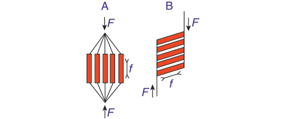
The benefit of pennation is that it allows for a greater packing of fibres into a given anatomical cross-sectional area. However, their range of movement is diminished because of the shortness of muscle fibres and oblique direction of pull.
Pennate muscles are located in positions requiring small but powerful movements, parallel muscles are located in positions requiring longer movements with less power.
Figure 20.47 The two arrangements of muscle fibres within a muscle. (A) Parallel arrangement. (B) Pennate arrangement. Double-headed arrows (f) indicate direction of force exerted by individual muscle fibres.
What is the role of the horizontal component of a pennate muscle?
The force of muscle action is resolved into two component forces – one acts in the line of pull and the other at right angles to it. This muscle force is essentially wasted, but not that much, and while contracting such muscles squeeze themselves, thus becoming more solid and compact. This is not in the standard orthopaedic textbooks! That’s what makes the basic science viva so feared! Trying to work this out from first principles during the viva is tough-going.
Can you draw me the structure of a muscle?
Epimysium wraps around an entire muscle. Perimysium subdivides each muscle into fascicles, bundles of 10–100 muscle fibres. Endomysium surrounds individual muscle fibres. These connective tissues form a continuous membrane around and within the muscle belly. The muscle cross-sectional s tructure/arrangement (Figure 20.48) is universal across all connective tissues (epi, peri and en doand includes ner ves,tendons and ligaments. The sarcomere drawing (Figure 20.49) deals with muscle ultrastructure and is more complicated to draw and explain.
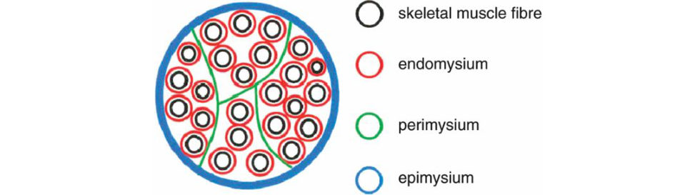
Actin – thin filaments have actin because if you’re active actin) you will be thin.
Myosin – o ands short and fat letters.
Figure 20.48 Candidate drawing. Cross-section of a skeletal muscle.
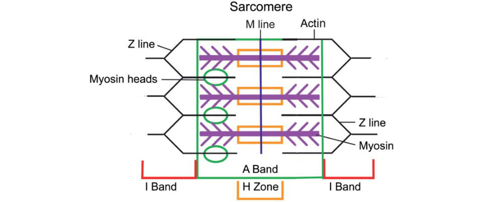
Figure 20.49 Candidate 20-second exam drawing of skeletal muscle sarcomere. Z line is where the thin filament (actin creates a zig-zag pattern. One sarcomere is Z-line to Z-line. In between the I bands is the A band (darker on the edges, just myosin present in the H zone).
What are the different types of skeletal muscles and can you draw them?
A. Muscles can be divided into: 1. Parallel Fascicles run parallel to the long axis of the muscle to provide a stronger pull (sartorius, rectus abdominis). 2. Fusiform 3. Pennate Fascicles are short. Attached obliquely to a central tendon that runs the length of the muscle. Produce more tension (more muscle fibres).
Unipennate (palmer interosseous), bipennate (rectus femoris) and multi pennate (deltoid) – to how many sides of the tendon do the fascicles attach?
4. Convergent (fan-shaped) Broad at origin converging to a narrower insertion, less pull than parallel muscle spec major).
5. Circular – fascicles arranged in a concentric ring. Act as sphincters, a ring around a body opening
(orbiculari soris).
Candidates need to be able to drawout the various types of muscles (unipennate, fusiform, strap) along with examples and be able to discuss the advantages of each type of muscle shape (Figure 20.50).
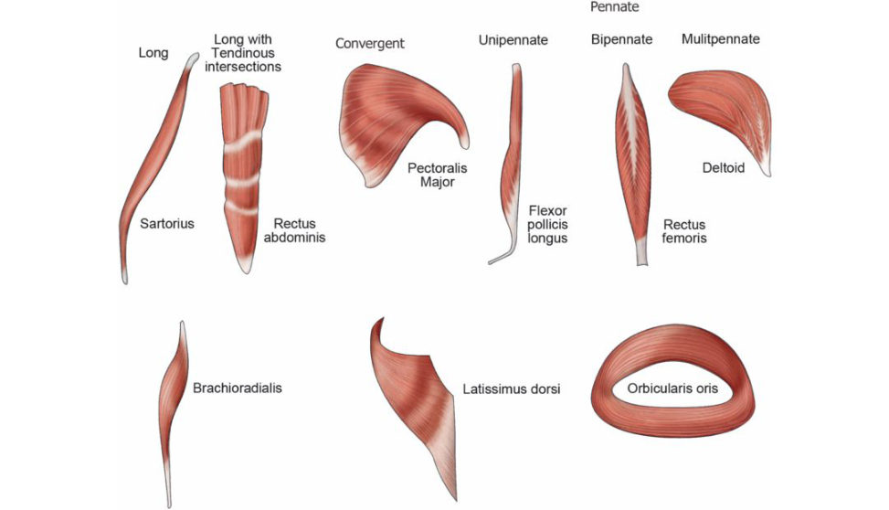
Figure 20.50 Muscle shape and fibre arrangement.
What is the hierarchical structure of muscle (Figure 20.51)?
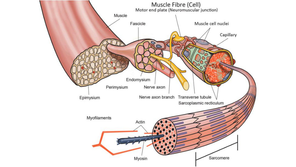
Figure 20.51 Hierarchical structure of muscle.
Sarcomere > myofibril > muscle fibre (single elongated muscle cell) (endomysium) > muscle fascicle (perimysium) > bundles of muscle fascicles > single muscle (epimysium) Terminology can be confusing. Bundles of muscle fascicles make up a single muscle = muscle bundles (fascicles) make up a single muscle. Bundles of 10–100 muscle fibres make up a muscle fascicle. A fascicle is the smallest unit of structure visible to the naked eye.
Draw the structure of striated muscle fibres including the myofibrils with light and dark bands, mitochondria, the sarcoplasmic reticulum, nuclei and the sarcolemma (Figures 20.52 and 20.53).
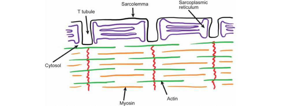
Figure 20.52 Candidate diagram. Sarcoplasmic reticulum arrangement in skeletal muscle.
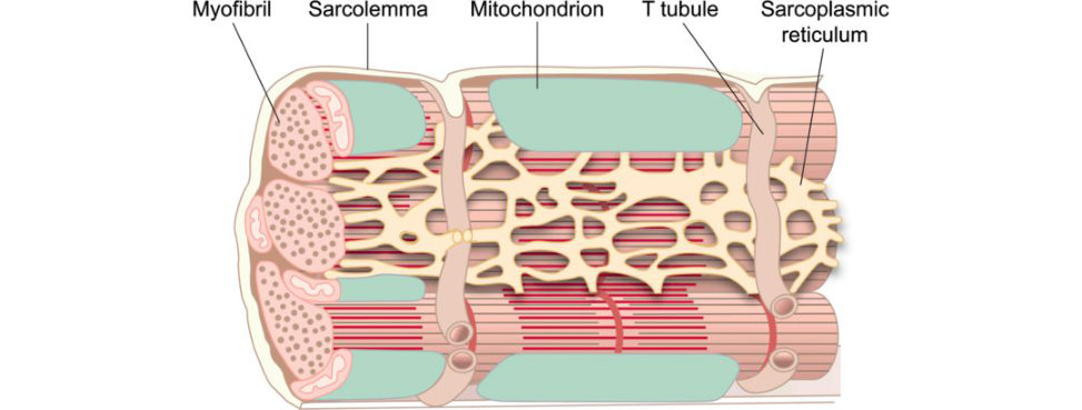
Figure 20.53 Sarcoplasmic reticulum arrangement in skeletal muscle.
The sarcomere is the smallest contractile unit of skeletal muscle. The sarcolemma is the cell membrane that surrounds the muscle fibre and has periodic invaginations that descend into the muscle cell forming transverse tubules (T-tubules). These T-tubules allow the depolarization that occurs during cell contraction to propagate deep into the cell. The sarcoplasmic reticulums tores calcium (Ca2+) in intracellular membrane-bound channels.
How does muscle move at a microscopic level, how do troponin, myosin and tropomysin interact?
Similar info to that asked for in oral examination question 2. This is one of the pivotal knowledge areas in the muscle section the T r & Orth curriculum sets out to test. Sarcomere contraction is regulated by troponin and tropomyosin. Troponin is closely associated with tropomyosin along the actin thin filament and serves as a regulatory protein for contracture. Troponin has three subunits, I, T and C.
Troponin I is inhibitory and is able to block actin –myosin interactions.
Troponin T enables binding of troponin and tropomysin.
Troponin C binds calcium.
When myoplasmic calcium concentrations are low, the troponin–tropomyosin complex is situated on the actin filament in a way that prevents actin –myosin cross-bridge formation ( Figure 20.54).
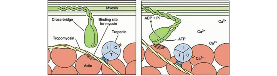
A rise in myoplasmic Ca2+ allows Ca2+ to bindwith troponin C. This binding causes a conformational change in the troponin I molecule complex that removes the troponin–tropomyosin complex from the actin binding sites. This change permits myosin–actin cr oss-bridge cycling.
When Ca2+ concentrations return to normal resting levels, troponin I reverts to its inhibitory conformation and the actin binding sites are again blocked from forming cross-bridges.
Figure 20.54 Cross-bridge. Troponin T (tropomyosin binding), troponin I (inhibitory protein) and troponin C (calcium binding). Binding of Ca2+ to the Tn Cunit of troponin exposes the myosin binding site on actin.
What about length versus strength of muscle contraction, and the gr aph, which shows this (Figure 20.54)?
Muscles generate the greatest force when at their resting (ideal) length, and the least amount of force when shortened or stretched relative to their resting length. Skeletal muscle fibre force production is defined in terms of myofilament overlap, i.e. in terms of sarcomere length. At optimal length, where actin –myosin interactions are maximal, muscle generates maximum force (region 2). As sarcomere length increases (region 3), force decreases owing to the decreasing number of interactions between actin and myosin myofilaments. At lengths shorter than the optimum region 1), force decreases owing to double interdigitation of actin filaments with both myosin and actin filaments from opposite sides of the sarcomere.
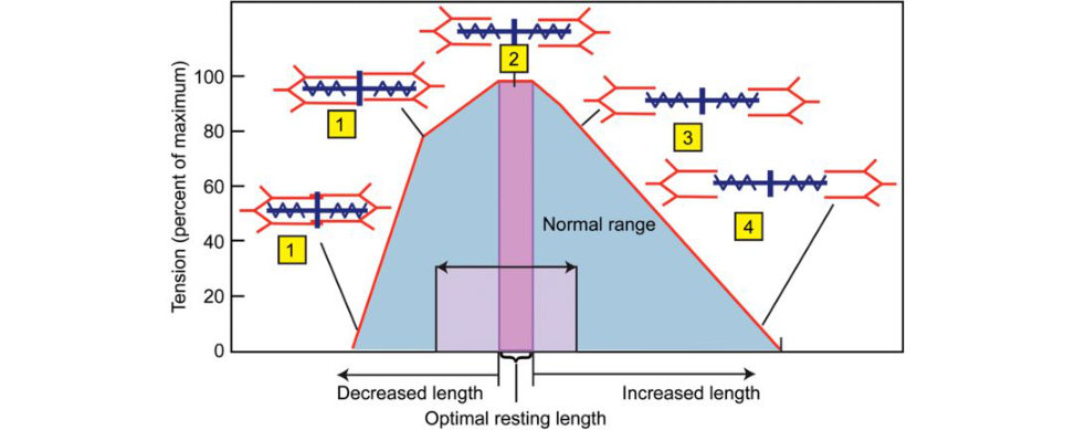
Figure 20.55 Length versus strength of muscle contraction.
What about the force–velocity relationship ( Figure 20.56)?
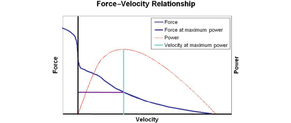
Figure 20.56 Force velocity curve of a skeletal muscle. Right of the vertical axis concentric contractions (the muscle is shortening), left of the a xis eccentric contractions (the muscle is lengthened underload);
power developed by the muscle in red.
The force–velocity relationship in muscle relates the speed at which a muscle changes length with the force of this contraction and the resultant power output (force × velocity = power). The force generated by a muscle depends on the number of actin and myosin cross-bridges formed; a larger number of cross-bridges results in a larger amount of force. However, cross-bridge formation is not immediate, so if myofilaments slide over each other at a faster rate the ability to form cross-bridges and resultant force are both reduced. At maximum velocity no cross-bridges can form, so no force is generated, resulting in the production of zero power (right edge of graph). The reverse is true for stretching of muscle.
The amount of force a sarcomere can exert, outside of the relative length at which it functions, is dependent on the speed the sarcomere is contracting.
Making the cross-bridges necessary for contraction takes time. When the filaments are being moved at a higher velocity, fewer myosin heads can bind to the actin filament sat a given time and as a result the total force is lower.
A higher speed during a concentric contraction results in a lower force. At speed 0, or an isometric contraction, the force is greater. When the contraction velocity turns negative and the sarcomere is stretched (eccentric contraction), the force a sarcomere generates increases even further. This can be explained by the force required to stretch passive structures and lengthen the muscle (Figure 20.57).
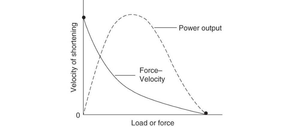
Although the force of the muscle is increased, there is no velocity of contraction and zero power is generated. Maximum power is generated at approximately one-third of maximum shortening velocity.
Figure 20.57 Force velocity curve with eccentric contracture. Force and velocity are inversely related such that at zero (0) velocity maximum force is generated, and at maximum velocity zero (0) force is generated.
5. Structure and function of tendons and ligaments#
Introduction#
Tendon and ligaments are complex connective tissues often grouped together as they share similar tissue composition and properties.
Candidates should, however, appreciate there are also significant functional and structural differences between these two tissues that the examiners may wish to discuss.
Tendon and ligament disorders are especially common, so it is important to have a clear understanding of their function in both health and disease.
Potential score 7/8 candidates should have something up their sleeve if the examiners probe into new treatment options and evidence for their therapeutic benefit.
Structured oral examination question 1#
Tendon structure and function. Leading onto a discussion about tendinopathy versus tendonitis.
What is the function of tendons?
Tendons attach muscle to bone and function to: Transmit tensile loads from skeletal muscle contracture to bone resulting injoint movement. Enable the muscle belly to be positioned a way from the joint so that bulky muscle bodies do not obstruct movement.
What are ligaments?
Ligaments are dense bands of collagenous tissue (fibres that span a joint and become anchored to bone at either end.
What is the function of ligaments?
Ligaments attach bone to bone and function to: Transmit tensile load from bone to bone. Provide joint stability by maintaining joint congruency. Limit freedom of movement by preventing excessive motion, being astatic restraint. Contribute to proprioception and position sense.
What are the differences in structure between ligaments and tendons (Table 20.5)?
If a fairly low-key viva start the generalized details contained in Table 20.5 should be sufficient. However, candidates are often probed in more detail about specific percentage differences in composition between tendons and ligaments (Table 20.6). Ligaments have a more random organization with a weaving pattern of collagen orientation compared to a more parallel collagen arrangement seen in tendons. They have a slightly lower collagen content compared to tendons but contain more ground substance.
What else?
Ligaments and tendons are composed of cells and extracellular matrix. Cells (fibroblasts) occupy around 20% of the total tissue volume, while the extracellular matrix accounts for the remaining 80%.
The extracellular matrix is composed of water (70%) and solids (30%).
The solid part of the matrix is mainly composed of collagen, but also ground substance and a small amount of elastin.
The collagen content is 70–80% in ligaments, slightly higher in tendons. Type I collagen is higher in tendons (95–99%) compared to ligaments (90%) and Type III collagen accounts for 1–5% in tendons and 10% in ligaments.
What functions does the ground substance perform?
The ground substance comprises hyaluronan, proteoglycans (decorin, biglycan, fibromodulin, lumican), structural glycoproteins and a wide variety of other molecules. The highly viscous and hydrophilic nature of the ground substance acts as a cement-like filling between the long, thin collagen molecules and provides the lubrication and spacing that enables collagen fibres to slide. It contains proteoglycan aggregates that bind most of the extracellular water of ligaments and tendons, making the matrix a highly structured, gel-like material.
What about elastin?
Elastin isnt usually present in ligaments to any large degree. The ligamentum flava and ligamentum nuchae are the two main exceptions. These ligaments contain large amounts of elastin.
How is collagen synthesized?
Collagen synthesis again! Candidates need to rehearse an answer based around the cellular collagen synthesis diagram. This can be quite detailed in some textbooks, but it is best to try and keep your answer fairly simply and low key. Collagen synthesis occurs in several stages with both intracellular and extracellular steps. The first stage in the synthesis of collagen is the formation inside the cell of mR NAf or each type of polypeptide alpha-chain. Messenger RNA translation initiates synthesis of polypeptide chains in the r ough endoplasmic reticulum. Subsequently, the signal peptide is cleaved off, lysine and proline amino acids are hydroxylate dand the hydroxylated amino acid residues are glycosylated. The resultant procollagen is packaged into secretory vesicles in the Golgi apparatus. On release into the extracellular matrix the ends of the procollagen are cleaved enzymatically by peptides to form tropocollagen fibrils. Several adjacent collagen molecules pack together (aggregate), overlapping by a quarter staggered array and appear as cross-striations under anele ctr on microscope.
Describe the structure of a tendon.
Tendons are composed of cells and extracellular matrix. The cells are fibroblasts and make up 20% of the total tissue volume. Most of the extracellular matrix is water (70%), but around 30% of the matrix is solid, comprising mainly type I collagen, ground substance and a small amount of elastin.
What about its hierarchical structure?
Although there are lots of different ways to draw this diagram there are two main illustrations that regularly appear in textbooks. Although both are quite similar there are also enough differences to cause confusion. We have included both for completeness sake (Figures 20.58 and 20.59). Our own preference is for the classic tendon structure described by Kastelic et al. (Figure 20.58).
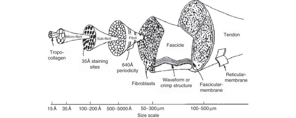
Figure 20.58 Classic tendon hierarchical structure by Kastelic et al.
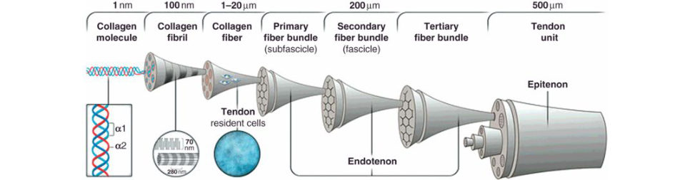
Figure 20.59 Alternative tendon hierarchical structure.
1. Classic tendon structure and description by Kastelic et al.33 Tropocollagen molecules assemble into microfibrils, these microfibrils into subfibrils and several subfibrils give rise to a collagen fibril, with a characteristic 65 nm periodicity visible in scanning electron microscopy.
Multiple fibrils combine to form a tendon fascicle, and fascicles, separated by the endotenon, join together to form the macroscopic tendon.
2. Alternative hierarchy structure of tendon.
Collagen molecules assemble into collagen fibrils with a characteristic 65 nm periodicity visible in scanning electron microscopy. Multiple collagen fibrils are packed into larger structures to form collagen fibres. Multiple collagen fibres form primary fiber bundles (subfascicle), groups of which form secondary fibre bundles (fascicle). Multiple secondary fibre bundles form tertiary fibre bundles, groups of which inturn form the tendon unit.
Primary, secondary and tertiary bundles are surrounded by a sheath of connective tissue known as endotenon, which facilitates the gliding of bundles against oneanother during tendon movement.
The endotenon is contiguous with the epitenon, the fine layer of connective tissue that sheaths the tendon unit.
What is a collagen fibre?
A collagen fibre is the smallest tendon unit that can be tested mechanically and is visible underlight microscopy.
Describe the structure of a molecule of collagen.
This question can be asked across a number of section 2 Tr & Orth topics such as bone, articular cartilage or even intervertebral disc. The structural unit of collagen is tropocollagen. Tropocollagen is formed in the fibroblast cell as procollagen that is then secreted and cleaved extracellularly to become collagen. It consists of three polypeptide chains each forming a left-hand helix. The chains are connected by hydrogen bonds and wind together to form a rope-like, right-handed superhelix, which gives the collagen molecule a rod-like shape. They can be divided into fibrillar and non-fibrillar collagens. Almost two-thirds of the collagen molecule consists of amino acid triplets (GLY-X-Y), where X is often proline andY is often hydroxyproline. The structure at the centre of the helix is so spatially restricted that only glycine, the smallest amino acid, can be accommodated.
What about cross-linking?
Tropocollagen molecules are stabilized and held together by cross-linking. Hydroxyproline is involved in hydrogen bonding (intrachain and interchain) between the polypeptide chains.
Tell me about ligament ultrastructure.
The ultrastructure of ligaments is similar to that of tendons, but compared to tendons the tensile strength of ligaments isless, there is a higher percentage of proteoglycans and water and they have a higher elastic content.
Ligaments exhibit non-linear anisotropic mechanical behaviour and under low loading conditions they are relatively compliant, due to recruitment of ‘crimped’ collagen fibres as well as to viscoelastic behaviours and interactions of collagen and other matrix materials.
What are the differences in structural collagen arrangement between ligaments and tendons?
In tendons, the collagen fibres are arranged completely in parallel, as they need to withstand large tensile loads in one direction only . In ligaments, collagen fibres are not arranged completely parallel like tendons; fibres are branched and interwoven. This is because although ligaments need to withstand large loads mainly in one direction, the y also need to withstand smaller loads in other directions. When unloaded the collagen fibres in both tendons and ligaments are ‘crimped’. This arrangement allows tendons and ligaments to be initially stretched without too much resistance. Ligaments exhibit non-linear anisotropic mechanical behaviour and under low loading conditions they are relatively compliant, due to recruitment of ‘crimped’ collagen fibres as well as to viscoelastic behaviours and interactions of collagen and other matrix materials.
You have mentioned viscoelastic behaviour what do we mean by this?
Creep, stress relaxation hysteresis and rate-dependent deformation Tendons are less viscoelastic when compared to ligaments.
How do ligaments fail? What is the reason for midsubstance rupture and bony avulsion?
The most common mechanism of ligament failure is rupture of a sequential series of collagen fibre bundles distributed throughout the body of the ligament and not localized to one specific area. Midsubstance ligament tears are common in adults while avulsion injuries are more common in children. Bony ligament avulsions usually occur between the unmineralized and mineralized fibrocartilage layers, as this is the weakest link in the ligament/bone complex. Table 20.5 Structural differences between tendons and ligaments.
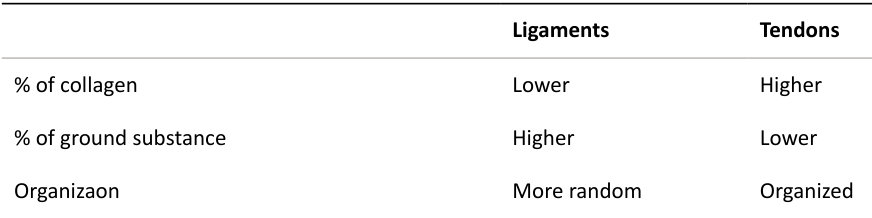
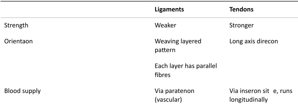
Sheathed avascular tendons via single vinculae (mesotendon) and diffusion
Table 20.6 Compositional comparison between tendons and ligaments.
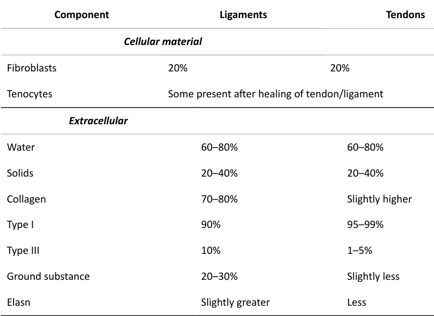
Structured oral examination question 2#
How do tendons and ligaments heal after injury? What factors affect healing?
After injury, a large number of growth factors and cytokines are released by the injured tendon and adjacent tissues, including in terleukins, vascular endothelial growth factor (VEGF), platelet-derived growth factor (PDGF), fibroblastic growth factor (FGF), transforming growth factor beta (TGF-β), connective tissue growth factor (CTGF), epidermal growth factor (EG Fand insulin-like growth factor 1 (IGF1). There are three phases of tendon/ligament healing (Table 20.7): 1. Inflammatory. 2. Proliferative. 3. Remodelling. In strict terms this is section 3 material, but there is almost always some sort of varying overlap between structure and function of connective tissue ( section 2 ) and pathology (section 3 ). Phase 1 is characterized by haematoma formation and the initiation of a rapid inflammatory response. A few days after the injury, phase 2 begins. In this phase, tendon fibroblasts synthesize abundant collagen and other extracellular matrix components such as proteoglycans and deposit them at the wound site. After about 6 weeks, the remodelling phase starts. This phase is characterized by decreased cellularity and decreased collagen and glycosaminoglycan synthesis. During this period, the repair tissue changes to fibrous tissue, this again changes to scar-like tendon tissue after 10 weeks. During the later remodelling phase, covalent bonding between the collagen fibres increases, resulting in repaired tissue with highest stiffness and tensile strength. Also, both the metabolism often ocytes and tendon vascularity decline.
Are there any new methods developed to improve tendon healing?
There have been attempts at biological augmentation of tendon healing.
Such as?
These include applying growth factors, singly or in combinations tem cells innative or genetically modified form, and biomaterials, alone or cell-loaded, at the site of tendon damage.
What is their basis for use?
Table 20.7 Cells and matrix changes associated with tendon healing.
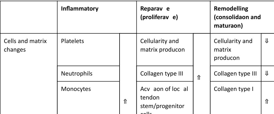
| Inflammatory | Reparative (proliferative) | Remodelling (consolidation and maturation) | ||||
|---|---|---|---|---|---|---|
| Cells and matrix changes | Platelets | ⇑ | Cellularity and matrix production | ⇑ | Cellularity and matrix production | ⇓ |
| Neutrophils | Collagen type III | Collagen type III | ⇓ | |||
| Monocytes | Activation of local tendon stem/progenitor cells | Collagen type I | ⇑ | |||
| Erthyrocytes | ||||||
| Circulation-deriv ed mesenchymal stem cells | ||||||
| Molecular changes | Interleukin-6 | bFGF | GDF-5, -6 and -7 | |||
| bFGF | GDF-5, -6 and -7 | IGF-1 | ||||
| IGF-1 | IGF-1 | TGF-β | ||||
| PDGF | PDGF | |||||
| TGF-β | TGFβ | |||||
| VEGF | VEGF |
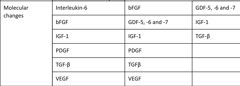
Growth factors
Tendon injury stimulates the production of a variety of growth factors at multiple stages in the healing process leading to increased cellularity and tissue volume. Increased expression of growth factors is particularly prominent in the early phases of healing.
Growth factors can be applied by local injection, percutaneously or operatively, or by implanting scaffolds or even suture material containing growth factors.
Mesenchymal stem cells
Mesenchymal stem cells can be applied directly to the site of injury or can be delivered on a suitable carrier matrix.
Structured oral examination question 3#
Tell me about ligament ultrastructure.
Ligaments are dense bands of collagenous tissue (fibres that span a joint and become anchored to bone at either end. The ultrastructure of ligaments is similar to that of tendons, but compared to tendons the tensile strength of ligaments isless, there is a higher percentage of proteoglycans and water and they have a higher elastic content.
What are the differences in structural arrangement of collagen between ligaments and tendons?
In tendons, the collagen fibres are arranged completely in parallel as they need to withstand large loads in one direction only . In ligaments, collagen fibres are not arranged completely parallel like tendons; fibres are branched and interwoven (Figure 20.60).
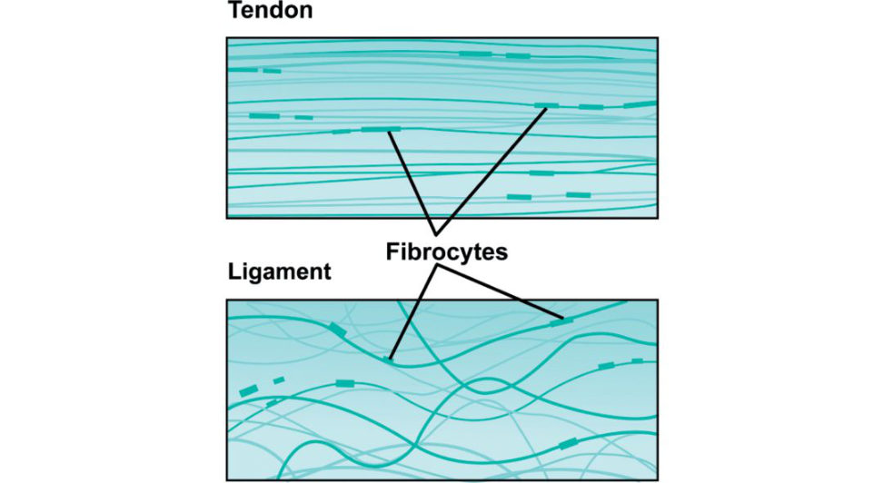
Figure 20.60 Collagen arrangement in tendon and ligament tissue Tendon: parallel bundles of collagen fibres. Ligaments: irregular wavy bundles of collagen fibres.
Why?
This is because although ligaments need to withstand large loads mainly in one direction, they also need to withstand smaller loads in other directions.
In any single layer the fibres lie parallel to each other, but in subsequent layers they lie in a different direction.
When unloaded the collagen fibres in both tendons and ligaments are arranged in a wavy
‘crimped’ pattern. This arrangement allows tendons and ligaments to be initially stretched without much resistance, increasing their capacity to absorb energy.
How do ligaments fail? What is the reason for midsubstance rupture and bony avulsion?
The most common mechanism of ligament failure is rupture of a sequential series of collagen fibre bundles distributed throughout the body of the ligament and not localized to one specific area. Midsubstance ligament tears are common in adults, while avulsion injuries are more common in children. Avulsion of ligaments usually occurs between the unmineralized and mineralized fibrocartilage layers as this is the weakest link in the ligament bone complex.
But why is this? What is the reason? CANDIDIATE : With tendons, ligaments and bones their stiffness increases with increasing strain – they are strain-rate sensitive Bone is more sensitive to strain rate than tendons or ligaments, so its stiffness increases more proportionally . Therefore, avulsion is more common at slow strain rates and tendon and ligament tearing is more common at higher strain rates. During slow strain rates, avulsion is common, while, as the strain rate increases, the bone becomes stronger than the tendon or ligament and so tearing becomes more common. So why do bony avulsions occur in children and midsubstance ligament tears occur in adults?
The ligament/bone junction is relatively weak in children resulting in a greater chance of avulsion. In adults the strength of the ligament/bone junction has increased more rapidly than that of ligament tissue so that the strength of the ligament/bone junction exceeds the ligament tissue leading to a greater chance of midsubstance tearing.
Structured oral examination question 5#
Draw the stress–strain curve of a ligament/tendon and describe its various parts as you go along.
There are several minor variations of the stress strain curve for tendons/ligaments seen in textbooks. Although this can become confusing, remember that the principles are more important than absolute specifics. There are, however, specific regions and points on the graph that do need to be understood. Although the stress–strain curves of tendons and ligaments are broadly similar, there are a few subtle differences. Despite this, they can often be viewed as similar materials sharing comparable features on the stress–strain curve. Therefore, clarify (if in doubt) whether you have been asked to drawout the stress–strain curve for a ligament, tendon or if the examiners want to focus on a curve demonstrating the composite features of both materials.34 Stress–strain curve for ligament There are four regions that are commonly used to describe the stress-strain curve (Figure 20.61). 1. Non-linear (toe) region. Collagen crimped: low stiffness; change in slope as collagen fibres straighten; modulus of elasticity is not constant; the ligament becomes stiffer as more fibres are recruited. The toe region represents ‘uncrimping’ of the collagen fibrils. Because it is easier to stretch out the crimp of the collagen fibrils, this part of the stress–strain curve shows a relatively low stiffness compared to the linear portion. T oe region ends at about 2% of strain when all crimpled fibres straighten. 2. Elastic linear region. Slope = stiffness = elastic modulus. Elastic modulus stabilizes. The ligament deforms in a linear fashion due to the intermolecular sliding of collagen triple helices. If strain isless than 4%, the ligament will return to its original length when unloaded; therefore, this portion is elas tic and reversible, and the slope of the curve represents an elastic modulus. 3. Progressive failure or yield region. At the end of the linear region there is early sequential failure of a few greatly stretched collagen fibres, causing a series of small force reduction (dips). The load/stress at which this occurs is called the yield point. As elongation continues microfibrils begin to tear/rupture and the curve bends towards the strain axis as stiffness is reduced.
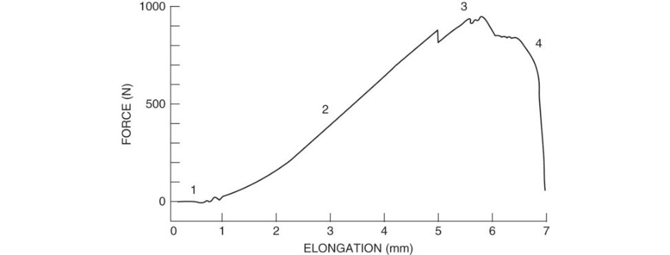
The ligament undergoes irreversible plastic deformation. The fibres that fail first will be those that were less crimped, while those that fail last will be those that were initially crimped and sow ere recruited last.
4. Complete rupture. When the ligament is stretched to more than 8% of its original length, macroscopic failure occurs, and the stress–strain curve falls quickly to zero. The ultimate tensile strength is the maximum load/stress that can be achieved before the ligament ruptures. Complete rupture (failure) occurs rapidly after ultimate tensile strength is attained.
What is the normal operating condition of the A CL within the knee?
During everyday activities (such as walking or light jogging) the ACL operates along the ‘toe region’ of the stress–strain curve. It is thought that ligaments are not generally loaded above a quarter of their ultimate tensile load during these everyday activities. The early part of the linear region is considered the upper operating range of the ACL during strenuous activities as might be experienced during pivoting while running. Loading of the A CL beyond the linear region which may occur with a bad football tackle or ski accident will result in ligament damage and possible rupture.
What about the stress–strain curve of a tendon?
The stress–strain curve (Figure 20.62) is very similar to that of ligaments except: 1. Toe region. This is much less prominent than in ligaments because fibres are more aligned. Waxy collagen fibres straighten out with a small increase in load. 2. Elastic (linear region. Fibres are straightened out and slope represents stiffness which is more orless linear. Elastic recovery at stresses < 4%. 3. End of linear region. The load value at this point is referred to asP lin incertain textbooks. Micro failure takes place after Plin and small force reduction (dips) occur in the curve. Corresponds to strains of 4–8%. Cross-links between collagen fibres fail. Collagen fibres slide past oneanother, irreversible changes such as tearing or permanent stretching of tendon. 4. Macroscopic failure. The ultimate tensile strength of the material is referred to as maximum load. Pm axin some textbooks. Once maximum load is surpassed complete failure occurs rapidly. Fibres recoil and blossom. Tangled bud at ruptured end.
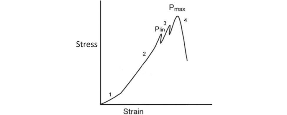
Can you draw me the stress–strain curve for the ligamentum flavum?
The ligamentum flavum has a high percentage of elastin fibres present so its stress–strain curve is completely different to that of the standard ligament/tendon curve (Figure 20.63). With tensile testing elongation of up to 50% occurs before the stiffness increases significantly. Beyond this point, the stiffness increases greatly with additional loading and the ligament fails abruptly with litile further load.
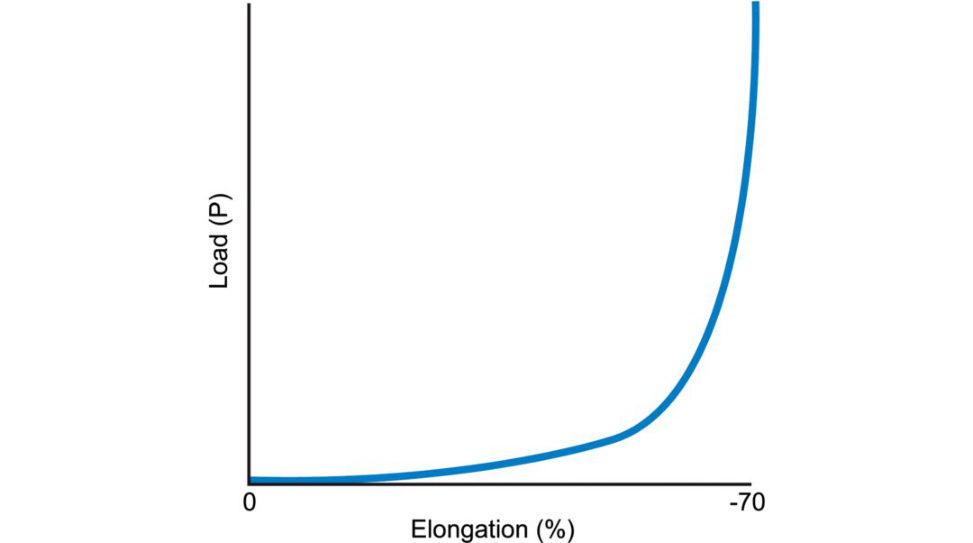
The elastic fibres allow the ligament to return to its original shape and size after the load has been released.
What is the difference between force/elongation curves and stress–strain curves?
Force/elongation curves are essentially the same ass tress–strain curves. Force/elongation curves involve mounting a specimen in a machine whereas stress–strain curves have been normalized with respect to specimen dimensions. In experiments, you measure load (force) and elongation(displacemen t). These are properties of the sample undertest that change with the size of the sample. For this reason, we often divide load by sample area to get strain and displacement by sample length to get strain. Now any derived quantity is independent of sample size and can be regarded as a true material property.
How do tendons receive their blood supply?
In general, tendons tend to have a sparse but adequate blood supply and low metabolic rate. Tendons can be divided into vascular or avascular tendons. Vascular tendons are surrounded by loose areolar connective tissue known as paratenon in which the blood supply reaches the tendon. In avascular tendons a true synovial sheath replaces the paratenon. The vascular supply to a tendon arises from three distinct areas: (1) musculotendinous junction; (2) osseotendinous junction; and (3) vessels from various surrounding connective tissue such as the paratenon, mesotenon and vincula. Vessels are generally arranged longitudinally within the tendon, passing around the collagen fibre bundles in the endotenon. The situation is more complicated insheathed avascular tendons in that the blood supply must enter the mesotenon in vincula that tether the tendon to its sheath incertain locations to supply one tendon segment. Avascular tendon areas receive nutrition via diffusion.
What is tendinopathy?
Tendinopathy is a multifactorial condition often related to a combination of micro trauma, excessive loading and normal ageing. It is characterized by pain, focal tendon tenderness, decreased strength and movement.
What about pathogenesis of tendinopathy?
The pathogenesis is poorly understood. It has been variously defined as a degenerative condition, a failure of the healing processor a region of diminished blood supply just above the tendon insertion. The role of inflammation int endinopathy is not clearly established. The exact relationship between tendinopathy and tendon rupture remains unknown. It is thought that tendinopathy could lead to tendon rupture.
What changes are occurring in the tendon?
Tendinopathy can be identified by the following histological characteristics collagen fibril disorganization, increased proteoglycan and glycosaminoglycan content and increased non- collagenous ECM, hypercellularity and neovascularization.
How do we treat tendinopathies?
First-line treatment involves non-steroidal anft-in flammatory medication for pain relief, physiotherapy with eccentric exercises (involves active lengthening of muscles and tendons) and steroid injections.
What about PRP injections, how do they work?
Platelet-rich plasma (PRP) is a blood derivative containing PRP. It is the plasma fraction of the blood containing concentrated platelets and high levels of growth factors, known to promote tissue healing.
What is the evidence for their use?
Current evidence suggests/My understanding of the literature is that PRP is useful for chronic degenerative tendinopathies such as lateral epicondylitis of the elbow or patella tendinopathy. There is no evidence to support the use of PRP in promoting tendon or ligament-to-bone healing in rotator cuff repair or ACL reconstruction. 35 The main points are that it should not be considered a first-line treatment but reserved for chronic tendinopathy refractory to standard non-operativ e management, such as physiotherapy and steroid injections.
Any other options?
Extracorporeal shock-wave therapy has some benefit in calcified tendinitis of the shoulder , and ultrasonography are other treatment options. St em-cell-based therapy may become available for use in the future.
What is the difference between tendinitis and t endinosis.
Terms can be misleading. Tendinitis suggests inflammation although inflammatory cells are often absent. Patients may experience localized pain, swelling, warmth and redness. Tendinosis is degeneration of the tendon. This can often be due to repetiv e microtrauma. Tendinopathy is typically used to describe any problem involving a tendon.
How do tendons and ligaments insert into bone?
There is direct and indirect attachment to bone.
Direct insertion in to bone is similar for tendon and ligament and consists of four zones.
Zone 1. Tendon. Parallel collagen fibres at the end of the tendon or ligament.
Zone 2. Uncalcified fibrocartilage Collagen fibres intermesh with unmineralized fibrocartilage.
Zone 3. Calcified fibrocartilage. Fibrocartilage gradually becomes mineralized.
Zone 4. Bone. Mineralized fibrocartilage merges into cortical bone.
This allows a gradual increase in the stiffness of the tissues other e is a lesser stress concentration effect at the insertion of a t endon/ligament into bone.
Otherwise, high stress levels will occur at the interface due to the difference in stiffness between the two materials. There is a gradual change in structure, composition and mechanical behaviour between tendon/ligament and bone. The mineralized cartilage tidemark forms deep interdigitations increasing the contact area and reducing the stiffness gradient between mechanically different tissues. This improves the ability of the unit to resist shear and tensile forces.
The continuous change in tissue composition from tendon/ligament to bone aids in the efficient transfer of load between the two materials.
With indirect insertion the deep la yer anchors to bone via Sharpey’s fibres.
Can you give me any examples of direct and indirect ligament insertion?
ACL direct and indirect superficial.
Figure 20.61 Stress–strain curve of ligament (ACL). The stress–strain curve is initially upwardly concave, but the slope becomes nearly linear in the prefigure phase of tensile loading.
Figure 20.62 Stress–strain curve of tendon tested to failure intension.
Figure 20.63 Stress–strain curve of tendon with large amounts of elastin (ligamentum flavum).
Structured oral examination question 6#
This is a coronal MRI through the thigh, can you tell me what has happened (Figure 20.64)?
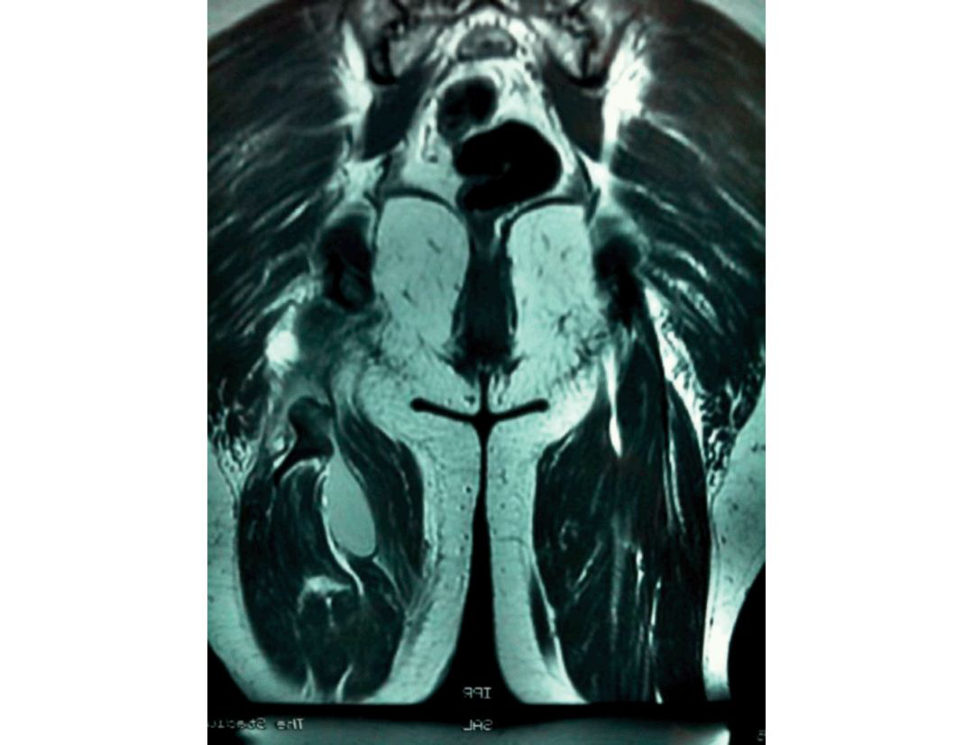
[I was not sure exactly what I was looking at initially!] There appears to be a tendon pulled off the bone, along with an associated haematoma. This is just using your basic understanding of MRI.
Appears to be, or actually is?
There is a definite avulsion of tendon from the bone. Be confident.
What’s the function of the bone attachment?
[I really didn’t know where to go, so I gave a very basic answer!] It represents an interface between bone and tendon.
But what is the function?
It allows a load to be transferred from muscle to bone and stores energy. [As you can see, I’ve not read much on this and gave short answers, so I didn’t get into trouble.]
What type of load?
Tensile?
What is the difference in general terms between ligaments and tendons?
[I started to talk about ligaments attaching bone to bone!]
Figure 20.64 Coronal T1 MRI image of thigh.
Mark 4 – poor fail.
No, I mean structurally!
[Finally I can get going now!] Collagen content is higher in tendons and can be up to 80–90% in extremity tendons. [Best answer – up to 99% of dry weight. Remember that tendons and ligaments are made up from 80% EC Mand 20% cells.]
What are the cells found in tendons and ligaments?
Fibroblast represents the vast proportion of cells, a t about 20% of the total mass.
You mentioned collagen, which is the common type?
Type 1 represents the most common type. Mark 5 – fail
That was alit ile guarded, give me a figure.
70–80 % (actually 90%) [I knew what was coming next!]
Tell me about type 1 collagen.
Type 1 collagen consists of three polypeptide chains, two alpha and one beta. [This was wrong, it’s two (alpha 1) and one (alpha 2). They combine to form a triple helix, which provides the tensile strength.] If you can’t talk at this stage you will fail this section, you must have a broad knowledge base. Mark 6 – pass
How does this structure retain its stability?
There is cross-linking which is allowed to occur due to hydrogen bonds.
Can you draw a picture of collagen in a ligament and tendon and explain how they differ?
I drew a longitudinal section basically with a well-aligned pattern for a tendon and more haphazard for a ligament. I explained that the less-parallel structure in a ligament means it takes tension in one direction but can also take stresses in other directions. In each la yer they are parallel, but in subsequent layers the collagen is at a slightly different angle.
Can you draw a schematic of this hierarchical structure?
[I talked about the layers as I drew, only got to the fibril and he was bored!] (Figure 20.65) Remember there are lots of different ways to draw this diagram, just use one and learn how to draw it and talk. It is principles not details which are important!
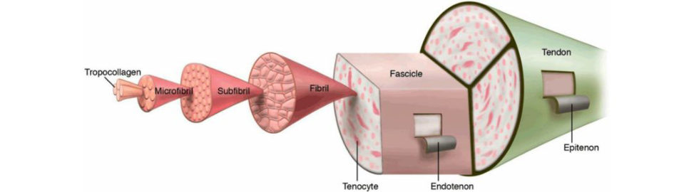
So, what makes up the extracellular matrix?
This essentially is a pr oteoglycan matrix, with plasma proteins and glycoprotein. These proteoglycans bind water and provide a gel-type matrix. The proteoglycans are glycosaminoglycans which bind to a protein core, linked to a hyaluronic acid chain which provides a very high molecular weight.
Figure 20.65 Anatomy of a normal tendon.
Mark 7 – good pass
Can you draw the stress–strain curve for tendons and ligaments and talk through it (Figure 20.66)?
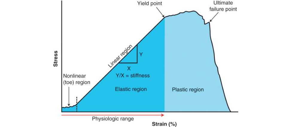
You can get this from any of the basic science books, just learn it! There are four major regions of the stress strain curve: (1) the toe or toe-in region, (2) the linear region and (3) the yield and (4) failure region. In physiological activity , most ligaments and tendons exist in the toe and somewhat in the linear region. These constitute a non-linear stress– strain curve because the slope of the toe-in region is different from that of the linear region. In terms of structure–function relationships, the toe-in region represents ‘uncrimping’ of the crimp in the collagen fibrils. Because it is easier to stretch out the crimp of the collagen fibrils, this part of the stress–strain curve shows a relatively low stiffness. As the collagen fibrils become uncrimped, then we see that the collagen fibril backbone itself is being stretched, which gives rise to a stiffer material. As individual fibrils within the ligament or tendon begin to fail, damage accumulates, stiffness is reduced and the ligament/tendons begin to fail. Thus, a key concept is that the overall behaviour of ligaments and tendons depends on the individual crimp structure and failure of the collagen fibrils.
Figure 20.66 Stress–strain curve for tendon and ligament.
Mark 8 – excellent Pass
This is an example of a question which is based on one specific topic, which is great if you know it well, but very bad if you don’t. This emphasizes the fact you must be able to talk to a basic level on all topics, or you may flounder very early on in the question. This question will be in your basic science viva and last
5 minutes.
6. Structure and function of the nervous system#
Introduction#
The difficulty with structure/function of the nervous system is that there is considerable overlap with section 3 (nerve injury and regeneration) and section 9 (electrophysiological investigations).
On top of this, the core material is quite dry and complicated but needs to be well understood before candidates would feel confident enough to deal with the viva questions the y are likely to be asked.
Structured oral examination question 1#
With reference to nerve conduction studies: what do you see indifferent types of injury (axonotmesis, neurotmesis, etc.)? The normal reference ranges? Please draw a cross-section of a nerve and label.
This is immediately bang on the money. Candidates need to know straightup their definitions of neuropraxia, axonotmesis and neurotmesis and then they will get hammered with th eNCS findings in each specific case. This is a way of grilling candidates about the VERY specifics of NCS while minimizing too much candidate preamble and chit-chat. These are unGoogleable questions. There are three main types of nerve injury: 1. Neuropraxia where the myelin sheath is dysfunctional. 2. Axonotmesis where the axon is damaged but the supporting connective tissues remain intact. 3. Neurotmesis where both the axon and its sheath are damaged. In clinical reality, nerve trauma usually leads to a mixture of all three to the fascicles within a nerve. However, there will usually be a predominance of one of these, which is important to predicting the prognosis. Neuropraxia Myelin is critical to effective signal transmission via saltatory conductance across the Nodes of Ranvier. In neuropraxia, there is focal oedema or breakdown of the myelin sheath which disrupts the nerve’s ability to conduct signals and can lead to slowing or ‘blocking’ of conduction. This type of injury usually arises from a mild stretch or crush injury. As myelin is formed by the Schwann cells, these tend to regenerate quickly, depending on the degree of disruption, from weeks to months. The axon itself is preserved and so there is no Wallerian degeneration and no secondary degeneration of muscle fibres. There are a number of hallmarks of neuropraxia on nerve conduction studies (NCS) and electromyography (EMG). NCS will show: Conduction slowing or even conduction block across the level of the lesion but should be normal below the level of the lesion. EMG will show: Electrical silence persisting after 3 weeks in a completely stunned state (there won’t be any fibrillations as the muscle fibre isn’t denervate dand there will be no voluntary activity duet o the severity of the signal block).
Single (or limited) motor units at high rates in isolation in a partiallys tunned state as the brain attempts to generate force via the remaining working fibres.
Axonotmesis
Here there is injury primarily to the axon and its myelin sheath, usually following a more severe crush, or even avulsion. However, the supportive connective tissues remain mostly preserved (endoneurium, perineurium, epineurium) and so the tubes that encapsulate the axons remain preserved.
Wallerian degeneration will occur distal to (and alit ile proximal to) the site of the lesion and will spread in an anterograde direction, i.e. peripherally over the coming days. Axon degeneration in the distal stump triggers a number of responses in the remaining Schwann cells which are the key to successful regeneration. First, they degrade myelin and signal macrophages to remove debris. Then, they proliferate and form Büngner bands which are bridging tubes as well as signalling molecules to atir act the regenerating proximal axon bud into them.
Prognostically, regeneration is likely to occur if the re-innervating fibres follow the pre-existing pathways formed by their original endoneurial tubes. When the distances involved in regeneration are long, then a number of factors may preclude successful re-innervation, particularly if the target muscle fibres have atrophied or fibrose din the meantime. Occasionally, regenerating axons may follow the wrong endoneurial tube and innervate the wrong fibres. Perhaps the most visible manifestation of this is
‘synkinesis’ seen in Bell’s palsy where apa tien t may try to smile and ends up blinking.
NCS will show:
Reduced amplitudes of sensory and motor fibres.
Relative preservation of conduction velocities (noting that some fast fibre dropout is often expected when there has been severe axonal loss leading to mild slowing).
EMG will show:
Positive sharp waves and fibrillations following 2 weeks in the upper limbs and 3 weeks in the lower limbs as thed enervated muscle fibres upregulate their acetylcholine receptors.
Reduced interference patterns (i.e. fewer motor units being recruited) for the degree of recruitment (i.e. volitionary effort to generate force) as there are simply fewer working axons to transmit the signal.
Neurotmesis
Here there is complete destruction of the nerve and surrounding supportive tissues, usually caused by serious injuries such as anatomical severance of nerve and/or extensive avulsion or crushing. The axon,
Schwann cell and endoneural tube is completely disrupted and the perineurium and epineurium have varying degrees of injury and disruption Prognosis in these situations is usually poor .
NCS will show:
Initial preservation of distal responses for the first 3–5 days for motor studies and 6–10 days for sensory studies until Wallerian degeneration reaches distal regions. The earlier impairment of motor fibres is now thought to relate to neuromuscular junction transmission failure.
Thereafter there will be absent sensory and motor responses.
EMG will show:
Where axonal loss is severe, there will be an immediate and complete lack of voluntary activity .
For the first few weeks there will only be electrical silence and then fibrillations will appear around
2 weeks later in the upper limbs and 3 weeks later in the lower limbs.
This differentiates this state from severe neuropraxia (see above).
Fibrillations will be abundant and of large amplitude in the first 6 months, and then diminish as the muscle tissue a trophies and/or fibroses.
What are nerve conduction studies?
Nerve conduction tests are used to evaluate the function of mot or and sensory nerves. An NCS may consist of the following: Compound motor action potential (CMAP). Sensory nerve action potential (SNAP). F wave. H reflex wave. Motor nerve conduction studies examine the conduction of a signal along the course of a peripheral motor fibre to its muscle fibres. Latency – the time interval between stimulus and the onset of a response. Amplitude – the maximal height of the action potential. Conduction velocity – speed of the fastest part of the impulse calculated by the time of the recorded impulse between two points of known distance. It is reduced by a reduction in myelin (e.g. external compression or demyelinating conditions).
Please draw a cross-section of a nerve and label (Figure 20.67).
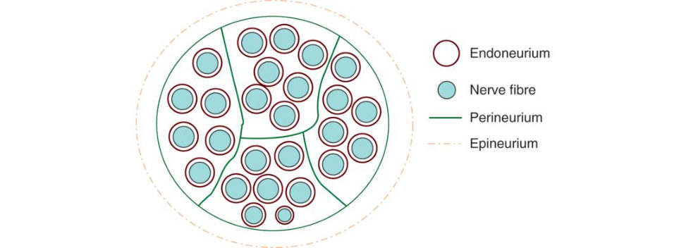
Figure 20.67 Candidate diagram. Cross-section of a nerve fibre.
Axons are grouped together in spatially arranged motor or sensory bundles called fascicles. Individual axons are surrounded by a connective tissue la yer, the endoneurium, and fascicles are separated by the perineurium. Groups of fascicles are contained within a peripheral nerve surrounded by a connective tissue la yer called the epineurium. The epineurium is the outermost layer of dense connective tissue surround inga peripheral nerve. The perineurium is the layer that covers individual fascicles and provides tensile strength. The endoneurium is the inner layer, which is mostly collagenous, that surrounds axons within fascicles and it nourishes and protects the axons.
Structured oral examination question 2#
Nerve action potential: explain the graph with relationship of membrane potential to sodium and potassium concentrations exchange pump, channels.
Neurons exhibit a lipoprotein cell membrane with a negative resting cell (around –70 mV), due to the voltage difference resulting from a high concentration of intracellular potassium (K+) and low concentrations of sodium (Na +) and chloride (Cl–) ions within the cells.
The concentration gradient is maintained by:
A metabolically active Na+/K+ exchange pump.
A lipid membrane that prevents the passage of water-soluble ions.
Donnan equilibrium. Irregular distribution of permanent ions across anim permeant membrane when a large impermeable organic ion is present on one side.
What is a resting potential?
A resting potential is a term used to describe the electrical potential across the membrane of a cell in its inactive unexcited state.
What is an action potential?
The whole basis of an action potential is based around a change in permeability to sodium and potassium due to opening/closing of voltage-gated channels in response to a stimulus ( Figure 20.68). When a neuron is stimulated some of the sodium channels on the membrane open. This allows sodium ions to go into the cell from the outside. As a result, the charge difference between the inside and outside of the cell starts to decrease. When the membrane potential increases to around –50 mV, most of the voltage-gated sodium channels rapidly open to allow Na+ to enter the cell, and the membrane potential spikes to more than 30 mV. The inside of the neuron becomes positively charged with respect to the outside. However, this open channel configuration is unstable and exists for only a fraction of ase cond before a second conformational change occurs with an inactivation g ate to block the sodium channel, thereby stopping the flow of Na+. For a few ms after closing, they cannot be open again (the refractory period). This limits the number of action potentials a neuron can experience. The rapid depolarization of the membrane also triggers the more slowly acting v oltage-gated potassium channels to open and allows K+ to exit the cell. The flow of K+ from the cell has a longer duration than the flow of Na+ into the cell and the cell begins to repolarize as soon as the sodium channels close.
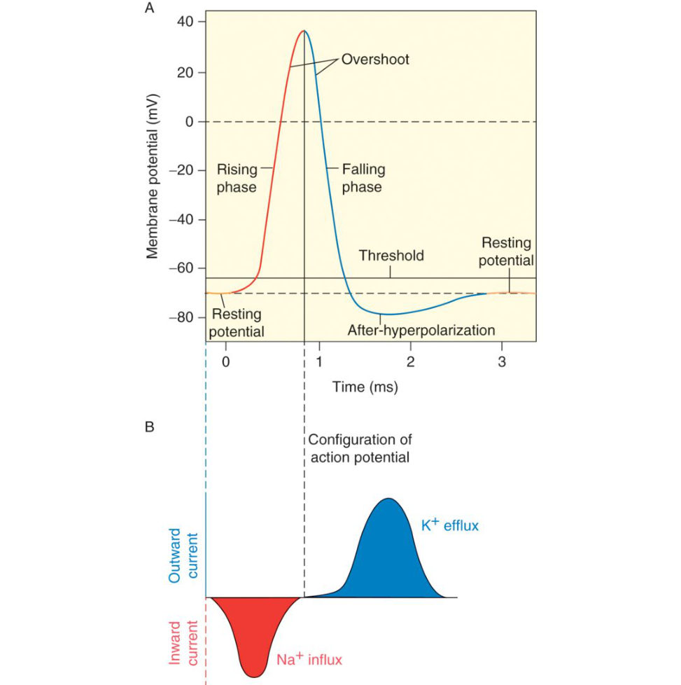
The electrical potential f alls to a level below the original resting potential of –70 mV
(repolarization).
The potassium channels eventually close and the sodium–potassium pump will continue to restore the neuron to its original resting potential.
What do we mean by the threshold stimulus?
The threshold stimulus is the minimum stimulus intensity needed to produce an action potential. A smaller stimulus (subthreshold) will not produce a stimulus However, summation of a number of subthreshold stimuli is some times sufficient to stimulate a response.
Figure 20.68 Action potential ‘hops’ from one non-myelinated region (Node of Ranvier) to the next (saltatory conduction). 1, Rest ing stage. 2, Depolarization stage. 3, Repolarization. 4, Hyperpolarization.
Structured oral examination question 3#
Nerve picture ... asked how action potential and muscle contraction produced ... Cross-bridge theory and power stroke .... These are gitis, dont waste them .... Wallerian degeneration ...
Can you drawout an action potential and t ell me what is going on as you go along (Figure 20.69)?
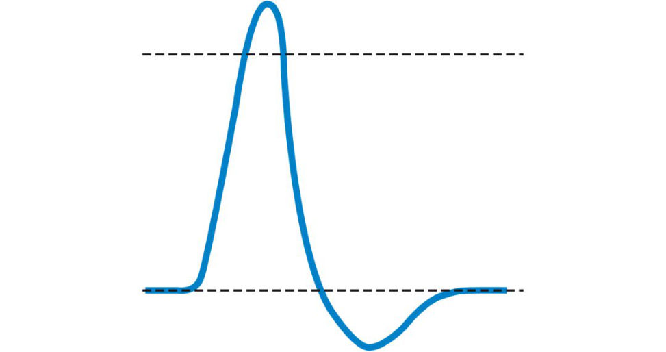
Neurons possess a membrane potential of –70 mV due to the voltage difference between the intracellular and extracellular space. This voltage difference is due to the high concentration of potassium ions [K+] and low concentration of sodium ions [Na +] and chloride ions [Cl–]. Action potentials are important for nerve signalling occurring as a result of rapid changes in membrane potential. The threshold stimulus is the minimum stimulus intensity needed to produce an action potential. An action potential is generated when a neuron is stimulated resulting in the opening of the Na channels and an in-rush of the Na+ ions into the cell.
Can you be more specific and point out some features of the diagram (Figure 20.70)?
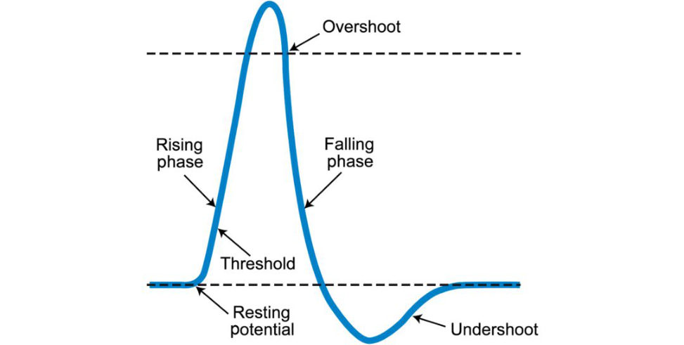
Point out: Threshold: Threshold is the membrane potential a t which enough voltage-gated sodium channels are open so that the relative permeability of the membrane is higher for sodium ions than it is for potassium ions. Rising phase/depolarization : When the inside of the membrane has a negative potential, there is a large driving force on sodium ions. Therefore, sodium rushes in through the open sodium channels, causing a rapid depolarization of the membrane. Overshoot: Because of the high permeability to sodium, the membrane potential g oes to a value that is close to the equilibrium potential for sodium (~ +55 mV). Falling phase/repolarization : First, the voltage-gated sodium channels inactivate Second, the voltage-gated potassium channels open (the delayed-rectifier potassium channels). The driving force pushes potassium out of the cell, causing the membrane potential to become negative again. Undershoot/hyperpolarization : The open potassium channels add to the normal resting membrane permeability to potassium, and drive the membrane potential closet o the equilibrium potential for potassium, thus hyperpolarizing the membrane.
Tell me about the refractory periods.
Refractory periods: the absolute refractory period is due to the inactivation of sodium channels. These channels cannot be opened again until the membrane potential is sufficiently negative to deactivate them. The relative refractory period is due to the hyperpolarization from the open potassium channels. This means that more depolarizing current is necessary to initiate another action potential.
What are the channels dependent on and how does this cause depolarization?
The channels are dependent on oxygen and ATP. There is depolarization of the membrane from the initial restings tate of –70 mV due to ionic conductance and the polarity across the cell membrane becomes positive.
What do you mean by ionic conductance?36
Conductance is the inverse of electrical resistance. If the conductance of the membrane to a particular ion is low, then the resistance to movement of that ion across the membrane is high. High conductance indicates that electrical charge moves easily through a membrane. The polarity across the cell membrane becomes positive This also triggers the opening of moreNa+ channels. The Na+ channels remain open for 1 ms before closing. The refractory period relates to the channels remaining closed for a few milliseconds and notable to reopen, thus limiting the number of stimuli to which a nerve can respond. Repolarization of the membrane results from the passage of K+ ions out of the cell through K+ channels.
What happens at the motor endplate?
The motor endplate includes the terminal portion of the nerve and the muscle membrane. There is a small gap known as the gap junction that separates the nerve from the muscle at the motor endplate. An action potential is propagated via the axon of the neuron to the nerve terminal. The presence of an action potential a t the nerve terminal triggers the opening of voltage-gated Ca2+. Ca2+ triggers the release of acetylcholine. This is the main neurotransmift er that acts on the motor endplate. It is stored in the presynaptic axon in membrane-encased compartments called vesicles. Acetylcholine diffuses across the synaptic cleft and binds with specific receptor sites on the motor endplate of the muscle cell membrane. This binding brings about opening of ion channels with large movements of Na+ into the muscle cell and smaller movements of K+ outward. This results in a depolarizing potential called an endplate potential spreading over the surface of the muscle fibre.
This potential triggers the release of calcium (from the sarcoplasmic reticulum), which elicits the movement of actin and myosin filaments, resulting in muscle contraction.
Acetylcholine is destroyed by an enzyme called acetylcholinesterase that inactivates acetylcholine by detaching it from its receptor and hydrolyzing it to acetate and choline.
What is the postsynaptic membrane?
The postsynaptic membrane is the specialized portion of the muscle cell membrane subjacent to the axon terminal, exhibiting a large number of folds that increase the surface area of the muscle cell in contact with the axon terminal.
Do you know of any diseases affecting the neuromuscular junction?
Myasthenia gravis. This is characterized by antibodies that bind to nicotinic acetylcholine receptors with resultingly sis of postsynaptic receptors.
How is a muscle contraction produced?
Depolarization in muscle cell plasma spreads to T-tubules and opens voltage-gated Ca2+ channels. This triggers the release of calcium ions (Ca2+) from storage in the sarcoplasmic reticulum (SR). The Ca2+ then initiates contraction, which is sustained by AT PAs longas Ca2+ ions remain in the sarcoplasm to bind to troponin, which keeps the actin-binding sites ‘unshielded andas longas AT Pis available to drive the cross-bridge cycling and the pulling of actin strands by myosin, the muscle fibre will continue to shorten to an anatomical limit.
What do we mean by the sliding filament mechanism of muscle contraction?
In the 1950s Huxley and Hanson discovered that skeletal muscles were composed of hexagonal latices of actin and myosin filaments and that muscle contraction resulted from relative sliding between the two filaments. Cross-bridge interaction between actin and myosin brings about muscle contraction. The thin filaments slide inwards over the stationary thick filaments.
What is a power stroke?
The power stroke describes a step in the cross-bridge cycle. For thin filaments (actin) to slide past thick filaments (myosin) during muscle contraction myosin heads must pull the actin a t the binding sites, detach, re-cock, attach to more binding sites, pull, detach, re-cock, etc. This repeated movement is known as the cross-bridge cycle. Binding: The active site on actin is exposed as calcium binds to troponin. The myosin head is atir acted to actin, and myosin binds actin a t its actin-binding site, forming the cross-bridge. Power stroke: During the power stroke, the phosphate generated in the previous contraction cycle is released. This results in the myosin head pivoting toward the centre of the sarcomere, after which the attached ADP and phosphate group are released.
Detachment: A new molecule of ATP attaches to the myosin head, causing the cross-bridge to detach.
What do we mean by Wallerian degeneration?
When a nerve is cut or crushed Wallerian degeneration occurs. The part of the axon separated from the neuron’s cell nucleus degenerates. Distal to the level of injury this involves the whole axon. Proximal to the injury retrograde (primary degeneration) occurs to the next Node of Ranvier. Macrophages ingest the fragmented myelin to provide a clean endoneural tube for advancement of regenerating axons. Wallerian degeneration is accompanied by marked proliferation of Sch wann cells and fibroblasts lining the endoneural tubes. Viva seems to be jumping around topics.
Figure 20.69 Candidate basic drawing of action potential.
Figure 20.70 Candidate basic drawing of action potential with labels.
Structured oral examination question 4#
Anatomy of spinal cord and cord syndromes. Candidate is shown an axial CT of C5 (Figure 20.71) with fracture with discussion of why you do not get cord injury at that level commonly.
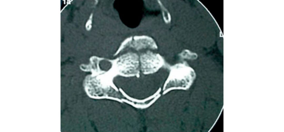
Figure 20.71 Axial CT image of C5 burst fracture.
Central cord syndrome (MUD-E)
Results from bleeding, infarction, or oedema to the central grey matter of the spinal cord.
Blood supply comes from periphery to centre.
Motor loss > Sensory
Motor loss affects Upper extremity > Lower extremity
Distal > Proximal
Commonly follows hyper-Extension Injury in an elderly patient with pre-existing spondylosis.
Pain and sensation affected.
Touch and proprioception unaffected.
Dissociative anaesthesia.
Hands and upper extremities are located centrally in corticospinal tract.
Finger and wrist motor function more affected than shoulder and biceps function.
Relatively good prognosis although full recovery rare.
Anterior cord syndrome
Preservation of posterior column-proprioception and vibration sense is intact.
Bilateral loss of motor function, light touch pain and temperature.
Brown–Sequard syndrome
Ipsilateral loss of motor function and pr oprioception/vibr ation.
Contralateral loss of pain and temperature (spinothalamic tract crosses over).
With a C5 fracture, why don’t you get cord injury at that level commonly?
The spinal cordis situated within the spine. The spine consists of a series of vertebral segments. The spinal cord itself has ‘neurological’ segmental levels which are defined by the spinal roots that enter and exit the spinal column between each of the vertebral segments. Spinal cord segmental levels do not necessarily correspond to the bony segments. Apa tien t with a burst fracture of the C5 vertebral body. The burst fracture will typically injure the C6 spinal cord situated at the C5 vertebra and also the C4 spinal roots that exit the spinal column between the C4 and C5 vertebrae. Such an injury should cause a loss of sensations in the C4 dermatome and weak deltoids (C4) due to injury to the C4 roots. Due to oedema (swelling of the spinal cord), the biceps (C5) may be initially weak but should recover. The spinal vertebral and cord segmental levels become increasingly discrepant further down the spinal column. For example, a T8 vertebral injury will result in a T12 spinal cord or neurological level. A T11 vertebral injury usually results in a L5 lumbar spinal cord level.
Structured oral examination question 5#
Photograph of cut section of median nerve shown at wrist all prepared for surgical repair.
What is this picture (Figure 20.72).
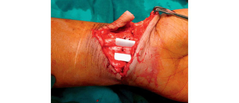
This is picture of a cut nerve at the wrist. The position of the nerve on the radial side of the hand is suggestive of median nerve transection.
What factors adversely affect recovery of a nerve after repair?
Much better to have a structured answer than a random generation off acts. List factors in terms of (1) patient, (2) injury, (3) surgical. Patient 1. Older age. 2. Systemic factors. Diabetes, alcoholism, smoking, rheumatoid arthritis, neur opathies, etc. Injury 3. High energy of initial injury. 4. Associated vascular or bony injury. 5. Crush or traction injury. 6. More proximal injury (increased timet o reach target organ). 7. Nerves that supply multiple sites with sensory and motor components. 8. Large gap. Surgical 9. Delay in repair (increased time for end-plate degeneration). 10. Repaired under tension. Excessive tension can cause breakdown at the area of repair. 11. Infection. 12. Need for nerve graft. If excessive tension present, use interposition autologous nerve gratis. 13. Quality of repair. It is important to try to suture only the epineurium and not pass the needle and suture through the fascicles, as this can create more damage and scarring, yielding a poorer result. Preservation of blood supply and accurate apposition of the fascicle.
What happens to a nerve when it is cut? How does it regenerate?
During the first few hours chromatolysis and swelling takes place in the cell body and nucleus. Oedema and swelling then continue in the axonal stump for the first few days. Within 2– 3 days Wallerian degeneration commences, which involves axonal and myelin disintegration both in an antegrade and retrograde direction Antegrade Wallerian degeneration then continues with Schwann cells and macrophage infiltration to remove cell debris, leaving only the basement membrane for about 3–6 weeks. Schwann cells then start to proliferate and organize guiding the axonal sprouts between the basement membranes of the two nerve ends. Nerve regeneration then begins on the columns of Schwann cells called Bunger bands. The proximal intact axon then sprouts a growth cone. The lamellipodia and filopodia cytoplasmic extensions allow the axon to explore the new environment and help in guiding the repair. Actin found in the axon allows elongation, within the tube. Growth continues a t the restricted rate of 1–3 mm/day, but simultaneously scar tissue interferes with growth.
Any new developments in nerve regeneration?
Sorry, no idea.
Figure 20.72 Median nerve laceration a t wrist.
Structured oral examination question 6#
Can you drawout a neuron for me?
Have an easy to drawout drawing already rehearsed from your exam preparation ( Figure 20.73). Candidates should be able to describe (talk through) the elements of the neuron as it is drawn.
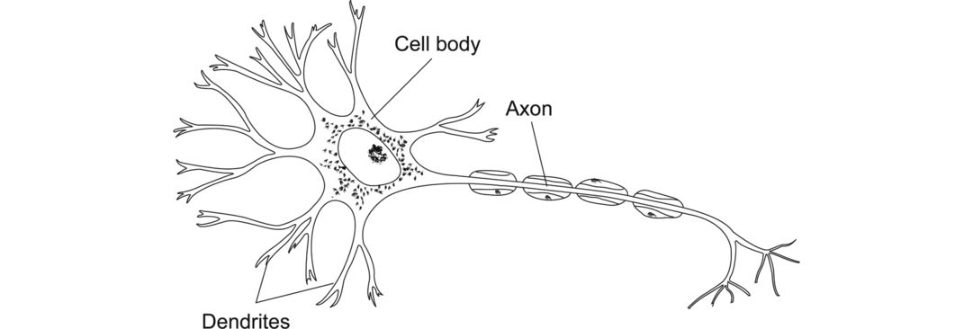
Figure 20.73 Candidate drawing of a neuron.
Neuron
The functional unit of the nerve.
Made up of a cell body and an axon.
Cell body
Gives rise to the axon and dendrites.
Contains most of the neuron’s organelles.
Dendrites
Thin branching extensions of a neuron that receive messages from other cell bodies and conduct impulses towards the cell body.
Sensory.
Axon hillock
Cone-shaped region of an axon where it joins the cell body, the region where the signals travel down the axon are generated.
Axon
The extension of a neuron.
The primary route of conduction to tissues.
Size of axon is between 0.2 and 20 μm.
Longest part of the nerve.
Glial cells
Anchor neurons and form myelin sheath.
In CNS, glial cells are:
Oligodendrocytes (make myelin astrocytes and microglia.
In PNS glial cells are:
Schwann cells.
Size of the nerve axon determines whether it will be myelinated.
Myelinated larger axons
Invaginated by one Schwann cell per axon internode.
Unmyelinated axons
Bundled together surrounded by one Schwann cell – no myelin.
Remak bundle.
Myelin
Lipid- and protein-rich multilaminar substance.
Laid down in the PNS to form a neurilemma.
No neurilemma is present around CNS myelinated axons.
Nodes of Ranvier
Gaps between Schwann cells along an axon.
Structured oral examination question 7#
Draw a cross-section of a nerve and nerve fibre.
[I talked about the three layers and the only four components of a nerve fibre I knew] (Figure 20.74).
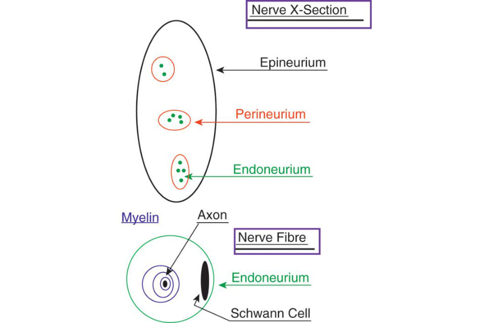
How are nerve injuries classified?
For a score 6 pass all you need to know is Seddon37 and Sunderland.38 Seddon classified the injury originally into three types andS underland defined these into six. Seddon defined neuropraxis as ionic block with possible segmental demyelinization (Sunderland 1), axonotmesis with axon severed but endoneurial tube intactS underland 2), endoneurial tube tornS underland 3) or only epineurium intactS underland 4). Finally, neurotmesis with loss of continuityS underland 5) or a combination of aboveS underland 6). At this point I could not remember exactly what the implications of all theS underland classifications were, so I did not offer them BasicallyS underland 1 and 2 are full recovery, 3 are incomplete, 4 is neuroma, 5 are none and 6 is unpredictable. I think the easiest way to get this down is to draw a box diagram and get used to filling it in.
If you cut the median nerve during surgery, what factors affect long-term results?
The age of the patient is the single most critical factor in sensory recovery after nerve repair and a good result is adversely affected by associated injuries to muscle, arteries, tendons and bone. In children, I believe you get good results in 50% and 40% in adults. The good results in children are actually 75% and 50% in adults, although this drops on a yearly basis year on year. The examiner either didn’t know himself or decided to leave this point. Just be a bit careful of using percentages, as they may ask where this information originates, which you may well not know.
What will you do if there is a 2-cm graft?
If there is going to be tension on the graft, I would either use a nerve graft or a conduit. [I don’t offer any further information a t the point as my knowledge is limited! Unfortunately, this strategy does not work very well.]
What nerve gratis are available for use?
Sural nerve, which has up to 20 cm, medial or lateral antebrachial cutaneous nerve.
What about nerve conduits?
I have never seen a nerve conduit, but they may allow up to 2 cm of nerve growth. [I had never seen a conduit graft, and knew virtually nothing about them, so I justified my answer. The examiner was happy with this and admift ed it would be unfair to ask anything further as I had not seen the procedure. Lucky escape.] A nerve conduit involves reconstruction of a gap defect by the placement of proximal and distal nerve stumps into a tubular repair construct. A nerve conduit provides a means to approximate nerve stumps within a biologically enhanced microenvironment, which minimizes fibrosis and the potential for ingrowth of external scar tissues. These products can provide directional growth cues and prevent dissipation of pr o-regenerativ e trophic and tropic factors away from the repair site. As a result, these constructs may reduce axonal escape or misdirection improve regeneration in to the distal nerve and enhance functional recovery. Tubular conduits also offer the possibility of avoiding nerve autograft harvest and thereby avoid the potential morbidity of that procedure.
Figure 20.74 Diagram of cross-section of a nerve and nerve fibre.
References
1. Better still, dont mention it.
2. Ellioft DS, Newman KJ, Forward DP, et al. A unified theory of bone healing and nonunion. Bone Joint J. 2016;98(7):884–891.
3. Tactical miscalculation from too long a coffee break and non-focused chitchat.
4. Urist MR. Bone: formation by autoinduction Science. 1965;150(3698):893–899.
5. Wall A, Board T. Bone: Formation by Autoinduction . In P Banaszkiewicz, D Kader (Eds.), Classic Papers in Orthopaedics. London: Springer; 2014.
6. This can use up a lot of precious time.
7. Much simpler for candidates to not mention them and just stick with the two above.
8. These contradictions make basic science unnecessarily complicated.
9. You could be given a diagram of articular cartilage or be asked to draw it. A score 8 candidate would be able to continually t alk away describing each layer (and fast as welldoing all the work with the examiners (usually) keeping silent, not interrupting and (hopefully) pleased the content is being covered. An excellent candidate with an excellent answer.
A score 6 candidate would discuss enough detail of each layer for a safe pass, but will need alit ile prompting from timet o time. A sc ore 4 or 5 candidate would struggle with naming the layers, structure and function and require a lot of prompting , giving the impression they hadn’t really learnt the subject.
10. If you are proactive you should be able to draw and talk at the same time, so practise this beforehand. If you are given a laminated diagram you have to get on the money straightaway and keep talking.
11. You are only going to be able to do this if you have already practised to perfection drawing out articular cartilage in 30 seconds flat.
12. The candidate was hesitant and needed to be prompted.
13. Dense collagen skin.
14. Depending on your interpretation.
15. The word ‘esoteric’ perfectly describes the basic science viva questioning.
16. All chondrocytes are not the same. If you grow cartilage in the laboratory and place the surface, deep and middle zone cells in the wrong place, they quickly move to the correct site.
17. You may be asked to drawout a proteoglycan.
18. Looks like a test-tube brush.
19. This is really a subtopic discussion rather than an isolated question.
20. Collagen synthesis and structure can easily take up 5 minutes of a viva, especially if a candidate’s answers are sluggish.
21. Testing clinical application of basic science.
22. If a candidate is poor at drawing diagrams they may need to compensate for this by more detailed diagram practice beforehand. In the big scheme of things, poor artistic ability shouldnt make a massive input into a candidate’s performance and/or mark. If a candidate has identified this weakness, practised drawing out the relevant diagram and perhaps more importantly is confident when delivering their discourse, this should be enough.
23. The viva usually develops in a slightly different direction than the previous two viva questions.
24. Andrews S, ShriveN, Ronsky J. The shocking truth about meniscus. J Biomech. 2011;44: 2737e40.
25. Smeathers J. Shocking news about discs. Curr Orthop. 1994;8(1):45–48.
26. Providing a convoluted answer.
27. If the examiners let you.
28. The question should not have been asked in its present form if at all. It should have been picked up with standard seting on the questions the night before the exam begins and discarded.
29. Neumann DA. Kinesiology of the Musculoskeletal System, Foundations for Rehabilitation , 2nd edition. Maryland Heights, MI: Mosby Elsevier; 2010.
30. Neumann DA. Kinesiology of the Musculoskeletal System, Foundations for Rehabilitation , 2nd edition. Maryland Heights, MI: Mosby Elsevier; 2010.
31. Coventry MB, Ghormley RK, Kernohan JW. The intervertebral disc: its microscopic anatomy and pathology: Part III. Pathological changes in the intervertebral disc. J Bone Joint Surg. 1945;27(3):460– 474.
32. Rudert M, Tillmann B. Lymph and blood supply of the human intervertebral disc: cadaver study of correlations to discitis . Acta Orthop Scand. 1993;64(1):37–40.
33. Kastelic J, Galeski AB aer E. The multic omposite structure of tendon. Conn Tiss Res. 1978;6(1):11– 12.
34. Essentially the f our regions of the curve (toe, linear, plastic and failure).
35. Fralinger DJ, Kaplan DJ, Weinberg ME, Strauss EJ, Jazrawi LM. Biological treatments for tendon and ligament abnormalities . A critical analysis review. J Bone Joint Surg Rev. 2016;4(6):e5.
36. This info is not contained in your average basic science orthopaedic textbook and requires a deeper level of knowledge to answer. This is why the viva can be difficult. As a candidate, you can’t really prepare for this type of question.
37. Seddon HJ. Three types of nerve injury. Brain. 1943;66(4):237–288.
38. Sunder landS. Advances in diagnosis and treatment of root and peripheral nerve injury. Adv Neurol. 1978;22:271–305.