Chapter 25 Tribology and biomaterials
Structured oral examination question 1#
Picture of THA with osteolysis shown.
This is an X-ray of a THA in an elderly patient complaining of pain. What do you see (Figure 25.1)?
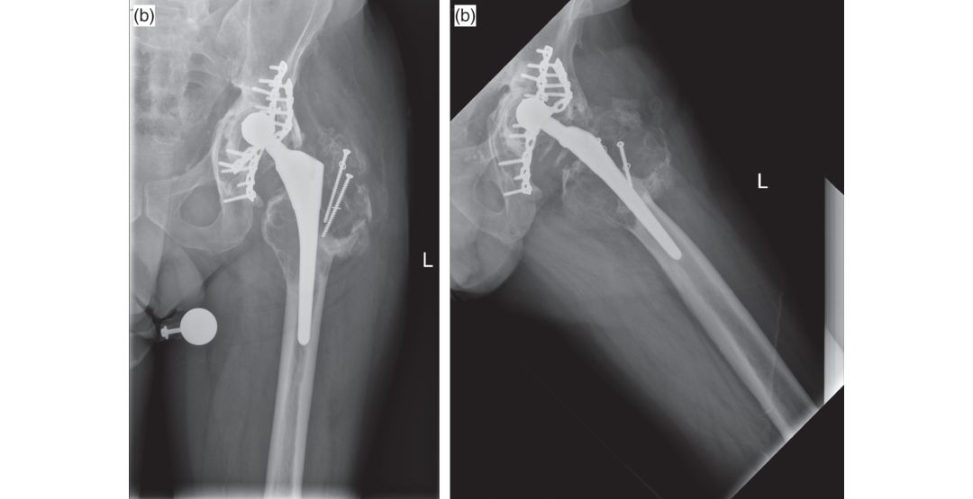
Figure 25.1a and 25.1b Anteroposterior (AP) and lateral radiographs of hybrid THA with areas of osteolysis around greater trochanter.
Plain radiographs of a reverse hybrid THA with a significant area of osteolysis in the greater trochanter.
What do you think caused this?
Potential causes are infection, poly w ear ...
Tell me about wear.
Wear is a progressive loss of material from the surface of a body owing to relative motion a t that surface generating debris.
What types and modes of wear do you know?
Well there are four modes of wear and various types of wear (I couldn’t remember how many). Mode 1: Wear from two articulating surfaces that are intended to rub together, such as the femoral head and the poly.
Mode 2: Wear from an articulating surface and a non-articula ting surface, such as the femoral head and the shell.
Mode 3: Third-body wear, which is particles coming between bearings.
Mode 4: Wear between non-articula ting surfaces such as the neck and the edge of the shell.
OK, how about types of wear?
There are mechanical and chemical. Mechanical includes abrasive, adhesive, fatigue, thir d- body and freting. Chemical includes corrosive and corrosive freting.
Do you know how we can measure wear?
There are linear and volumetric wear which are two methods of measuring wear. Volumetric wear measures the volume of material lost in cubic millimetres per year or per million cycles. Linear wear measures the penetration of the component into the other and is measured on X-rays. For example, the bigger the femoral head the greater the volumetric wear and the smaller ...
Are you sure? Poly has changed over the last 50 years. So, tell me then, what’s the ideal size of a femoral head?
[I was waiting for him to ask me this question, I had some papers in mind and drawings.] McKee and Farrar [1] used large heads and had minimal dislocation but significant wear, Charnley [2,3] initially used size 41.5-mm diameter femoral heads but had massive wear and early failure. He then used 22.25-mm femoral heads and reduced his wear, but bearing in mind that polyethylene quality wasn’t like the ones we have these days, highly cross-linked poly. There are various factors that increase wear in a THR, they include thickness < 6 mm, malalignment of components, young patients men and activity levels.
OK, let’s move on, how does osteolysis happen?
It’s a histiocytic response to wear debris.
OK, what does that mean?
It’s a cascade of events that ends with osteoclast activation. Its tarts when the macrophages are activated by the debris. They release osteolytic factors like TNF-alpha, osteoclast-activ ating factor and interleukins. This activates osteoclasts and cause osteolysis. Osteolysis causes micromotion of the prosthesis which then causes further debris. Also, as the patient walks they release the debris into the effective joint space causing further inflammatory response and lysis [4].
References#
1. McKee GK, Watson-Farrar J. Replacement of arthritic hips by the McKee–Farrar prosthesis. J Bone Joint
Surg Br. 1966;48:245–259.
2. Charnley J. Surgery of the hip-joint: present and future developments. Br Med J. 1960;1:821–826.
3. Charnley J. Arthroplasty of the hip. A new operation . Lancet. 1961;1:1129–1132.
4. Ing hamE, Fisher J. The role of macrophages in osteolysis of total joint replacement Biomaterials.
2005;26(11):1271–1286.
Structured oral examination question 2#
Tell me about synovial fluid.
Synovial fluid is produced by the synovial membrane in the joint. It is a dialysate of blood plasma without the cloting factors, haemoglobin or RBC. It contains hyaluronic acid, lubricin, proteinase collagenases and prostaglandins. The main function ist o lubricate articular cartilage and nourish it through diffusion. It exhibits non-Newtonian flow characteristics.
Tell me about lubrication.
Lubrication is when a film of lo wer shear strength is present between two bearing surfaces, reducing friction. There are two main hypotheses surrounding joint lubrication in synovial joints.
What is friction?
The resistance of two surfaces to slide against each other.
Tell me about the two hypotheses surrounding joint lubrication in synovial joints.
The first is fluid-film lubrication, in this hypothesis the joint surfaces are separated by a fluid film which fully supports the applied load, preventing contact between the surfaces. The minimum thickness of the fluid film must exceed the surface roughness of the bearing surfaces in order to prevent asperity contact. The secondis boundary lubrication. In this situation, the bearing surfaces are in contact but separated by a boundary lubricant of molecular thickness, which prevents excessive bearing friction and wear. In boundary lubrication the load is carried by the surface asperities rather than by the lubricant. It is thought that fluid-film lubrication dominates in synovial joints. However, realistically, both fluid-film and boundary lubrication occur in synovial joints, depending on the specific joint in question and the particular type of loading applied.
Do you know the lambda ratio?
This is the ratio of fluid film thickness to surface roughness, in fluid film it’s 3 and in boundary it’s less than 1.
Can you tell me about the different types of fluid-film lubrication?
There are various types of lubrication, the y include hydrodynamic lubrication (HD), elastohydrodynamic (EHD), micro-elastohydrodynamic (MEHD), squeeze film, weeping and boosted lubrication. In hydrodynamic lubrication the reis no contact between the joint surfaces. The surfaces are separated by a thin fluid film which supports the applied load. In simplistic terms, the movement of the joint surfaces in parallel with oneanother creates a thin, wedge-shaped fluid film between the surfaces which prevents them from contacting oneanother .
A model that is more likely in synovial joints is elastohydrodynamic (EHD) lubrication. In this model, the cartilage is not considered to be rigid, as it is in the previous model; rather it is elastic and deformable. In EHD lubrication elas tic deformation of the bearing surface enlarges the area of the surface and traps pressurized fluid. This inturn increases the capacity of the fluid film to carry load and decrease stress within the cartilage.
A modification of the elas tohydrodynamic model of lubrication is micr o-elastohydrodynamic lubrication (MEHD). The micr o-elastohydrodynamic lubrication model assumes that the asperities of articular cartilage are deformed under high loads. This smoothes out the bearing surface and allows a fluid film to be created which is sufficient for fluid-film lubrication.
Tell me about weeping lubrication.
Weeping lubrication happens when cartilage is compressed – tears of lubricant fluid are generated from it. Contrary to boosted lubrication where the water is compressed into the cartilage leaving behind a concentrated fluid pooled with hyaluronic acid in the joint.
What are asperities?
These are the projections from the articular surface; the taller they are, the rougher the surface, also increasing friction.
OK, what types of lubrication happen in prosthetic joints?
Well, it is believed that boundary lubrication predominates but mixed conditions do occur . Lubrication occurs through a pseudosynovial fluid that is created over the surfaces. Different surfaces have different affinity to the lubricant, this is called wettability and can be measured by the angle of contact at the edge of the drop of the lubricant applied to that surface. For example, ceramics are more hydrophilic than metals, thus they have improved lubrication and lo wer friction ( Figure 25.2).

Figure 25.2 Lubrication [I drew this picture at the same time].
OK, so what else decreases friction in the joint?
The cartilage.
So, tell me about cartilage.
Cartilage is a highly differentiated connective tissue made of cells extracellular matrix (EC Mand water. The cells are chondrocytes and ECM includes fibres, collagen (mainly Type II), elastin, proteoglycans, cartilage oligomeric proteins (COMP) and cartilage matrix protein (CMP).
If you have answered all questions and the y have time the y might ask you a question to fill the 5-minute time. Cartilage is a very common question and you need to know it by heart. I used the above definition (cells, matrix and water) to answer questions on bonecartilage, meniscus, ligaments and tendon and just changed the cell component and the ECM (i.e. it is made of water, cells and matrix). This definition helped met o relax and start building up for that station.
Structured oral examination question 3#
Bone
What makes up bone?
Bone is a highly differentiated connective tissue made up of cells (10%) and extracellular matrix 90% (ECM). The cells are: osteoblasts, osteoclasts, osteocytes, bone lining cells. The ECM contains inorganic and organic matrix, the inorganic matrix includes calcium hydroxyapatite and osteocalcium phosphate. The organic matrix includes collagen Type I, proteoglycans, non-collagen matrix proteins (osteocalcin, osteonectin, os teopontin), growth factors and cytokines. Bone is the primary reservoir of calcium, and also contains the haematopoietic marrow and plays a mechanical role in supporting the bodys tissues. It can be divided into woven bone which is immature bone with randomly organized collagen fibres and no lamellae making it weak but flexible. It’s found ingrowing bone and pathological bone. Lamellar bone is mature bone with organized layers and its structure is arranged according to the stress on the bone. It is found at the periosteal surfaces. Cortical compact) bone forms the cortex of long bones. Also in flat bones it comprises 80% of the adult bone.
Can you draw cortical bone for me (Figure 25.3a)?
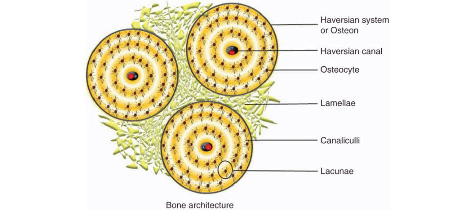
Figure 25.3a Drawing of cortical bone.
Which cell forms the majority of the bone cells?
The osteocyte forms approximately 90% of bone cells, it forms from osteoblasts. It controls the calcium and phosphorus metabolism responding to chemical, mechanical and electrical stimuli.
Tell me about the osteoclasts.
Osteoclasts are large multinucleated giant cells; they differentiate from haematopoietic precursors. They resorb bone within pits or depressions known as Howship’s lacunae. When laying on bone they have a contact area called ruffled borders which increase their surface area.
Bisphosphonates work on the ruffled borders.
I was drawing something like this as I talked (Figure 25.3b).
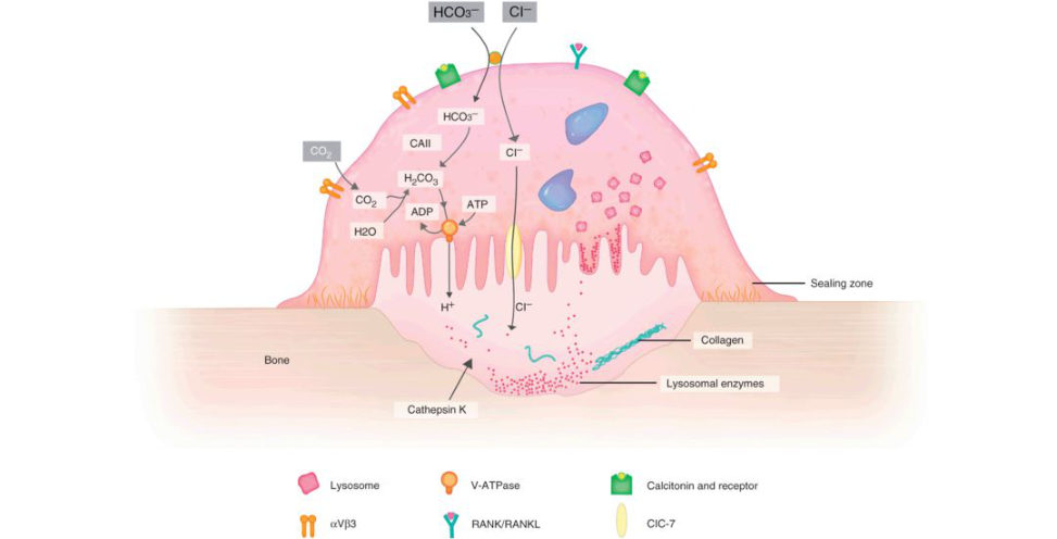
Figure 25.3b Osteoclast.
How does the blood reach it?
Bone receives 5–10% of cardiac output. There are various systems that supply it: nutrient artery system (high-pressure), metaphyseal–epiphyseal system and periosteal system (low-pressure). The nutrient artery enters mid diaphysis and divides into ascending and descending branches supplying the inner two-thirds of the cortex. All three systems are interconnected, and the direction of flow is centrifugal (inside to out).
How is bone metabolized?
A complex interplay and interaction of various hormones, growth factors and cytokines regulate plasma calcium and phosphate levels. They include vitamin D, PT Hand calcitonin.
How does vitamin D regulate calcium?
Vitamin D is either taken through diet or activate din the skin by ultraviolet light. Vitamin D enhances the absorption of calcium and phosphorus from the small intestine and enhances osteoclast resorption from bone. In the kidneys it causes increased calcium retention and phosphate excretion ( Figure 25.3c).
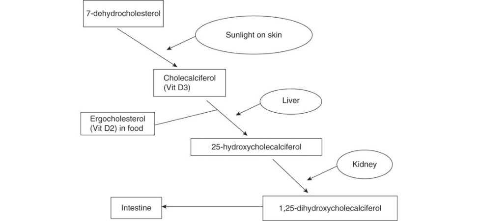
Figure 25.3c Vitamin D metabolism.
Structured oral examination question 4#
Bone gratis
So, you have apa tien t with a tibial plateau fracture on your operating t able and you find a big bony defect that needs filling. How do you approach that?
Bearing in mind this is a basic science station I went straight for the kill and mentioned bone graft.
What types of bone gratis do you know?
There are autogratis, allo gratis and z enogratis. Also , there is demineralized bone matrix (DBM), synthetic, bone morphogenetic protein (BM Pand stem cells. AUTOGRAFT utilizes bone obtained from the same patient receiving it. It can be harvested locally or from a distant site like the iliac crest. It has all the good properties of a gr aft, it isos teoconductiv e, osteoinductiv e and osteogenic. It can be cancellous, cortical or vascularized. Cortical provides structural support whereas cancellous is used for its osteogenic properties. ALLOGRAFTS are obtained from a different patient than the one receiving it. They are taken from cadavers or living donors such as femoral heads. They are available indifferent methods depending on their processing and preservation, deep fr ozen–70°C, freeze dried –169°C and fresh. They are not as good as autograft as the y have osteoconductiv e properties only and some os teoinductiv e properties.
What would you use for your case?
Well it depends on the individual case, but I would prefer to use autograft as it has no immunogenicity, no risks of disease transmission and is cheap, but it has donor site morbidity whereas allogratis have no donor site morbidity, but are slow to incorporate ...
Ahhh, what’s graft incorporation?
It is the process by which invasion of the graft by the host cells and the graft is then replaced either partially or completely by host bone or rejected. This happens in stages: 1. Inflammations timula ted by the necrotic debris. 2. Osteoblast differentiation from precursor cells. 3. Osteoinduction where osteoclasts and blasts are stimulated. 4. Osteoconduction: new bone starts to form. 5. Remodelling, which continues for years.
So, we know how an autograft is taken, do you know the process of allograft donation and bone banking?
Yes, living donors are consented and in case of cadavers as longas there is lack of objection from next of kin then it can be harvested.
The donors get screened for comorbidities and bloods are taken. They are screened for history of intravenous drug abuse, malignancy TB, steroids. Bloods for hepatis B & C , HIV, syphilis and Rhesus status.
The graft g oes through processing of cleaning then preservation. The gr aft gets debrided from unwanted tissue treated with ethanol/antibiotic soak s/irradiation and then preserved either fresh, fresh frozen or freeze-dried.
OK, quickly tell me about the synthetics and what you are going to use for this tibia?
(I realized I have told him all about bone graft but not t old him what I will use). Synthetics are commercially available products. The main constituents are either calcium sulphate, calcium carbonate, calcium triphosphate or hydroxyapatite. I will use calcium phosphate derivative because Russell et al. (JBJS Am 2008) performed an RCT of autograft vs. calcium phosphate cementin tibial plateau fractures and showed significantly reduced rates of subsidence, also Buckley published similar results in 2009 in calcaneal fractures.
References#
1. Russell TA, Leighton RK. Comparison of autogenous bone graft and endothermic calcium phosphate cement for defect augmentation in tibial plateau fractures. A multic entre, prospective randomized study.
J Bone Joint Surg Am. 2008;90(10):2057–2061.
2. Johal HS, Buckley RE, Le IL, Leighton RK. A prospective randomized controlled trial of a bioresorbable calcium phosphate paste (alpha-BSM) in treatment of displaced intra-articular calcaneal fractures. J
Trauma. 2009;67(4):875–882.
Structured oral examination question 5#
THA stem design
What are these (Figure 25.4a)?
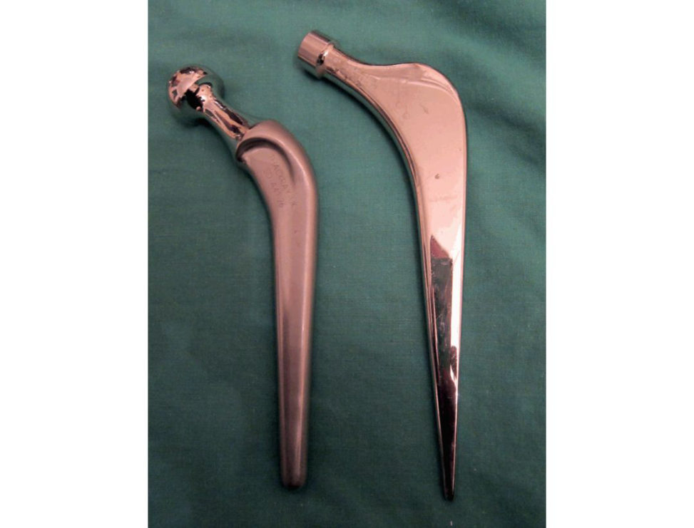
Figure 25.4a Exeter and Charnley femoral stem.
The hip replacement on the left is an Ex eter hip replacement and the one on the right looks like a Charnley hip replacement.
OK. What are the differences between the two?
The Exeter hip replacement is a collarless, polished double-taper cemented hip replacement. By contrast, the Charnley has a collar to prevent subsidence and is not as smooth.
By what biomechanical principles are they supposed to work?
The Exeter femoral stem works by utilizing a t aper slip design and controlled subsidence. It is implanted within a cement mantle and the highly polished nature and the taper allow controlled subsidence of the stem within the cement mantle. The stem will subside on average 1.3 mm in the first 2 years, followed by a period of stability. This subsidence seals off the cement stem interphase, preventing fluid flow leading to loosening. The viscoelastic properties of cement allow for creep to occur. As the stem is loaded the taper engages into the cement, producing radial hoops stress which is distributed to the bone interface minimizing stress shielding. The theory is that it loads all the bone.
By contrast, the Charnley stem works in a composite beam principle, relying on achieving mechanical interlock at the bone–cement interface and sufficient adhesion to achieve force transfer.
The load on the femoral head is transmift ed through the stem to its tip and then to the cement and bone below it [1].
What material are they made of?
The Exeter stem is made of stainless steel alloy (Orthinox), it has high strength with ductility plus it is resistant to corrosion. The Charnley stem is made of stainless steel alloy as well (Ortron).
Which one will you use in your practice and why?
I will use the Exeter stem based on the theories I mentioned before; also it has excellent survivorship in the NJ Rand a recent publication by the Exeter team showed the survivorship at 22 years was 99.0% and excellent preservation of bones tock at 20–25 years [2].
What is cement?
Cement is a synthetic polymer of methyl methacrylate (PMMA). PMMA is viscoelastic and the mechanical properties change as a product of time. The important factors that affect cement over time are creep, stress relaxation and fatigue. It is made from mixing a liquid and a powder. The liquid contains: the monomer, methylmethacrylate; binding agent, butylmethacrylate; activator, dimethyl-paratoluidine; and a stabilizer, hydroquinone. The powder contains: the polymer, polymethylmethacrylate; an initiator benzyl peroxide; contrast agent and antibiotics. Cementin a hip replacement acts as a grout. It functions to fill the defect in between the stem and the bone, act for load transfer, allows modulus matching between implant and bone, and in the case of the taper slip stems, allows controlled subsidence of the stem. It is strong in compression and weaker under tension and has a low elastic (Y oung’s) modulus compared to metal and bone.
Can you draw the relative moduli on a graph and compare it with other materials used in Orthopaedics (Figure 25.4b)? What is 3, what is 2, what is 11?
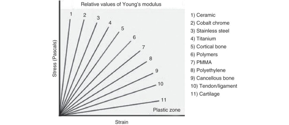
Figure 25.4b Diagram stress/strain curve and Young’s modulus.
Figure 25.4b is important as it tests a candidate’s ability to appreciate where Young’s modulus of bone exists when compared to other implanted materials or connective tissue.
What are the design principles behind the use of uncemented joint replacements?
The uncemented joint replacement can be put in in two ways. The first is a press-fit design, the secondis a line-to-line fixation. In the press fit, the femoral canal is prepared to match the orientation of the eventual stem, but the broach is slightly smaller than the stem. When the stem is inserted it is therefore a tight fit. As it is inserted, the viscoelasticity of the bone allows the slightly larger stem to be inserted and then the stem is gripped by the bone. Theoretically, no additional fixation is required. In line-to-line fixation, the bone is prepared to be the same size as the eventual implant, e.g. an uncemented cup. A cup the same size as the prepared acetabulum is inserted but, in this case, additional fixation, such as screws, is frequently required.
What can you tell me about the surface finish of the implant?
The surface finish allows either bony ingrowth or ongrowth. Ingrowth surfaces include sintered beads, fibre mesh and porous metals. Ongrowth surfaces are created by grit blasting or plasma spraying. Ingrowth requires a pore size between 50 and 400 μm, and the percentage of voids within the coating should be between 30% and 40% to maintain mechanical strength. Most modern cementless implants rely on bone ongrowth of a plasma-sprayed surface or are a hybrid of ingrowth and ongrowth.
Do you know anything about the design of the pores?
The porosity of the stem should not be greater than 50% otherwise there is the possibility of the pores shearing off.
How do stems fail?
The cemented stems fail in four modes: 1a. Pistoning: stem in cement. 1b. Pistoning: stem in bone. 2. Medial stem pivot. 3. Calcar pivot. 4. Cantilever bending.
Do you know any name associated with this classification?
Sorry. Gruen’s mode of cemented femoral stem failure.
Reference#
Gruen TA, McNeice GM, Amstutz HC. ‘Modes of failure’ of cemented stem-type femoral components: a radiographic analysis of loosening. Clin Orthop. 1979;141:17–27.
And the uncemented stems?
Delamination of the HA coating , there is also a mechanism known as flag-staffing. If a relatively small stem is inserted in apa tien t with strong cortical bone which is stiffer than the implant, then a microenvironment is created allowing for excess stress through the implant, resulting in loosening.
References#
1. ShenG. Femoral stem fixation. An engineering interpretation of the long-t erm outcome of Charnley and Exeter stems. J Bone Joint Surg Br. 1998;80(5):754–756.
2. Petheram TG, Whitehouse SL, Kazi HA, et al. The Exeter Universal cemented femoral stem at 20 to 25 years: a report of 382 hips. Bone Joint J. 2016;98B(11):1441–1449.
Structured oral examination question 6#
Articular cartilage
What do you see (Figure 25.5)?
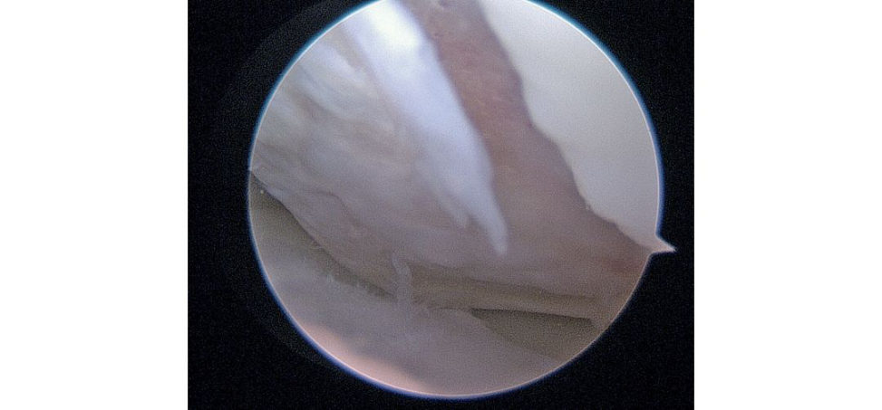
Figure 25.5 Knee arthroscopy picture focal area of cartilage loss.
This is a picture of a knee arthroscopy showing a grade IV cartilage defect.
What is cartilage?
Cartilage is a highly differentiated connective tissue made of cells extracellular matrix (EC Mand water. The cells are chondrocytes, and the ECM includes fibres, collagen (mainly Type II), elastin, proteoglycans, cartilage oligomeric proteins (COMP) and cartilage matrix protein (CMP). There are three types of cartilage hyaline cartilage, fibrocartilage and elastic cartilage.
Draw a cross-section of cartilage.
I drew all layers, superficial, middle and deep, arcades of Benninghoff, the tidemark and calcific zone. The examiner was waiting for me to draw the tidemark. This is the standard cartilage picture present in the usual textbooks.
What is the importance of the tidemark?
The tidemark is the boundary between the calcified and uncalcified cartilage made visible by histological staining. Clinical importance is that above the tidemark its avascular and nutrition depends on diffusion, and below it it’s vascular.
What is the importance of water in cartilage?
70% of articular cartilage is water; 30% of it exists between the collagen fibres and this is determined by the negative charge of the proteoglycans (PG) which lie within the collagen matrix. Because the PG are bound closely the closeness of the negative charges creates a repulsion force that must be neutralized by positive ions (hydrogen ions in water) in the surrounding fluid. The amount of water present depends on the amount of PG and the stiffness and strength of the collagen network. The PG aggregates are basically responsible for the turgid nature of the cartilage and in articular cartilage they provide osmotic properties needed to resist compressive loads.
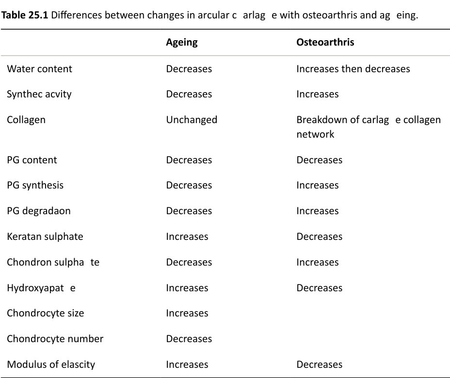
What happens in osteoarthritis?
The collagen network is disrupted either by trauma or an increase in degradation enzymes concentration – this allows the proteoglycans to atir act more water and softens the articular cartilage thus reducing its Young’s modulus of elasticity and its ability to bear load. At this stage you will have to draw the table showing the difference between ageing an dOA (Table 25.1).
OK, so how will you treat this defect? [Going back to the arthroscopy picture.]
This is grade IV on the Outerbridge Arthroscopic Grading System. Using the hookas a reference, it measures 1.5 cm × 1 cm; as it is below 4 cm2 then I will treat this with microfracture.
How would you do that?
The goal is to allow access of marrow elements into the defect to stimulate the formation of fibrocartilage.
I would prepare the defect until I achieved stable vertical walls and the calcified cartilage layer was removed. Then using a bone awl, I would make multiple perforations through the subchondral bone
3–4 mm apart, looking for the fat droplets to confirm that I have penetrated far enough.
Postoperatively patients are allowed protected weight-bearing with full range of movement of the knee.
If that defect was larger than 4 cm2 then I may consider osteochondral autograft or mosaicplas ty;
however, I will refer this patient to a knee surgeon expert in this field. I’m also aware of other methods such as autologous chondrocyte implantation (A CI) and matrix-associated autologous chondrocyte implantation (MA CI).
Would you do an ACI?
Well, NICE does recommend it, but it has strict criteria and it has to be done at a tertiary centre expert in this field so no I wouldn’t, I will refer it on. www.nice.org.uk/guidance/ta477/chapter/3-Commift ee-discussion#conclusion
Structured oral examination question 7#
Fracture fixation
Tell me about your approach to the fixation of this fracture (Figure 25.6a).
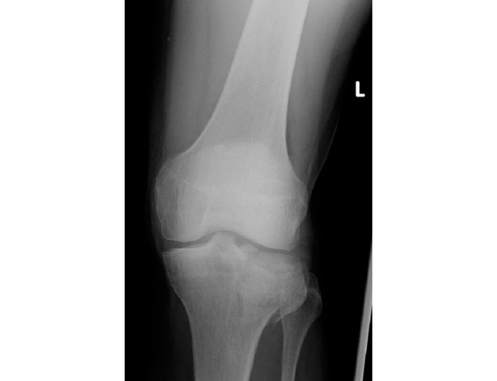
Figure 25.6a Anteroposterior (AP) radiograph of the left knee demonstrating tibial plateau fracture.
First, I would assess the patient the soft tissue and then consider the fracture. I would start with ATLS ...
Yes, yes, there are no tricks in this question everything is fine with the patient this is a question about the biomechanics of fracture fixation.
Right, OK, this is a plain radiograph of a comminuted displaced, intra-articular fracture of the tibial plateau. The principles regarding the fixation of this fracture are anatomical reduction and absolute stability of the articular block and then the restoration of length, axis and rotation of the lower limb by attaching the articular block on to the shaft of the tibia by utilizing a bridging plate and relative stability.
Tell me about how you would achieve a stable fixation of the articular surface.
I would open the joint and reduce the articular fragments under direct vision. I would apply a periarticular clamp to hold the fragments reduced and to compress the fragments. If necessary, I would use K-wires as a temporary fixation device. I would then use 6.5-mm partially threaded cancellous screws to act as both a subchondral raft and also to compress the fracture fragments and achieve absolute stability and primary bone healing.
Why do you want to achieve absolute stability?
As it is an intra-articular fracture, the healing of the bone should be by primary bone healing without the formation of callus. In order for this to happen there has to be no movement at the fracture site. The lack of movement means that the osteoclasts involved in the remodelling of bone will essentially ignore the fracture site and proceed directly across the fracture site using cuting cones, therefore healing by primary bone healing without the formation of callus.
How does a screw work?
A screw converts rotational movement into longitudinal advancement. With a lag screw, the screw is partially threaded therefore as the distal threads bite in the fracture fragment, the proximal smooth barrel can slide over the proximal fragment and so as it is tightened the distal fragment is pulled towards the proximal and compression of the distal piece against the proximal cortex occurs.
What are the parts of the screw?
It is easy to draw and illustrate (Figure 25.6b).
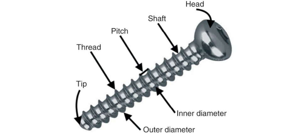
Figure 25.6b Design features of orthopaedic screw.
You said you will use a cancellous screw. What is the main difference between a cancellous and acor tical screw?
Pitch, core to outer diameter and tip. Pitch: closely spaced threads incor tical screws with smaller pitch compared with deeply cut and widely spaced threads in cancellous screws. Core to outer diameter: cancellous screws have deeper threads, making them better at pull-out strength.
Tip: cortical screws are blunt.
You mentioned you will use your plate in bridging mode, what other modes do you know?
But iress, bridging, compression, tension band, neutralization.
So why would you use this one in bridging mode?
There is a large amount of comminution in the metaphysis; therefore, it is impossible to compress the fracture fragments. If the other features of length, axis and rotation are correct then the fracture would be expected to heal by secondary bone healing with callus formation.
What do you understand by Perren’s strain theory?
Strain is defined as change in length over original length. The theory suggests that the mechanical environment (strain) governs the type of tissue that is laid down between the fracture fragments. Granulation tissue can tolerate 100% strain, fibrous tissue is formed when there is up to 17% strain, fibrocartilage 2–10% and lamellar bone at 2%. As the strain decreased at the fracture site a stiffer tissue is produced.
Describe the stages of fracture healing.
The stages are haematoma and inflammation, soft callus (1 week to months), hard callus (1– 4 months) and then remodelling (years). In the first stage, haematoma forms and provides key cells such as macrophages, key inflammatory cytokines (IL1, IL6, TGF-β) are secreted and eventually haematoma is replaced with granulation tissue. Next phase: soft callus starts to form, providing a bridging role and depending on the mechanical environment, as we mentioned before, osteoblastic differentiation or chondr oblastic differentiation happens. Soft callus is converted to woven bone (hard callus) via the process of endochondral ossification. With progression of this process collagen will change from II to I. Once the fracture has united the final phase of remodelling happens where hard callus is remodelled from woven bone to hard, dense, lamellar bone by the process of osteoclastic resorption followed by osteoblastic bone formation.
What factors influence fracture healing?
Two key factors are involved, mechanical environment and biology. I explained the mechanical environment earlier. The biology is affected by systemic and local factors. Systemic such as smoking, diabetes, age and nutrition. Local factors such as open or closed injury, high mechanism, bone loss, site and type of bone.
Structured oral examination question 8#
Osteoporosis
Could you define osteoporosis for me please?
In 1993 WHO defined osteoporosis as a systemic skeletal disease characterized by low bone mass and deterioration in the microarchitecture, leading to enhanced bone fragility and a consequent increase in fracture risk. In practice, it is defined on the bases of bone density assessed by a DEX Ascan T score. It is considered normal when the bone mineral density is within 1 standard deviation of the mean peak bone mass of a healthy 25-year-old. Osteopenia when it is 1–2.5 below the mean and osteoporosis when it is> 2.5 below the mean. Osteoporosis can be classified into primary type 1, primary type 2 or secondary. Type 1 is most common in women after menopause. Type 2 is senile osteoporosis and occurs after the age of 75 and is seen in both sexes. Secondary may arise at any age; it’s equal in women and men and is a result of a chronic predisposing medical problem or disease, like prolonged steroid use.
How does a DEX Ascan work?
DEX Ascan is dual-energy X-ray absorptiome try used to assess bone mineral density (BM DIt involves the use of two X-ray beams of different energies passed through the neck of femur or lumbar spine (L2–L4). It provides a value in g/cm2. The femoral neck is the preferred site because of its higher predictive value for fracture risk: Kanis and Gluer 2000 [1], Kanis et al. 2008, level 1a Evidence [2]. The result is then compared to a sex- and race-matched BMD of a healthy young adult population (25–30) producing a T score. The Z score refers to the BM Din relation to an age-matched population.
This is a DEX Ascan report (Figure 25.7), how would you interpret it?
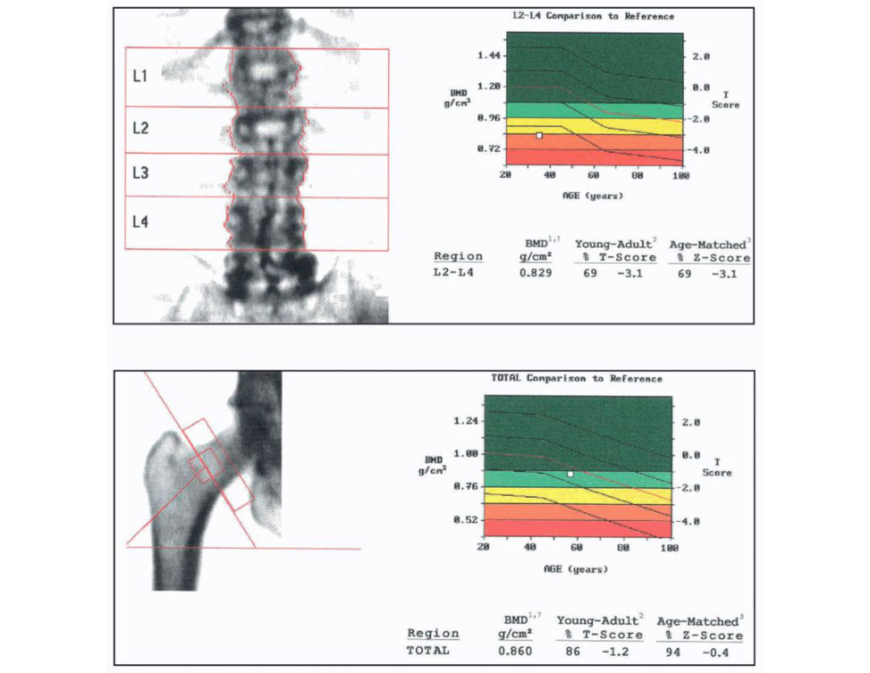
Figure 25.7 DEX Ascan report for proximal femur and lumbar spine.
You are expected to know how to interpret a DEX Ascan report. So, you need to familiarize yourself with one. It will be either of a hip or lumber spine or both. Simplifying it, I just concentrated on the T score and explained that the risk of fracture approximately doubles for each standard deviation decrease inT score. It depends on what the score is, you then can define it as normal/osteopenia or osteoporosis. In my station it was apa tien t with osteoporosis. Four important numbers to look at in the report: 1. The percentage of normal bone density for the patient age. 2. The percentage of bone density compared with normal young adults. 3. Z score. 4. T score.
How would you manage this patient?
Prior to obtaining the DEX Ascan I would have performed an initial work up which includes detailed history, examination and bloods. In the history I will be looking for risk factors including age, sex, hormonal problems, chronic illness, family history of hip fractures, medication lifestyle risk such as smoking, alcohol and diet.
I would also use the FRAX tool which computes the 10-year probability of hip fracture or a major osteoporotic fracture.
Treatment will be according to the NOGG [3] and NICE guidelines [4], dividing it into lifestyle changes and pharmacological treatment.
If the patient is at risk or has confirmed OP, then I would provide them with information regarding lifestyle changes. Lifestyle measures to improve bone health include increasing the level of physical activity , stopping smoking, reducing alcohol intake to ≤ 2 units/day, reducing the risk of falls and ensuring adequate dietary calcium intake and vitamin D status.
(www.shef.ac.uk/FRAX)
How much calcium and Vit D do you give them?
In postmenopausal women and older men (> 50 years) at increased risk of fracture a daily dose of 800 IU cholecalciferol should be advised. In postmenopausal women and older men receiving bone-protectiv e therapy for osteoporosis, calcium supplementation should be given if the dietary intake is below 700 mg/day, and vitamin D supplementation considered in those at risk of or with evidence of vitamin D insufficiency.
Which pharmacological treatment would you choose?
Alendronate 70 mg once-weekly or Risedronate are first-line treatments in the majority of cases. Treatment review should be performed after 3 years of zoledronic acid therapy and 5 years of oral bisphosphonate treatment.
How do bisphosphonates work?
They can be divided into nitrogen-containing (Alendronate) and nitrogen-lacking (Etidr onate). They all share the common P-C-P backbone which makes them resistant to metabolism. The nitrogen group inhibits protein prenylation with an end result of loss of guanosine triphosphatase (GTPase) formation, this is needed for ruffled border formation and cell survival. The nitrogen-lacking bisphosphonates are metabolized into a non-functional adenosine triphosphate (ATP) analogue, which induces cell apoptosis.
References#
1. Kanis JA, Gluer CC. An update on the diagnosis and assessment of osteoporosis with densitometry.
Commift ee of Scientific Advisors, International Osteoporosis Foundation. Osteoporos Int. 2000;11:192–202.
2. Kanis JA, McCloskey EV, Johansson HO den A, Melton LJ, 3rd, Khaltaev N. A reference standard for the description of osteoporosis. Bone. 2008;42:467–447.
3. National Osteoporosis Guideline Group (NOGG). 2017.
4. NICE guidelines:
Compston J, Cooper A, Cooper C, et al. Guidelines for the diagnosis and management of osteoporosis in postmenopausal women and men from the age of 50 years int heUK. Maturitas. 2009;62:105–108.
Compston J, Bowring C, Cooper A, et al. Diagnosis and management of osteoporosis in postmenopausal women and older men int heUK: National Osteoporosis Guideline Group (NOGG) update 2013. Maturitas.
2013;75:392–396.
Structured oral examination question 9#
Antibiotic prophylaxis
Which antibiotic do you use for infection prophylaxis in arthroplasty?
Our hospital policy recommends cefuroxime 1.5 g at induction provided that the patient is not allergic to penicillin Alternatively, teicoplanin and gentamycin.
How does cefuroxime work?
Cefuroxime is a beta lactam antibiotic; it inhibits the cr oss-linking of polysaccharides in the cell wall by blocking the transpeptidase enzyme Penicillins also work in a similar action.
Could you tell me about other antibiotics you know and their mechanism of action?
In general, I divide the antibiotics by their mechanism of action to antibiotics working on cell wall synthesis, protein synthesis (50S subunit and 30S subunit), nucleic acid synthesis (Figure 25.8).
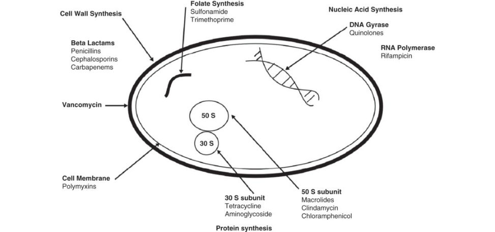
Figure 25.8 Simple bacterium (I drew a simple bacterium which is available in most books).
How do the bacteria develop resistance?
Bacteria can develop resistance either by intrinsic resistance or by acquired resistance to antibiotics. Bacteria can develop the ability to hydrolyze the antibiotic using beta lactamase, or through genetic mutations, like MRSA. Other bacteria can alter the cell wall permeability or create a biofilm barrier.
How does MRSA develop resistance?
MRSA develops resistance through a gene called mecA, it produces the enzyme penicillin- binding protein 2a (PBP2a). This enzyme prevents the normal enzymatic acylation of antibiotics. EAMINER : What is your local trust policy regarding screening and eradication of MR SA? We have a routine preoperative screening involving nasal, groin and axilla swabs for Staphylococcus aureus; 20% of patients are staph carriers and 5% are MRSA carriers. Patients must have three negative samples before considering joint arthroplasty.
Carriers are treated with nasal mucopirocin and 4% chlorhexedine bath for 5 days, re-swabbed and repeat if still positive.
How do you prevent infections in your department?
There are various steps performed to prevent infection in our depart mentI divide them into preoperative, peri operative and postoperative measures. PREOPERATIVE : screening for MRSA, ulcers and UTI. Admission on day of surgery to ring-fenced wards. Optimization of co morbidities, especially diabetes. PERIOPERATIVE : laminar flow theatres, minimize theatre traffic, prophylactic IV antibiotics within 1 hour of incision, antibiotic-loaded cement shaving at time of surgery, good hand-washing technique, draping with disposable drapes and ioband, opening of sets within the laminar flow, efficient surgery, good haemostasis and sound wound closure and avoid hypothermia throughout the procedure. POSTOPERATIVE : minimize dressing changes, encourage a culture of infection control, for example using alcohol gel and bare below elbows. Minimize unnecessary transfusions, postoperative antibiotics; however, this is debatable depending on departmental policies, early mobilization and physiotherapy, optimal medical management, timely but safe discharge.
Structured oral examination question 10#
Theatre suite
How would you design a theatre suite?
When designing a theatre suite, the first important aspect is theatre location, it needs to beclose to related facilities such as ITU , A&E, Surgical wards, and Radiology. The internal structure is usually thought of in terms of four zones that are intimately linked: Outer zone: reception (and r est of hospital). Clean zone: reception to theatre doors. Aseptic z one: theatre itself. Disposal zone: sluices (Figure 25.9). These zones will be connected with corridors that should not cross-contaminate each other. The design of the theatre itself should be based around the table, with careful attention to lighting , temperature, humidity and ventilation. Lights should be satellite with 40,000 lux at the wound site. Temperature and humidity is controlled via thermostats and the ventilation system. Optimum temperature for the patient is 25°C and for the staff is 19°C. We aim to create a microclimate for the patient to prevent hypothermia by using Bair huggers, warmed mat iress and fluids ...
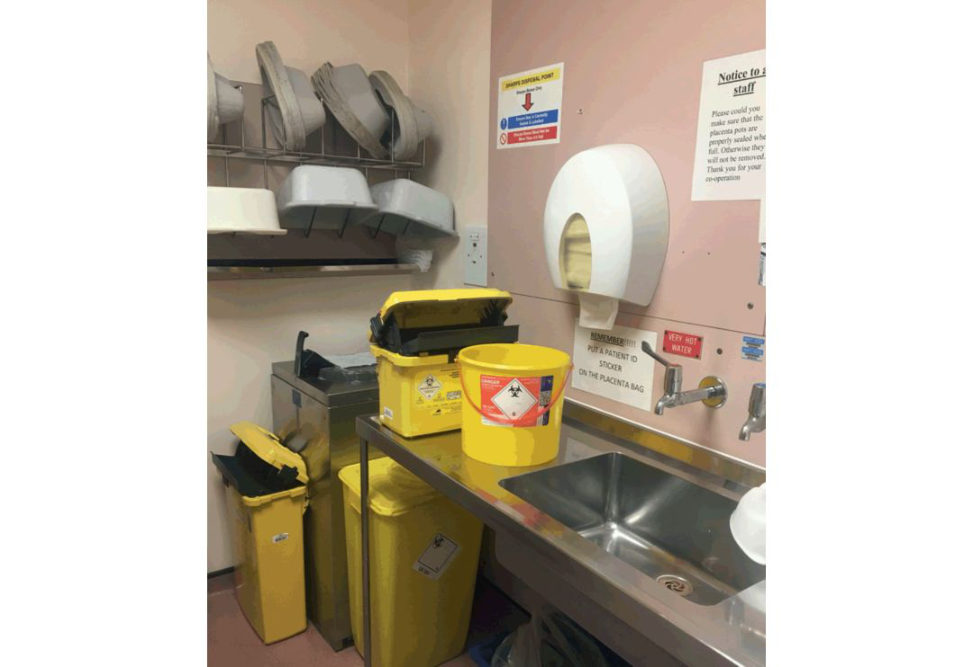
Figure 25.9 Disposal zone. Four zones: outer zone, clean zone, aseptic z one and disposal zone. The purpose of having zones is to have defined boundaries.
Which ventilation system is used in your department?
We have theatres with a laminar flow ventilation system for elective arthroplasty cases and a plenum type for all other cases.
What is laminar flow?
It is a system that produces an entire body of air flowing in one direction with uniform velocity and along parallel flow lines. Flow rate is 0.3 m/s. There are three types of airflow: horizontal, vertical and ex-flow.
Tell me about the ex-flow Howarth system
[I had no idea, the only thing I could remember was a trumpet type of airflow. I rescued myself by saying I’m more familiar with the vertical laminar flow]
OK, tell me about the vertical laminar flow.
Vertical laminar flow was introduced by Charnley and is the most common type used. It creates a room within a room, with panels from the ceiling to 2 m from the floor. Airflows through aH EPA filter in the ceiling and directed towards the operative field. HEPA stands for high-efficiency particulate air filters. It filters particles of 0.5 μm with 99.9% efficiency.
Does it filter viruses?
Umm, no.
What are the advantages and disadvantages of horizontal vs. vertical laminar flow?
Horizontal is easier to install, but positioning of staff and equipment is restricted because of the sideways flow of air. Vertical laminar flow does not restrict staff and equipment as longas they are within the marked areas; however, if they stand within the periphery of the laminar flow area they may deflect contaminated air towards the wound.
What are the sources of infection theatres?
Patient surgeon, instruments and airborne. We shed microorganisms, also when we talk ...
How do you measure bacterial contamination in the air?
By slit samplers called Casel lait draws in air over 1 minute which passes overculture plates, which are then incubated over 48 hours and colonies formed are counted. It is measured in laminar flow theatre by colony forming units per cubic metre (CFU m3). Acceptable CFU values for a laminar flow theatre are < 20 CFU in periphery and < 10 in the centre.
Do you think laminar flow prevents joint infection?
I believe it is a combination of multiple factors. Results of the MRC trial in 1982 (Lidwell et al.) published in BMJ showed that deep sepsis in arthroplasty was reduced significantly by the use of antibiotic-loaded cement prophylactic IV antibiotics peri operatively, laminar flow and body exhaust suits [1]. Charnley also showed a reduction in deep infection with the use of vertical laminar flow in combination with other techniques [2]. On the other hand, a review of the New Zealand joint registry questioned the use of laminar flow and space suites as it showed that the rate of revision for early deep infection was not reduced by using laminar flow and spacesuits [3].
References#
1. Lidwell OM, Lowbury EJ, Why teW, Blowers R, Stanley SJ, LoweD. Effect of ultra clean air in operating rooms on deep sepsis in the joint after total hip or knee replacement: a randomised study. Br Med J.
1982;285:10–14.
2. Charnley J. Postoperative infection after total hip replacement with special reference to air contamination in theo per ating room. Clin Orthop Rel Res. 1972;87:167–187.
3. Hooper GJ, Rothwell AG, Frampton C, Wyatt MC. Does the use of laminar flow and spacesuits reduce early deep infection after total hip and knee replacement? The ten-year results of the New Zealand Joint
Registry. J Bone Joint Surg Br. 2011;93(1):85–90.
Structured oral examination question 11#
VTE
How do you set up a VTE protocol in your department?
I would set up a protocol to assess all elective and trauma patients. I would have two pathways, one for elective patients and one for trauma. The elective patient pathway starts when listed in clinic identifying correctable risks that can be addressed preoperatively. Risk assessment will be performed using a published tool or guidelines such as the NICE guidelines to balance their risks of VTE against bleeding. They will be stratified in to a low-risk group or a high-risk group. The low-risk group will be offered mechanical prophylaxis only and the high-risk group will be offered both mechanical and pharmacological prophylaxis suitable for them. During their stay they will have a continuous reassessment protocol at senior review or if their clinical condition changes.
What are the current guidelines for elective hip replacement?
The guidelines will be changing in 2018 and aspirin will be considered; however, the current guidelines suggest starting mechanical prophylaxis at admission, either anft-embolism stocking or intermift ent pneumatic compression devices. This should continue un til the patient no longer has significant reduced mobility or for 4 weeks. Provided there are no contraindications for pharmacological prophylaxis, prophylaxis should start after surgery with either LMWH or Rivaroxiban. Continue prophylaxis for 28–35 days.
What are the guidelines for total knee replacement and how do they differ from total hip replacements?
It is the same protocol, but the duration is shorter 10–14 days.
OK, what is the new guideline that is coming up?
For elective hip replacement. In the new guidelines it is recommended to offer VTE prophylaxis to people undergoing elective hip replacement surgery. Choose any one of: LMWH (for 10 days) followed by aspirin (for 28 days). LMWH (for 28 days) combined with anft-embolism stockings (until discharge). Rivaroxaban. Consider anft-embolism stockings until discharge from hospital if pharmacological interventions are contraindicated in people undergoing elective hip replacement surgery.
And for knees?
Offer VTE prophylaxis to people undergoing elective knee replacement surgery. Choose any one of: Aspirin (for 14 days).
LMWH (for 14 days) combined with anft-embolism stockings (until discharge).
Rivaroxaban.
Consider intermift ent pneumatic compression if pharmacological prophylaxis is contraindicated in people undergoing elective knee replacement surgery. Continue un til the person is mobile.
Is there a role for chemical prophylaxis inpatient’s lower limb immobilization?
The new guidelines suggest considering pharmacological VTE prophylaxis for people undergoing foot or ankle surgery, in particular: Those requiring immobilization (for example, arthrodesis or arthroplasty). When total anaesthesia time is greater than 1 hour. When the patient risk of VTE outweighs their risk of bleeding.
What is the mechanism of action of LMWH, aspirin and Riv aroxaban?
Aspirin is a cyclo-oxygenase (Cox) inhibitor. It inhibits prostaglandin production preventing platelet aggregation. It also prevents formation oft her omboxane A2, which is a prothrombotic agent secreted by platelets. LMWH : forms a complex with antithrombin 3, but this complex selectively inhibits factor 10a only. RIVAROXIBAN : direct action on factor 10a. If you have time you can draw the intrinsic and extrinsic pathways and explain where each drug works (Figure 25.10).
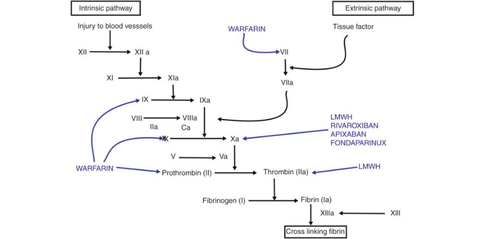
Figure 25.10 Intrinsic and extrinsic cloting pathway .