Chapter 11 Lower limb trauma II
Will Eardley, Mohammed Al-Maiyah and Patrick Williams
Introduction#
Alexander Suvorov would have done well in the trauma viva section of the FRCS Tr & Orth. Two citations atiribut ed to him underpin the approach to the exam: Train hard, fight easy and He who is afraid is half beaten. Approach and strategy is everything and this comes from a combination of practice and knowledge acquisition. It is a time-depende nt chess match where every move will be undertaken in a specified time, but in a sequence that is out of your control. Keep this analogy as you attempt different clinical scenarios. It is not only knowing the subject that is important, but also imparting it in an appropriate fashion, flexibly so that you can tell the examiners what they want to hear.
Remember, the examiner does not know you and is basing the standard of your knowledge and patient care on the words that leave your mouth. What they don’t hear, they can’t score you on. The examiners want a safe and sensible approach of the generalist, not eminence-based pracft ce of someone you may work for.
Treat each question as a chess g ame that is going to last five minutes.
Structured oral examination question 1#
A 35-year-old male lost control on a bend and came off his motorcycle yesterday; he has been fully resuscitated and has an isolated closed injury of the knee (Figure 11.1).
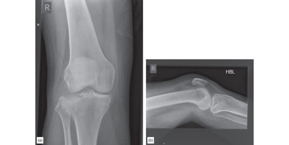
Figure 11.1a and 11.1b Anteroposterior (AP) and lateral radiographs of right knee demonstrating tibial plateau fracture.
Minute 1
What do you see? Here the next minute belongs to the candidate and you can take it whichever way you want to. However, there are essentials to be covered. In the first 30 seconds you are expected to comment on the following: Site of radiograph and its suitability – also always ask for two views if only one is given. Adult or paediatric skeleton. General features: fracture of the proximal tibia with depression of the lateral tibial plateau. In the next 30 seconds the candidate is expected to comment (without any prompt from the examiner) on the exact nature of the injury, such as Schatzker III fracture with more than 10 mm depression in the articular surfaces, comminuted, concern about the fracture going through the tibial spines and whether the medial side is intact. The candidate can end these 30 seconds by saying they will assess the soft -tissue envelope, the distal lower limb (palpating the distal pulses and providing a documented assessment of the named nerve function), ensure that a full tertiary survey has been performed and then plan further management of the fracture.
Minute 2
How would you investigate further?
A computed tomography (CT) scan to evaluate the fracture pattern as this helps to plan surgery, particularly with regard to approaches to the fracture and the philosophy of implant choice. (The examiner is then likely to produce slices of the CT.) Don’t get carried away at this point. Check the scan is of the same patient and make a basic description of what you see (coronal/sagift al/axial slices, demonstrating ...). It’s vital, in order to score points, that you comment on this constructively, i.e. how what you see may influence your approach/fixation. This is what we actually do – the CT slices are not presented as an abstract diagram, they should be used in your answer to demonstrate that you are used to interpreting them and how they influence your management. End by stating that you would of course discuss the findings of the scan with the patient and use it in the informed consent process. You should be at this stage by 90 seconds. Punctuation of the viva is important and helps you stay calm. Having done all this, take a breath and pause. Then, offer to discuss treatment options. Do NOT plough straight in with your plate of choice.
What are the treatment options?
[Take the next 30 seconds to describe operative and non-operativ e options in the generality. This must be based on the particular patient (recent alignment of consenting processes) and it is critical here that the information will be given to the patient clearly regarding the impact of differing treatment strategies on that particular patient with that particular injury.] Non-operativ e management would not normally be suitable in this patient This is due to the articular segment depression, which will impact on overall stability of the joint in addition to the articular congruity and impact on long-term function, aswell as the wish to restore joint congruity and stability and avoidance of the generic negative aspects of non-operativ e management (such as restricted mobility, prolonged periods of non-weight-bearing, blood clots, etc.). Any operative treatment discussion must be put in the context of the soft tissue envelope and it is important to state that this will influence your decision-making, particularly with regard timing of surgery.
Minute 3
At the two minutes mark you should have commift ed yourself to offering the patient operative intervention. Before the examiner asks, offer your treatment because it bugs them to keep asking again and again what you will do. Stick with the principles.
The principle of treatment is to restore the articular surface, stabilize and hold the fracture in such a fashion to allow early mobilization. The aim of the treatment is to have a mobile, pain-free and functional joint.
The options of surgical treatment include director indirect reduction, percutaneous or open fixation augmented with plate osteosynthesis or external fixation. Before being prompted, suggest your preferred option, which in the authors’ opinion is indirect reduction using acor tical window in the proximal tibia, restoration of articular surface with a raft of screws, augmented with abut iress plate. Suggest at that stage you will do assessment under X-ray control for a ligamentous stability and if needed an arthroscopic assessment.
Minute 4 (yes, you are still going ...)
The examiner can then take the viva along two routes.
What is abut iress plate?
A plate applied perpendicular to the force that is trying to resist. It is one of the modes of plate uses, along with compression, bridging and tension band, for example.
What is the role of knee arthroscopy?
It is potentially of use in three areas. One, to assess the reduction of the articular surface. Second, to ensure soft tissues (lateral meniscus) are not trapped in the fracture. Third, to assess intra-articular ligament damage. (Be clear to state that pressure pumps are not to be used in order to avoid iatrogenic compartment syndrome due to extravasation of fluid, as well as the f act that you will use a bladder syringe through the arthroscopy cannula to washout the haemarthrosis before viewing the joint – this gives the examiner the impression that you have done the procedure before.)
What surgical approach will you use?
Anterolateral approach with the skin incision being longitudinal and if needed, a reverse L- shaped incision inside. The incision is curved anteriorly over Gerdy’s tubercle and is extended distally, 1 cm lateral to the anterior border of the tibia Proximally the iliotibial bandis incised inline with its fibres and the fascia over tibialis anterior divided and elevated bluntly from the tibia distally.
What about bone graft?
I would prefer to use an impacted cancellous femoral head allograft. I realize that cancellous autograft harvested from the iliac crest is probably the gold standard. However, this procedure involves making a separate incision over the iliac crest to obtain the graft, which may result insignificant postoperative pain, neuro/vascular injury, haematoma, infection fractures and cosmetic concerns.
Anything else you can use?
Injectable calcium phosphate bone cement can be used as abut iress in articular cartilage depression. It is thought to reduce the risk of subsidence of the fracture fragments occurring by maintaining articular congruency until the fracture heals.
What does the literature say?
There is some evidence to support the use of bone graft substitutes to fill fracture voids, but a lack of level I evidence.
Minute 5
With one minute left and if the examiner is talking about rehabilitation and weight-bearing status, you know that you are probably winning. Talk about graduated range of motion, protected weight-bearing and the concept that true non-weight-bearing is very difficult for patients and protected weight-bearing ‘as able’ depending on patient compliance is what is actually going to happen.
Warning: be prepared for an X-ray of metalwork failure with the screws cutout into the articular surface. Stay calm. Assess the patient clinically, radiologically (including CT), rule out infection, soft -tissue problems, patient compliance and then proceed from the start, take out metalwork, align the articular surface, stabilize the fracture and mobilize again, often as a staged process. Key to this is proper work-up and identification of what went wrong and why. It is important not to repeat the same mistakes twice.
What will you tell the patient about long-term outcome?
The reported incidence of post-traumatic radiographic osteoarthritis of the knee following tibial plateau fractures varies from 25% to 45%. Not all patients however, are symptomatic. The outcome in tibial plateau fractures is more about restoring the mechanical axis rather than accurate reduction of the joint surface. Wasserstein et al. reported that regardless of operative fixation, sustaining a tibial plateau fracture requiring surgery increases the likelihood of TK Aby 5.3 times. 1 Older patients and those with a more significant fracture were more likely to need TKA.
Evidence base
A 2015 Cochrane review commented that there was insufficient evidence to recommend a specific method of fixation or bone defect replacement technique. They did comment that the evidence does not contradict the idea of minimizing soft -tissue dissection and avoiding donor site morbidity. A review by the
EFORT group in 2016 agreed with the above, but also commented on the use of TK Ain older patients.
McNamara IR, Smith TO, Shepherd KL, et al. Surgical fixation methods for tibial plateau fractures.
Cochrane Database Syst Rev. 2015;9:CD009679.
Prat-Fabregat S, Camacho-Carrasco P. Treatment strategy for tibial plateau fractures: an update. EFORT
Open Rev. 2016;1(5):225–232.
Wasserstein D, Henry P, Paterson JM, Kreder HJ, Jenkinson R. Risk of total knee arthroplasty after operatively treated tibial plateau fracture: a matched-population-based cohort study. J Bone Joint Surg.
2014;96(2):144–150.
Scoft CE Davids onE, MacDonald DJ, White TOKe ating JF. Total knee arthroplasty following tibial plateau fracture: a matched cohort study. Bone Joint J. 2015;97(4):532–538.
Structured oral examination question 2#
A 79-year-old woman fell in her garden. She is generally quite independent, has a history of angina which is well controlled and likes meeting her friends a t the local social club every Wednesday.
Minute 1
Please comment on the radiograph (Figure 11.2).
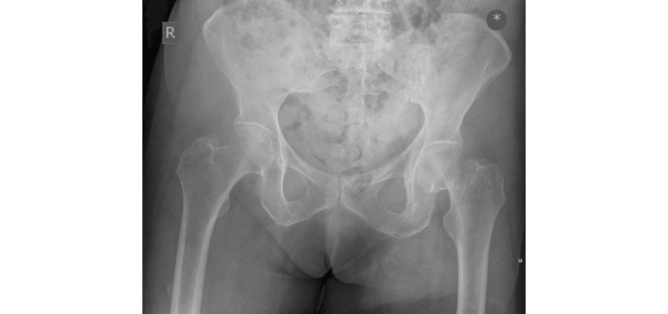
Figure 11.2 Anteroposterior (AP) pelvis radiograph demonstrating intracapsular fractured left neck of femur.
In the first 30 seconds you are expected to comment on the site of radiograph, its acceptability and the general findings it demonstrates.
The pelvic radiograph shows a displaced left-sided intracapsular neck of femur fracture in the presence of early degenerative changes of the hip joint. (Always ask for the lateral radiograph.)
How will you manage this patient?
I would like to assess the whole patient The degree of mobility prior to injury, comorbidities, ‘red flag’ features for any pathological lesions, drug history, cause of fall and appropriate investigations. This will include clinical examination of the patient including the left lo wer limb.
Minutes 2 and 3
She has well-controlled angina and is otherwise independent.
Operative treatment is preferred in this patient group to avoid complications of non- operative management. This will involve a discussion around arthroplasty, either hemiarthroplasty (HA) or total hip arthroplasty (THA). My choice is THA using a well-proven cemented prosthesis provided the patient meets the NICE guidelines of being fit for anaesthesia, not cognitively impaired and able to mobilize independently pre-injury.1
Why do you prefer THA rather than hemiarthroplasty? It is more expensive!
A THA has a better functional out come than HA and has better survivorship results. My choice will be a cemented tapered polished stem of long-term proven results with a cemented, highly cross-linked polyethylene cup using a relatively large head. There are data from a BMJ systematic review which suggest better functional out come and lower re-operation rates in those patients treated with THA. Recent NICE guidelines endorse such practice in a selected population, which includes mentally alert patients with good pre-injury mobility levels and who are relatively healthy. This patient ticks all the criteria and will benefit from THA. My practice ist o use a relatively larger head, such as 32 mm or 36 mm, to counter the increased risk of hip dislocation. 2,3 Surgical technique should focus on the correct orientation of components, good soft -tissue balancing , restoration of hip offset and equalization of leg lengths. Postoperative management continues with aggressive rehabilitation including early mobilization with full weight-bearing and repatriation to place of usual abode. It also includes addressing any underlying bone abnormalities such as osteoporosis, risk assessment for falls and nutritional deficiency. Ideally, the management should be carried out by a multidisciplinary team. With regards to price and impact on quality of life, THA is considered more cost-effectiv e.3
You keep mentioning NICE guidelines. What is a NICE guideline?
NICE clinical guidelines are recommendations for the care of individuals in specific clinical conditions or circumstances within the NHS.
So why do we use them?
NICE guidelines can be used to develop standards to assess the clinical practice of health professionals and can also be used in the education and training of health professionals. They are based on the best available research evidence.
What’s the problem with using NICE guidelines?
NICE guidelines do not replace a surgeon’s knowledge and skills, they are only guidelines to help a surgeon make an informed decision.
Minute 4
She arrives at 1800 to your ward. When will you undertake the surgery?
The surgery should be undertaken as soon as safely possible and ideally within 36 hours.4 It should not be rushed in the middle of the night; however, if the patient is fit for anaesthesia then the aim is for surgery on the next morning list with all the theatre staff, kit and consultant cover available. It is important to optimize any correctable medical causes prior to surgery. This should be undertaken in an objective and efficient manner to avoid ‘unnecessary’ delay.
Minute 5
The examiner can talk about the higher risks of complications of THA in this patient group compared to matched elective controls (9% vs. 4%). This includes a higher risk of dislocation (7% vs. 1%), leg length discrepancy, cement pressurization side effects such as cement reaction, higher medical complication rate
(32% vs. 6%) and higher mortality rate. Length of hospital stay is also increased.
Evidence base
This topic is so common that, yes, you do need to know some numbers:
1. CG 124 – Hip fracture: management (1.6.3).
2. Injury volume 47, issue 10, October 2016, pp. 2144–2148: 7% dislocation rate compared to 1%.
3. NIHR HTA volume 15, issue 36: significant increased risk of early dislocation a t 1 year (RR 3.98) for
THA compared to HA and statistically significant increased risk (RR 2.4) for all follow-up periods up to
13 years.
4. NIHR HTA volume 15, issue 35: cost per QALY $1960 for THA.
5. Geriatr Orthop Surg Rehabil. 2014;5(3):138–140.
Structured oral examination question 3#
Minutes 1 and 2
This 49-year-old lady fellon some steps. Her left foot is very painful, bruised, swollen and she can’t weight-bear. The junior doctor went to see her in the Emergency Department, but he is not sure what the problem is, what do you think? (Figure 11.3.)
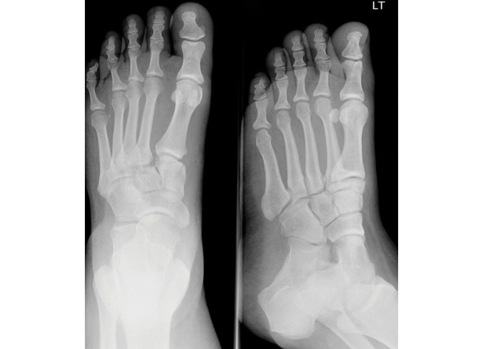
These are anteroposterior (AP) and oblique radiographs of the left foot. There is a diastasis between the base of the first and second metatarsals; features suggestive of ‘Lisfranc’ tarsometatarsal fracture dislocation. The reis a small avulsed fragment of bone in that interval. This is an avulsion fracture and could be from the insertion of the Lis franc ligament (medial cuneiform– second metatarsal) into the base of the second metatarsal (‘fleck sign’). Normal alignment on the AP view is demonstrated by examining the lateral borders of the first and second metatarsals, which should lineup with the lateral borders of the medial and lateral cuniforms respectively. The oblique internal rotated view also demonstrates that the medial border of the fourth metatarsal lines up with the medial border of the cuboid.
OK, how will you manage this patient?
I would start with the patient assessment as a whole, following the ATLS (Airway and protect cervical spine, Breathing, Circulation, Disability, Exposure and environment control) protocol with a focused history including mechanism of injury, patient general condition, co morbidities, allergies, smoking status as well as occupation and le vel of function. I will carryout an examination of the footnoting: Soft tissues welling, pain and ecchymosis. Pain on passive abduction/pr onation. Dorsalis pedis pulse if palpable. Compartment syndrome can be a feature of these injuries and I will include this in my differential. Following assessment, my initial management includes analgesia, elevation and splinting using a below-knee backslab. On admission to hospital I’ll arrange for regular, serial examination to detect compartment syndrome.
What would you do if the radiographs were inconclusive in diagnosing this condition?
I would arrange further imaging including oblique and lateral view weight-bearing radiographs if this can be tolerated by the patient. I would arrange a computed tomography scan that should pickup any subtle or occult fractures. MRI scan is useful in allowing direct visualization of the Lisfranc ligament itself, but I would discuss the MRI request with an experience musculoskeletal radiologist beforehand as images can sometimes be difficult to interpret.
Figure 11.3 Anteroposterior (AP) and oblique radiographs, left foot.
Minute 3
How do you treat Lisfranc tarsometatarsal fracture dislocation?
This depends on the severity of injury to both the bones and soft tissues and the degree of displacement of the fracture. There is a role for non-operativ e treatment for an undisplaced stable injury with a cast for 6 weeks with non-weight-bearing and regular clinical and radiological review. However, in the presence of subluxation or dislocation, then accurate reduction ands table fixation is essential. In this case, I would consider open reduction and internal fixation with screws and maybe plating , as required. In the case of a comminuted fracture, then primary arthrodesis of tarsometatarsal joints may be considered, although I would have a full discussion of the treatment options with the patient and record the outcome in the notes. I would use a dual dorsal incision approach. The first incision is performed between the first and second metatarsals to address the first and second TMT joints. The incision is centred over the TMT joint. The second incision is between the third and fourth metatarsals at the same level.
Minute 4
What prognosis will you give this patient?
This is a serious injury with potentially a poor out come. Post-traumatic osteoarthritis occurs in over 50% of cases, even if operatively treated with open reduction and internal fixation Residual pain and a stiff foot is a not uncommon complication of this injury Early identification of thein jury is key – up to 20% of tarsometatarsal joint complex injuries are missed on initial examination. The patient must be informed about the length of the recovery period and implications on lifestyle and work in the future.
Minute 5
If this patient develops compartment syndrome, then how would you manage it?
There is no clear evidence regarding the management of presumed compartment syndrome in the foot. In the BOAST guidelines regarding compartment syndrome, it is stated that there is no clear consensus on optimum management. I would discuss the case with the patient and if possible gain a second opinion from a consultant colleague. In general, for low-energy injuries, I adopt a low threshold for decompression in the foot. For high-energy injuries such as following motorcycle trauma or in the intubated patient, my threshold to intervene would be even lower.
How would you manage the injury if the soft tissues around the foot were very swollen with significant disruption of the bony anatomy but no compartment syndrome?
In this situation a prompt reduction of these injuries improves the alignment and relieves the pressure to the surrounding soft tissues, a voids the potential for skin necrosis, helps avert the development of a compartment syndrome, prevents compromise to the neurovascular structures and allows a safe waiting period to be undertaken until the swelling has decreased, re- epithelialization of blisters has occurred and ‘wrinkling of the skin’ has been noted. I would be concerned about just applying a back-slab and waiting for the swelling to improve as this may leave amal aligned midfoot that may be difficult to reduce once the swelling has subsided.
So, what are you going to do?
I would use an external fixator, applied to one or both sides of the foot to reduce the bony injury and allow the soft tissues to setile before definitive surgery.
Evidence base
The main area of controversy around Lisfranc injuries is whether to treat them with ORIF or ORIF with primary arthrodesis. A systematic review by Smith et al. demonstrated a higher rate of hardware removal in patients undergoing simple ORIF. There was no difference between the groups in terms of overall complication rates or PROMs data. ORIF with primary arthrodesis is particularly relevant for purely ligamentous injuries. This relates to the prolonged healing time of a ligamentous injury compared to a bony injury. ORIF has shifted more towards using bridging plates rather than cortical lag screw fixation and
K-wires. For further evidence base look through the review article by M. Clare.
Smith N, Stone C, Furey A. Does open reduction and internal fixation versus primary arthrodesis improve patient outcomes for Lisfranc trauma? A systematic review and meta-analysis. Clin Orthop Rel Res.
2016;474(6):1445–1452.
Clare MP. Lisfranc injuries. Curr Rev Musculoskel Med. 2017;10(1):81–85.
Structured oral examination question 4#
A 33-year-old roofer fell 20 feet when scaffolding collapsed under him, landing on his feet and sustaining an isolated injury to his heel.
Minute 1
This is a radiograph of his foot and ankle (Figure 11.4a). What are your thoughts?
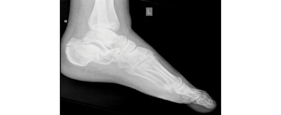
This is a lateral radiograph of the left foot. It shows a displaced comminuted intra-articular fracture of the calcaneus with reduced calcaneal height, flattening or even reversal of Bohler’s angle, increased angle of Gissane and a fracture of the calcaneal tuberosity. Regardless of the hindfoot trauma, the patient has had a significant fall, so initially I would assess the pa tientas a whole following ATLS protocol and screen for potential associated injuries. Vertebral compression fractures (10–15% of cases), fracture of proximal femur, knee (tibial plateau), ankle (pilon fractures) and other foot injures (contralateral calcaneum) must be looked for and excluded.
Figure 11.4a Radiograph left lateral foot.
Minute 2
Assume that there is no other injury. How would you manage this closed calcaneal fracture?
My management plan can be broken down into initial resuscitation followed by further investigation and planning for definitive treatment. Initial management includes analgesia, splinting , foot elevation and monitoring for compartment syndrome of the foot. The key is managing the soft -tissue envelope, which may require cryotherapy and use of foot pumps to reduce swelling. I would organize a CT scan to assess the fracture personality and plan definitive treatment. Patient factors such as comorbidities (diabetes and peripheral vascular disease) should be considered as well as smoking status, occupation and other functional demands.
This is the CT scan you requested (Figure 11.4b), what can you see and what would you do next?
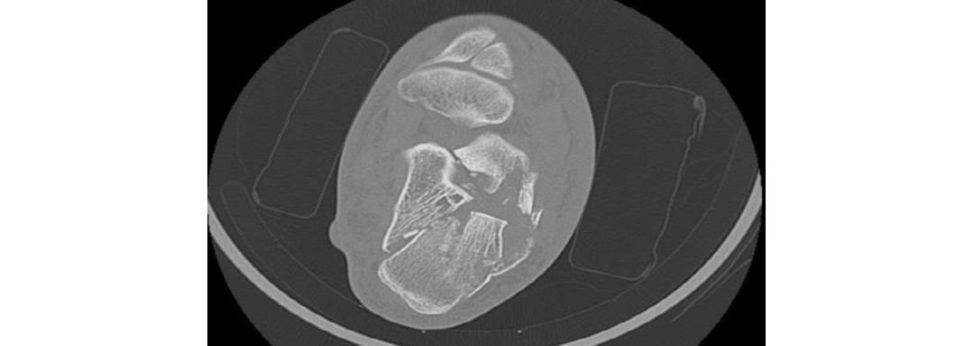
This CT scan axial section demonstrates shortening, varus deformity and considerable comminution. The reis a large sustentacular fragment, depressed middle fragment and blowout of the lateral wall. It also shows considerable heel widening.
Do you know any classification systems for calcaneal fractures?
The Sanders classification is a C T classification based on the number of articular fragments seen on a coronal view at the widest point of the posterior facet (Figure 11.4c). Type 1: Undisplaced posterior facet (regardless of number of fracture lines). Types 2, 3 and 4 are displaced fractures. Type 2: One fracture line (two-part intra-articular fracture). Divided into three subgroups on the basis of fracture line localization. Fracture line is lateral in Type 2 A fractures, central in Type 2B and medial in Type 2C fractures. Type 3: Two fracture lines in the posterior facet (three-part intra-articular fracture). Type 4: Comminuted fracture with more than three fracture lines in the posterior facet (four or more fragments). I would discuss treatment options with the patient including open reduction and internal fixation once the soft -tissue envelope is suitably resuscitated. I would base my decision on the fracture pattern, soft -tissues tatus and patient factors. This fracture pattern will benefit from surgical intervention, but it will depend heavily on several patient factors including smoking, occupation, comorbidities and the expectations of the patient.
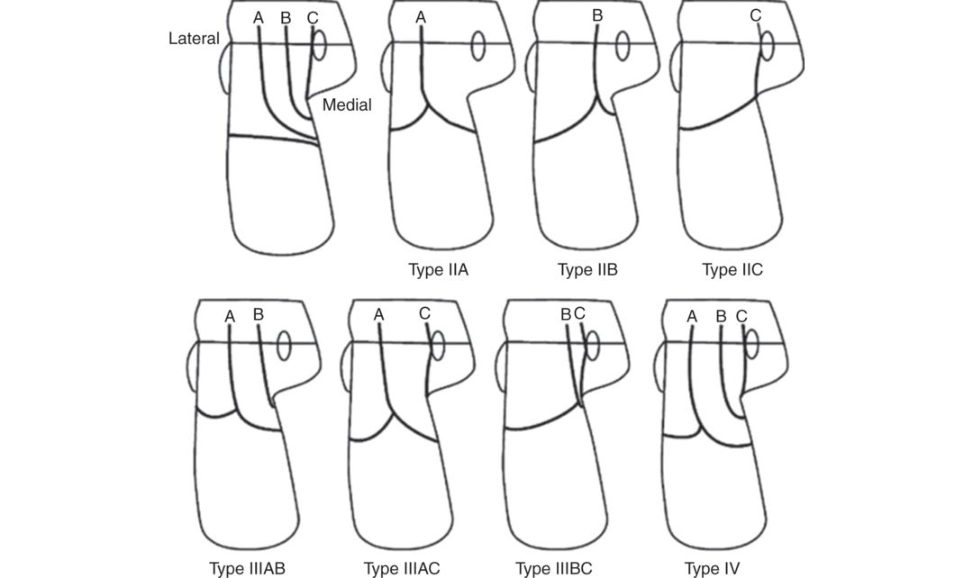
Figure 11.4b CT scan axial view left foot demonstrating calcaneal fracture.
Figure 11.4c Saunders classification of calcaneal fractures.
Minute 3
Following discussion with the patient you have decided to proceed with internal fixation. What are the aims/goals of surgery?
The aims of surgery are restoration of articular congruity while restoring calcaneal height, length and heel width and minimizing soft -tissue complications.
How will you fix the fracture?
I would take full informed consent, in particular concentrating on the risks and benefits of both operative and non-operativ e management. The patient will be undergeneral anaesthesia with prophylactic antibiotics, tourniquet and in the lateral decubitus position with fluoroscopy control. I would use an L-shaped lateral incision halfway between fibula and Achilles tendon avoiding damage to the sural nerve. I would employ full-thickness flaps by taking the incision down to the bone and use bent K-wires as retractors. I would take off the lateral wall, manipulate the fracture fragments to restore the length and height of the calcaneum as well as correction of varus deformity, reconstruct the articular surface and then reapply the lateral wall. I would use K-wires for temporary stability and then fixation using a fragment specific plate. My preference is a low-profile lateral calcaneal plate, the size of which depends on the patient calcaneus and I would contour the plate prior to application. The key is to capture the sustentacular fragment under fluoroscopy. Postoperatively the patient would mobilize non-weight-bearing for 6 weeks followed by a further 6 weeks of partial w eight-bearing.
What are the complications from surgery?
Complications include wound dehiscence, osteomyelitis, pos t-traumatic osteoarthritis, increased heel width, subtalar stiffness peroneal tendinitis, sural nerve injury, persistent heel pain, scar hypersensitivity , tarsal tunnel syndrome and CRPS.
If the wound got infected, how would you deal with it?
I would want to prevent direct extension to bone causing osteomyelitis. This needs aggressive antibiotic therapy and a low threshold for radical debridement. I would attempt to keep in place the plate and screws but would remove the metalwork if the infection was not setiling. Soft - tissue coverage with local or free flap involving the plastic surgeons should be considered if the wound is very large. Occasionally amputation may be needed.
The patient complains of pain.
There are many causes of pain which include subtalar incongruity, penetration of screws into the subtalar joint or arthritis Lateral pain may be caused by lateral impingement or peroneal tendinitis Anterior pain from talar neck impingement or scar tissue. Sub talar osteoarthritis may require a subtalar arthrodesis.
What prognosis will you give for this patient?
A calcaneal fracture is a significant injury with high incidence of long-term pain and disability. There is about a 40% chance the patient will have long-term chronic pain after a significant intra-articular fracture.
Evidence base
Be careful. Treatment of calcaneum fracture is still a controversial issue and atir acts a lot of debate.
A key paper to know is the Griffin article reporting the results oft heUK Heel Fracture Trial.2 This was a multic entre, pragmatic, randomized control trial which demonstrated no difference inpatient reported outcomes between operative and non-operativ e management of intra-articular calcaneal fractures.
However, it is important to note that patient selection was based on the idea that patients could be managed by either method. Those patients with clear indications for surgery (see above) were not included and so should still betaken on their own merit.
Another key paper is Buckle yet al.3 In this multic entre Canadian trial over 300 patients with displaced calcaneal fractures were evaluated comparing operative vs. non-operativ e treatment. The authors found that without stratification of patients functional results were the same with either non-operative or operative care.
When they looked at subgroups of patients they found that those receiving workers compensation had a worse outcome in general. Women fared better after surgical reduction, as did patients who:
Were not receiving workers’ compensation.
Were less than 29 years old.
Had a less severely displaced fracture.
Had a light workload.
Had an anatomic reduction.
In a later study, they noted that the overall cost of care of patients was less with surgical care than non-surgical management due to the need for additional surgery for fusion and for the higher disability cost from a longer period of missed work in the non-operativ ely managed group of patients.
Structured oral examination question 5#
A 21-year-old motorcyclist is involved in a road traffic accident. He is fully conscious, alert and following a global assessment using the ATLS protocol, it is revealed that this is an isolated, closed injury (Figure 11.5).
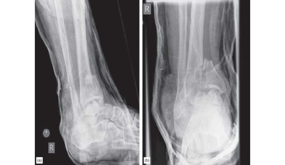
Figure 11.5a and 11.5b Anteroposterior (AP) and lateral radiographs, right lower leg.
Minute 1
Tell me how you would manage this injury.
These are anteroposterior (AP) and lateral views of the distal tibia and ankle joint. This is a complex intra-articular multi fragmentary fracture occurring as a result of high-energy trauma. There is a fracture of the distal tibia involving the ankle joint with articular impaction and comminution extending into the metaphysis, a fractured fibula, a disruption of the syndesmosis and possibly a fractured talus. I will take a concise history and perform a focused examination. I would enquire about smoking, alcohol consumption, a history of diabetes or peripheral vascular disease, etc. which are important risk factors for soft tissue (and bone) healing. Examination would particularly assess the s tate of the soft -tissue envelope looking for any skin damage, contusion and fracture blisters. I would also obtain tibial shaft and knee radiographs of the affected limbI will ensure the patient is comfortable and splint the limb, realigning the foot into a better position relieving pressure on the skin to avoid any skin necrosis. I will perform a thorough neurovascular assessment of the involved limb including palpation of the posterior tibial and dorsalis pedis artery as well as examination of capillary refill to check for an adequate vascular supply. I would obtain radiographs of the post splinted leg to check for adequacy of reduction and commence serial assessment for compartment syndrome. Isolated closed injury is coded language to say the examiners just want you to focus on the management of this fracture. No need to mention A TLS. Ruedi and Allgower have classified these injuries into three types: Type 1 Non-displaced fracture cleavage of ankle joint. Type 2 Displaced fracture with minimal impaction or comminution. Type 3 Explosive fracture with significant articular comminution and metaphyseal impaction.
How does this classification system help you?
The Ruedi–Allgower classification system is based on the severity of comminution and displacement of the articular surface and offers a rough guide to management.
Minute 2
My principle of managing this case is: ‘span–scan–plan’. I would prefer a staged management approach for this fracture rather than going for early ORIF. Early ORIF in the face of compromised soft tissues will lead to an increased risk of infection wound dehiscence, a poor overall clinical outcome and in a worst case scenario may lead to amputation. It is generally thought that a staged management protocol of span, scan and plan is the gold- standard method (first-line intervention) to deal with complex intra-articular fractures, especially in the presence of bruised, swollen, compromised soft tissues. I would consider early involvement of the plastic surgeons if the soft -tissue envelope was very badly compromised and especially so if the fracture was open. Span: I will placean external fixator in order to reduce and hold the fracture. This will allow correction of length, restoration of alignment and rotation and allow soft -tissue resuscitation and monitoring. This will also allow us to arrange timely definitive surgery. Scan: Following initial stabilization, computed tomography scanning will provide more details of the fracture type and pattern. A CT after EF will illustrate the overall alignment of the tibia, help identify the main fracture fragments, location of fracture lines, amount of articular impaction and comminution. The sc an usually influences the surgical approach chosen for definitive fixation. Plan: using the CT scan I can then plan the definitive treatment in detail; approach, how to fix fragments, what implant to use, timing of surgery, taking consent from the patient and ensuring all equipment, staff and company representatives are available.
Minute 3
When are you going to fix this fracture?
This is a serious and challenging injury to manage. The soft -tissue envelope needs to be resuscitated until it is in areason able condition (this may take up to 10–14 days to setile). Definitive surgery should be planned on a defined dedicated trauma list involving a surgeon with an interest in managing these complex fractures in order to achieve the best possible outcome.
How are you going to fix this fracture?
The principles of fixation of an intra articular fracture are anatomical reduction, interfragmentary compression and absolute stability at the fracture site to allow early mobilization. This is a generic statement that, while correct, isn’t using viva time efficiently. It is better for candidates to be more fracture-specific if possible. My goals of treatment are: Re-establishment of articular congruency. Correction of an y mechanical malalignment. Management of any bone loss. Reduction of the risk of soft -tissue complications. Early restoration of motion. Ruedi–Allogower specifically recommended four key operative principles: Plating the fibula to length. Articular reconstruction. Bone gratiing of metaphyseal defects. Medial but iress to the tibia to prevent a late varus deformity. The approach would be tailored dependent on fracture configuration as corroborated by CT scan prior to and with fluoroscopy during surgery. Looking at the fractures in the radiographs provided, I would favour an anterolateral approach that will allow me to reduce the pilon fracture, and approach the fibula as well as the talus. I would aim for anatomical reduction of the pilon fracture under direct vision and stabilize it with an anterolateral plate and then address the fibula and talus on their own merits. The fracture of the talus may well necessitate extending the approach and a release of the ATFL through a subperiosteal approach. I would anatomically reduce the talus fracture and stabilize it. I would make sure any loose debris is removed from the joint.
Anything else?
Sorry?
Anything else about the fracture pattern?
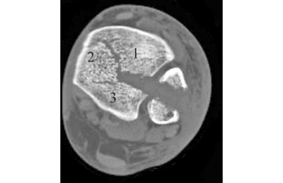
No, sorry. For score 7/8 candidates. There are three classic articular components of a pilon fracture that can be identified on axial CT scan. These are anterolateral (Chaput fragment), medial and posterolateral (Volkmann fragment) (Figure 11.5c).
How would you perform an anterolateral approach to the ankle?
The patient should be supine with antibiotics given and a thigh tourniquet applied. This incision is centred at the ankle joint, parallel to the fourth metatarsal distally, and parallel to and between the tibia and fibula proximally. Dissection through the skin and subcutaneous tissues should proceed sharply with maintenance of full-thickness skin flaps. Because the anterior compartment muscles arise from the anterior fibula, the incision is usually not extended more than 7 cm above the ankle joint. Distally, the incision can extend as far as the talonavicular joint. Care is taken not to damage the superficial peroneal nerve which lies directly beneath the skin. This nerve crosses the surgical incision proximal to the ankle joint. It should be identified, mobilized, and protected throughout the surgical procedure. The fascia over the anterior compartment of the distal tibia is then incised sharply , beneath the superficial peroneal nerve. Distally, the extensor retinaculum is incised, and the anterior compartment tendons are all retracted medially. Proximally, the entire anterior compartment musculature, including the peroneus tertius, can then be mobilized and retracted medially. These muscles and tendons are usually easy to mobilize from the underlying anterior tibiofibular ligament, the periosteum of the distal tibia, and the joint capsule. For score 8 candidates:
How will you fix the fragments?
The articular surface is visualized, and the impacted fragments reduced under direct vision. Reconstruction of the plafond proceeds from posterior to anterior with provisional fixation of fracture fragments using temporary K-wires, and small fragment screws (cannulated and partially threaded) then applied to secure definitive stability. Occasionally if fracture reduction is difficult a distractor can be used to aid fixation. A locking plate is then applied to the distal tibia.
Any other methods?
A circular frame with limited minimally invasive internal fixation is a possible option, but I have no experience with this method of fixation.
Figure 11.5c Classic articular components of a pilon fracture.
Minutes 4–5
How would you counsel the patient and their family regarding the outcome of pilon fracture?
These injuries represent a high-energy axial insult to the lower limb that leads to severe joint comminution, impaction and a large zone of injury. There are considerable risks of complications affecting both bone, joint and overlying soft tissues Wound breakdown and skin necrosis may lead to late sinus formation or osteomyelitis associated with infected metalwork. Delayed union, non-union, infected non-union and malunion can occur. Post-traumatic osteoarthritis may require arthrodesis or arthroplasty.
What is Hawkins’ sign, is it a good or bad sign?
It is the appearance of osteopenia in subchondral bone of the talar dome on the AP view, 6– 8 weeks following fracture of the neck of talus. The Hawkins’ sign is a good indicator of talus vascularity following fracture – it is therefore a good sign. It indicates that healing will occur without avascular necrosis.
Evidence base
The evidence base here is fairly disparate with no major RCTs. The general consensus is to use the ‘span–scan–plan’ approach. Fixation can be affected with either a frame or plate osteosynthesis and there is liti leto say one gives a better outcome than the other.
Structured oral examination question 6#
A 50-year-old lady is a front seat passenger involved in a head-on road traffic collision. In the Emergency
Department she is diagnosed with dislocation of her native right hip.
Minute 1
What will be your initial management?
A native hip dislocation is a marker of a high-energy injury and I would assess the patient along ATLS principles utilizing an ABCDE approach. I will look to exclude any associated injuries. I would clinically examine her lower limbs looking at alignment, position and neurovascular status in particular that of the sciatic nerve. With the mechanism of injury caused by impact of the dashboard on the knee the hip dislocation is likely to be posterior often with an associated acetabular rim fracture or femoral neck fracture.
Any clue as to whether it is posterior or anterior clinically?
With a posterior dislocation the leg would be shortened and internally rotated while with an anterior dislocation the leg is flexed shortened, externally rotated and abducted. Anterior dislocations of the hip are rare.
It appears to be an isolated injury with paraesthesia in the sole of the foot; however, motor function is intact. How will you take it from here?
A traumatic hip dislocation is a surgical emergency because of the risks to the vascularity of the femoral head, dangers of chondrolysis as well as pressure effects on the surrounding soft tissues, especially neurovascular structures. The paraesthesia in the foot is an indication of pressure or traction affecting thes cia tic nerve. I would arrange for the patient to go to the operating theatre urgently. I would get a CT scan done provided it does not delay transfer to the operating theatre; and of course, inform theatres, anaesthetic t eam and the ward. I would order baseline bloodtests including blood group and save. I would take informed consent for a closed or open reduction undergeneral anaesthesia.
Minute 2
There is delay in geting the C T scan and you take her to the operating theatre. How will you reduce the hip?
I would attempt closed reduction undergeneral anaesthesia. I would position her supine on the table with the table height as low as possible. I would request the anaesthetist to use full muscle relaxant to make it easier to reduce the hip. I would stand on the side of the dislocated hip and have the image intensifier come from the opposite side. My assistant will be on the opposite side towards the headend of the patient to stabilize and hold down her pelvis at theA SISs when I attempt manipulation. I would screen the hip first before attempting reduction to exclude a neck of femur fracture and also assess the acetabulum using Judet views. If it is posterior dislocation, I would apply gentle traction on the hip (inline) and then gradually flex the hip and the knee, maintaining traction. Reduction usually occurs with an audible ‘clunk’ and I would check reduction under the image intensifier and also check once again for any associated fractures. Bigelow’s technique is with hip flexed to 90°, the affected leg is placed in an adducted and internally rotated position. While an assistant stabilizes the pelvis with downward pressure, traction is applied inline with the femur while abducting , externally rotating and extending the affected hip.
You manage to reduce the hip and get this radiographic image (Figure 11.6a). What are your thoughts?
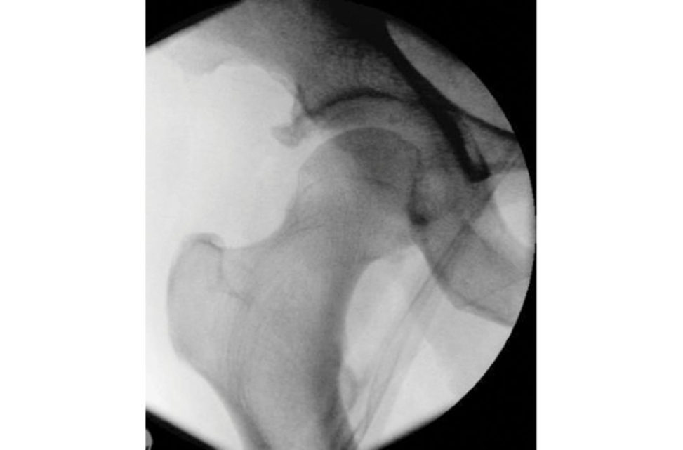
In the next 30 seconds you are expected to comment on the name of the patient site of radiograph and the exact nature of the injury. In this image-intensifier view of the right hip, the femoral head appears to be in the acetabulum but is incongruent; in addition, I would like to confirm this on lateral view. The femoral head is inferiorly subluxed and there appears to be a bony fragment in the hip joint superiorly and another one inferiorly. There is one more fragment on the superolateral lip of the acetabulum. The bony fragments are most likely to be from the acetabulum; however, femoral head fragments need to be ruled out.
Figure 11.6a Image intensifier (II) image right hip.
Minute 3
How will you assess this hip further?
Per operatively I would screen the hip in AP and lateral views as well as get Judet views to assess the anterior and posterior walls and columns. In addition, I would carefully assess the hip for stability by screening the hip through a range of motion. I do not want to re-dislocate the hip and cause any further hip damage. A CT scan will be useful to delineate this further, if it has not already been done.
You get a CT scan done in the morning and this is one of the sections ( Figure 11.6b). What do you think?
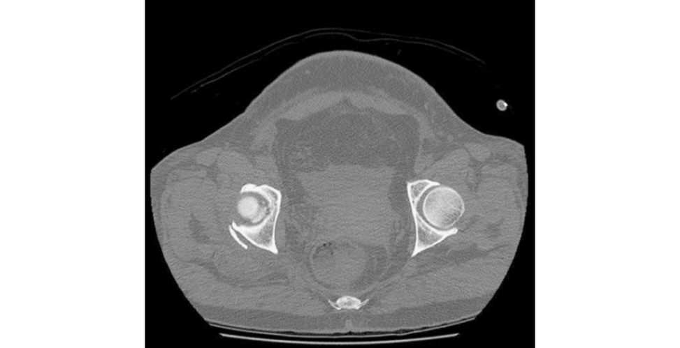
[Note: just comment on what you have rather than ask for more images!] In this axial section of the pelvis a t the level of the hip joints, I note that the femoral head on the injured side is at a different height to the opposite hip. There is a bony fragment trapped in the hip joint as well as a bony fragment lying posterior to the hip. This may represent a fracture dislocation with compromise of the acetabular wall posterosupeiorly. I would, however, need to study the whole CT sequence to ascertain the extent of damage. After obtaining a postreduction C T if I wasn’t in an MTC I would discuss the images with the regional acetabular and pelvic reconstruction unit and get their advice.
What about an MRI scan of the hip?
While MRI will demonstrate labral tears and soft -tissue anatomy it has not been shown to be beneficial in the acute evaluation and management of hip dislocations.
How will you deal with the bony fragment in the hip joint?
This depends on a number of factors including the exact original site of the fragment, the size of it, the integrity of the weight-bearing dome and the stability of the hip. The options for a bony fragment trapped in the hip joint are to remove it or to retrieve and fix it. If the fragment is quite small and does not affect the hip stability or the weight-bearing dome, then it can be removed arthroscopically. However, if it compromises the weight-bearing area of the hip or stability then I would retrieve it and fix it. It will have to be an open procedure, although reports of arthroscopic intervention have been published. (Note: If the candidate does not have sound hip arthroscopy knowledge, then the safe option is open procedure and stay clear of hip arthroscopy.)
Figure 11.6b CT axial view pelvis.
Minute 4
You find that it is the posterosuperior lip of the acetabulum. Which approach will you use to fix the fracture?
The approach depends on where the bony fragment is arising from. If it is posterosuperior or posteriorI would use a Kocher–Langenbeck approach to the hip. I would position the patient on the fracture table in lateral decubitus. In the posterior approach to the hip I would respect the blood vessels supplying the femoral head and therefore would not takedown the quadratus (medial circumflex femoral artery). I would incise the short external rotators at least 1.5 cm from their insertions to again avoid damage occurring to the medial circumflex MCFA. I would retract the gluteus medius superiorly, identify the capsule and dissect superiorly to identify the fractured rim of the acetabulum. I would be careful with the sciatic nerve as it may be closer than realized in the operating field duet o the distorted anatomy and could also be bruised from the original injury. I would reduce the fragment anatomically under image-intensifier screening and secure it with 2–3 partially threaded cannulated screws, making sure that the screws do not penetrate the hip joint. If the fragment was large I would consider using a posterior plate one-third small fragment tubular or 3.5 mm reconstruction plate. If an anterior approach to the hip is needed either an ilioinguinal or Stoppa approach can be used.
Minute 5
What are the risks of posterior dislocation of the hip?
Immediate complications are sciatic nerve injury (10% with posterior dislocation fractures, and haemorrhage. Intermediate risks are chondrolysis, postreduction neurological damage, avascular necrosis, intra-articular loose bodies heterotopic ossification, hip instability. Late complications are hip pain and post-traumatic osteoarthritis.
What other injuries are associated with this injury pattern?
This is determined by the direction of forces and may include patella fracture, PCL rupture, femoral shaft fracture, femoral neck and head fractures. Note: The candidate is smiling as the examiner has runout of questions on his crib sheet!
Evidence base
The evidence base here is largely based on expert opinion and there are no major RCTs.
Structured oral examination question 7#
A 78-year-old lady fell out of her bed while visiting friends in another part of the country and sustained this proximal femur fracture. She is in reasonably good health and was independently mobile, able to care for herself and do her own shopping.
Minute 1
What can you see (Figure 11.7a.)? What are the treatment principles?
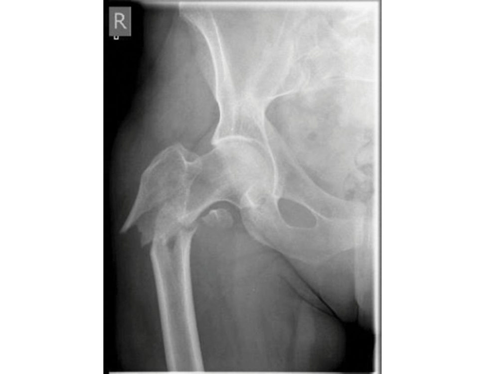
An AP radiograph of the right hip showing a reverse-obliquity inter-trochanteric fracture with subtrochanteric extension. The lesser trochanter is proximally displaced with associated loss of the posteromedial but iress. I would like to see a lateral view; however, based even on the AP view, it is an unstable fracture pattern. My management for this patient would start with thorough assessment and optimization of her general condition. W e need to consider the possibility of pathological fracture, although the available radiograph shows no evidence of that. I would get adequate radiographs of the full femur. Provided she is fit for surgery, I would aim to treat this fracture operatively and will do this within 36 hours of admission. I would use a cephalomedullary device to fix this fracture. I would do so as a load-sharing implant with a large diameter nail with a shorter, medialized lever arm will afford an enhanced biomechanical environment for this challenging fracture.
Figure 11.7a Anteroposterior (AP) radiograph of right femur demonstrating in ter-trochanteric fracture.
Minute 2
I agree. This lady was treated elsewhere initially with an extramedullary device. She presents 4 months down the line when you are on call with this complication, can you explain what happened (Figure 11.7b and 11.7c)?
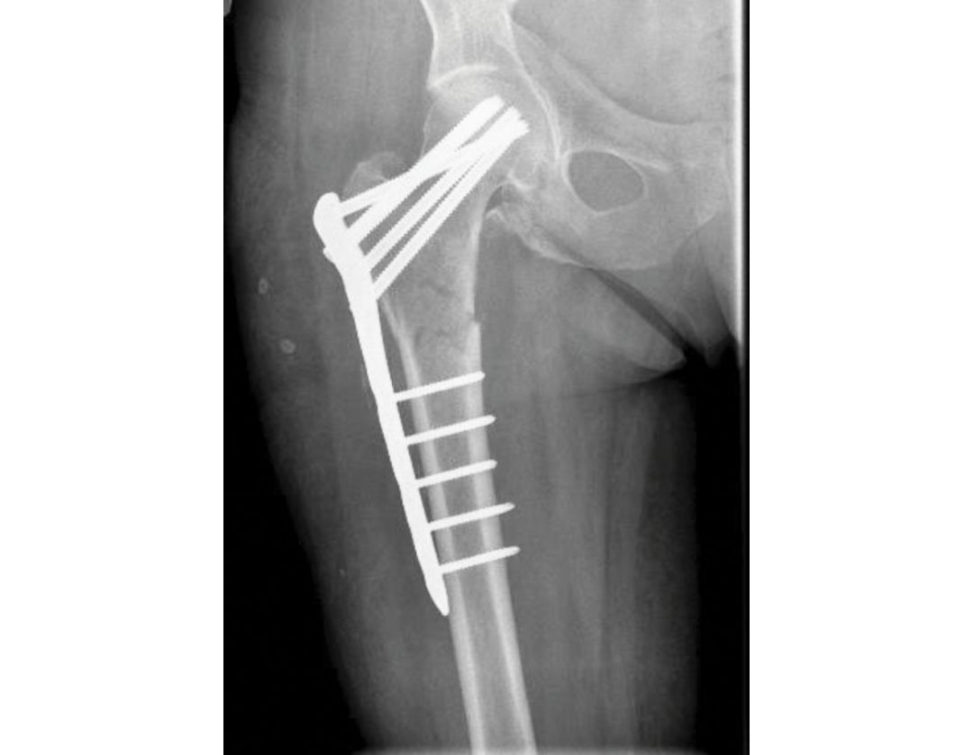
This lady was treated with a fixed angled locking plate. Two elements of fracture care are perhaps responsible for the implant failure – biomechanics and biology. Looking at the postoperative radiograph, there is a gap at the fracture site, especially on the medial side. The fixed angled device has been used with locking screws with five screws on either side of the fracture, which will make it a very rigid construct with a very high strain focused across a small fracture working length. This will minimize any micro-motion necessary for callus formation. On the other hand, there is a fracture gap and lack of compression, which will preclude primary bone union. This has resulted in delayed union/atrophic non-union at the fracture site. Essentially this fracture is in a surgical ‘no man’s land’ with none of the requirements of either primary or secondary bone healing. The implant has been under constant biomechanical load, which had led to the fatigue failure of the implant-grade steel. In this particular design the reis a stress riser at the junction of the last proximal locking hole and tapered part of the plate, which dictates the failure point in the implant. In addition, the plating device is applied on the lateral aspect of the femur, increasing the lever arm for the moment of force as compared to a cephalomedullary device, which further puts the fixed angled plating device in this position a t a biomechanical disadvantage. In this type of fracture, a cephalomedullary device has better biomechanical stability.
Figure 11.7b Anteroposterior (AP) radiograph, right femur, with fixed locking plate in situ.
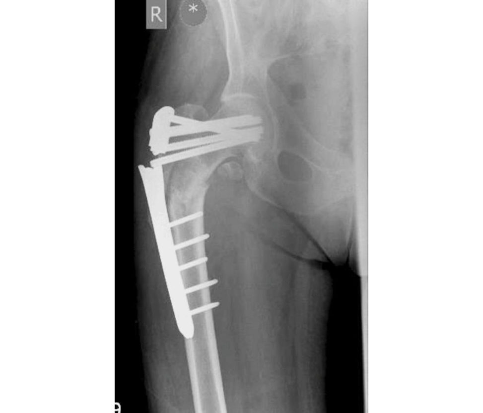
Figure 11.7c Anteroposterior (AP) radiograph, right femur, demonstrating hardware failure, 4 months postoperative.
Minute 4
You fixed it with this nail. What do you think about your check X-ray (Figure 11.7d)?
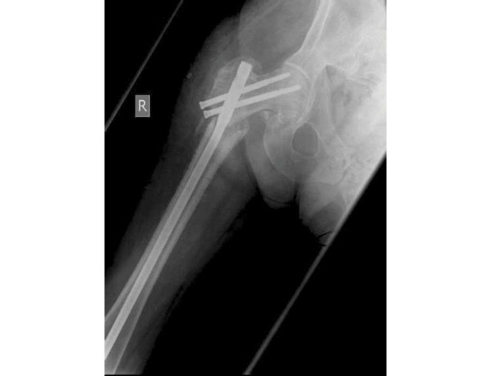
I fixed this?! I’m surprised as there are several fundamental issues with this construct. The screws in the proximal fragment are a bit superior to where I’d normally like them to be. The screws are also not absolutely parallel, and I’ll study my lateral radiographs carefully to make sure that the screws have not missed the nail while transiting in to the head. There is translation of the fragments and the femoral shaft is inv arus. None of these features are ideal. In addition, the nail is probably undersized, it looks relatively small for the canal with only limited isthmic fit. Keep out of the politics of criticizing an y suboptimal fixation, especially in the exam. Keep it factually straight down the line with neutral comments.
Figure 11.7d Anteroposterior (AP) radiograph demonstrating non-union femoral fracture.
Minute 5
How will you follow-up this patient?
I would follow-up this patient with clinical reviews and serial radiographs. I would start her weight-bearing as able, ensure she is not on non-steroidal anft-in flammatories or bisphosphonate treatment, counsel against smoking and do serial radiographs 6 weeks apart. If there is no callus formation a t 6 months, I would consider exchanging the intramedullary nail.
Evidence base
A 2014 Cochrane review suggested that there is very poor evidence surrounding cephallomedullary nails and more evidence is required comparing these devices to sliding hip screws.
Queally JM, Harris E, Handoll HHG, Parker MJ. Intramedullary nails for extracapsular hip fractures in adults. Cochrane Database Syst Rev. 2014; Issue 9.
While the mechanical benefits of the intramedullary devices may not lead to improved outcomes in patients with a simple intertrochanteric fracture, the more complex unstable patterns such as four-part inter-trochanteric or reverse oblique fractures may be better managed with cephallomedullary nails.
Be prepared to discuss the biomechanical differences between CMN and DHS.
Structured oral examination question 8#
A 72-year-old lady, fully independent with good health, was hit by a car when she was walking on a kerb.
She was brought to hospital with these two injuries. She was assessed following ATLS protocol, fully resuscitated and her injuries were splinted (Figure 11.8).
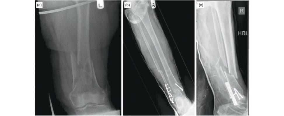
Figure 11.8a, 11.8b and 11.8c Anteroposterior (AP) radiograph, left femur, demonstrating supracondylar fracture femur, and AP and lateral radiographs, right lower leg.
Minute 1
What your thoughts about this patient management? Do you have any concerns?
This 72-year-old lady has multiple high-ener gy injuries. Although she was enjoying good health prior to this accident, I would be concerned about her physiological response to the trauma. Elderly patients have a limited physiological reserve when compared to younger patients, so she needs to be closely observed, kept well hydrated and her general condition optimized before definitive treatment. Fractured long bones should be stabilized as early as possible for many reasons: pain relief, to reduce the metabolic response to trauma, to allow for early mobilization and rehabilitation aswell as a decreased incidence of complications associated with recumbence. The anaesthetic t eam and orthogeriatricians should be involved early in the plan for her treatment.
Minute 2
What implants are you going to use to fix these fractures?
For the left femur fracture it is an unstable, multi fragmentary, supracondylar fracture with femoral shortening. The aim is to reduce the fracture for length, alignment and rotation and then stabilize using a relative stability device. To do this I may need to use a femoral distracter for temporary reduction and then stabilize with either a nail or a plate. Personally, I would use a fixed- angle plate with locking options and a minimally invasive technique. Failure in varus can be a problem for these fractures. Regarding the tibia fracture it is a distal third fracture that is periprosthetic in nature due to the presence of pre-existing implants from an ankle fixation. I need to employ a technique with minimal soft -tissues tripping and one that enables early weight-bearing. As with the femur I want to achieve reduction for length, alignment and rotation before stabilizing. To this end I would choose an antegrade, intramedullary, reamed tibial interlocking nail. If the soft tissues were compromised or the canal too narrow to allow nailing, I would consider using a circular frame.
Minutes 3–4
OK, have look on this radiograph (Figure 11.8d) and explain to me the technique the surgeon used and what the principles of such technique are.
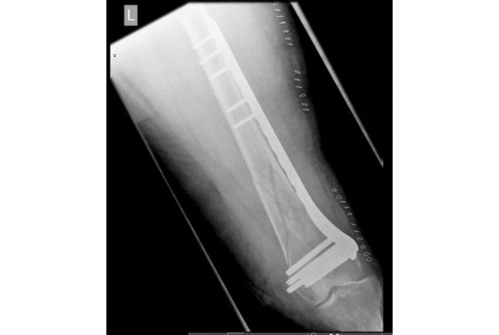
The AP radiograph shows a multi fragmentary fracture of distal diaphysis/metaphysis of the femur that has been stabilized with a fixed-angle plate, in bridging mode. Examining the skin staples, I can infer that closed indirect reduction and a less-in vasive technique was used. This technique was introduced to decrease soft -tissue disruption and preserve blood supply. Length, alignment and rotation of the bone have been restored. Baumgaertel et al. introduced the concept of biological plating and proved that indirect reduction and bridge plating was superior to direct fragment reduction and anatomical fixation in respect to bone healing.4
Can you explain why the surgeon put screws on either ends of the plate and missed the middle?
The surgeon intended to increase the working length of the implant (the distance between two points on either side of the fracture where the bone is fixed to plate or nail). This produces even distribution of forces over a long segment and decreases (shares) stress between the fracture and the implant.
Figure 11.8d Anteroposterior (AP) radiograph, left distal femur with locking plate in situ.
Minute 5
You mentioned circular frames – can you tell me the principles of their use?
Circular frames consist of fixation elements such as tensioned wires or half pins attached to rings on either side of the fracture. This forms proximal ring blocks and distal ring blocks. Segments of frame can be moved in terms of angulation rotation, translation and length. Their use has particular w orth with poor soft tissues and small segments . They may be definitive or temporary and may also be used in isolation or hybrid with other techniques such as limited ORIF. They allow immediate weight-bearing and therefore are beneficial in the elderly and patients with limited compliance (Figure 11.8e).
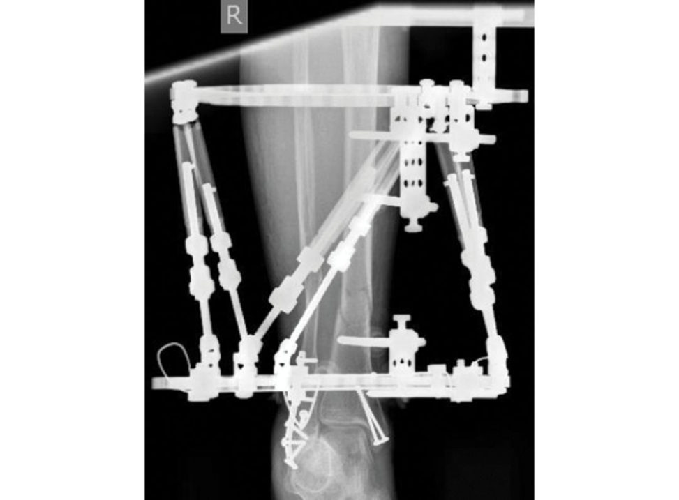
Figure 11.8e Anteroposterior (AP) radiograph, right distal tibia with circular frame in situ.
Structured oral examination question 9#
A 29-year-old female horse rider fell off her horse; she has been fully assessed in the Emergency department and has an isolated closed injury of the foot.
Minute 1
What are your thoughts (Figure 11.9a)?
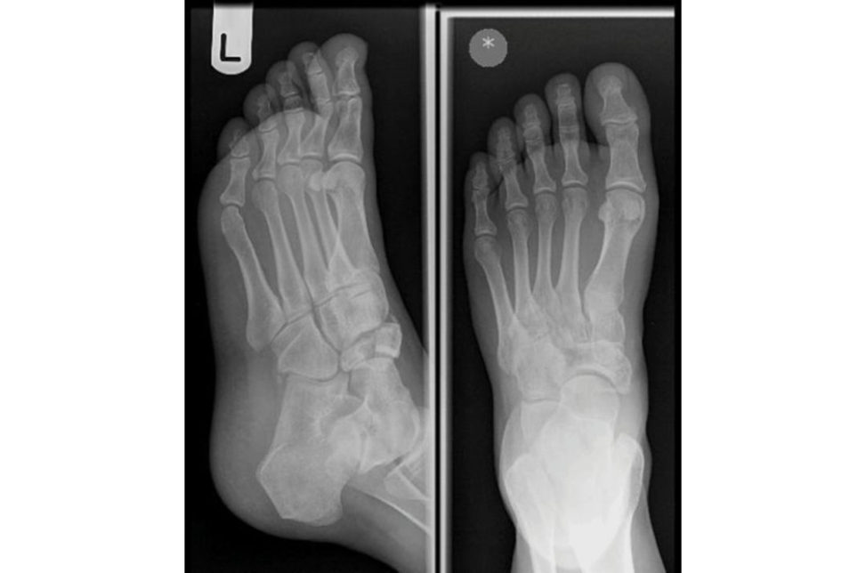
The radiographs of the left foot, AP and oblique, show a displaced, comminuted fracture of the body of the navicular. There is overlap of the midtarsal bones and I can’t exclude fractures of any other tarsal bones. The alignment of the foot is still maintained and there is no varus or valgus deformity. Given the high degree of ligamentous stability of the navicular this is a serious, high-energy injury. I need to assess the pa tientas a whole. I would secure a relevant focused history, clinical examination in general of the patient and in particular of the foot, ruling out compartment syndrome, any neurovascular damage and assessing the soft -tissue envelope of the foot. I would then request further imaging, the modality of choice being a computed tomography scan. I would initially tr eat the injured foot in abacks lab, with strict elevation and in termift ent cryotherapy, adequate analgesia and serial monitoring for any evolving compartment syndrome. Navicular body fractures are often associated with other injuries of the midtarsal joint often with varying disruption of the talonavicular and cuneonavicular joints. Sangeorzan et al. classified these injuries into three types (Figure 11.9b). This is probably worth knowing, although there is a move away in the exam from pure didactic learning of classification systems tousing a classification system as a guide to treatment. It is a useful guide towards difficulty of fracture reduction and eventual clinical outcome. Type 1 The fracture line splits the navicular into dorsal and plantar segments. No dislocation occurs. Type 2 These are the most common injuries. The fracture traverses from dorsal lateral to plantar medial across the body of the tarsal navicular. The major fragment is dorsal medial, with a smaller, often comminuted plantar–lateral fragment. There is usually medial forefoot displacement. Type 3 The fracture is characterized by comminution of fragments and significant displacement. The medial fragment is the major one and the medial border of the foot is often disrupted at the cuneonavicular joint. There is often lateral displacement of the foot. This injury carries the worst prognosis.
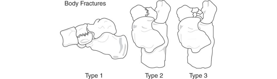
Figure 11.9a Anteroposterior (AP) and lateral radiographs, left foot.
Figure 11.9b Sangeorzan et al. classification oft alar body fractures.
Minute 2
This is the scan you requested, what do you see and how would you manage it (Figure 11.9c and 11.9d)?
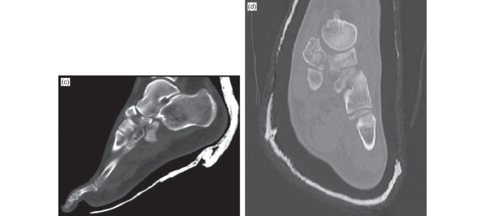
These are coronal and sagift al sections of the C T scan, and they confirm the radiographic findings of a displaced fracture of the body of navicular bone with comminution. It is an unstable displaced intra-articular fracture and I would therefore favour operative intervention rather than non-operativ e. The principles of management are to restore the articular surface, stabilize and hold the fracture to allow early mobilization. The aim of the treatment is to have a mobile, pain-free and functional joint. However, sometimes that is not possible due to severe comminution of the articular surface, in which case I may consider primary fusion of the talonavicular joint. I would discuss the findings, management options, aims of the treatment as well as potential complications with the patient and ensure balanced informed consent is secured before proceeding. I could only proceed once the soft -tissue envelope is satisfactory, however. Fractures with less than 2 mm articular displacement, no midfoot instability and noloss of bone length can be managed non-operativ ely. Non-weight-bearing cast for 6–8 weeks. In general, all navicular body fractures with >2 mm displacement require ORIF.
Figure 11.9c and 11.9d CT scan, coronal and sagift al sections of left foot.
Minute 3
Can you tellus about any possible complication associated with this case?
There are immediate, early and late complications. Immediate complications are in the peri operative period and include iatrogenic injury to structures, compartment syndrome and anaesthetic problems. Early complications include infection nerve injury (branches of superficial and deep peroneal nerves), vascular injury (dorsalis pedis); and late complications include non-union and loss of medial longitudinal arch support, painful talonavicular joint, post-traumatic osteoarthritis, as well as avascular necrosis and collapse. Late talonavicular fusion may be required as a salvage procedure.
What are you going to tell the patient?
With a good reduction most have a reasonable prognosis, but few are normal. Sangeorzan et al. found that the type of fracture and the accuracy of the operative reduction correlated directly with the final clinical result.5
Minute 4
Why does non-union and avascular necrosis occur in this fracture?
The navicular, similar to the talus, has a large articular surface area and for blood supply it relies on the radial arcade of vessels arising from the dorsalis pedis and medial planter arteries, and this could be injured either at the time of the fracture or during surgery, which could lead to AVN, non-union and/or collapse of the bone resulting in a painful midfoot.
What surgical approach are you going to use?
I would use a medial longitudinal approach, between the tibialis anterior and tibialis posterior tendons with minimal dissection to preserve the remaining blood supply as much as possible. The patient should be positioned supine, I would use a pneumatic thigh tourniquet with IV antibiotics given before inflation and the radiographer should be ready with II in theatre with relevant radiological requests made beforehand to reduce any potential for theatre delays. [This is all a bit waffly but it is covering the basics. Probably not scoring any real marks but avoids complete silence.] It is important to directly visualize the articular surface to ensure accurate reduction of the articular surface. I would stabilize the fracture with cannulated screws from lateral to medial; however, the eventual configuration of screws will depend on the fracture pattern. I would avoid extensive periosteal stripping over the dorsal navicular surface as this may disturb the tenuous blood supply of the central third portion of the body . Sometimes ase cond anterolateral incision is needed to help with reduction of a significantly displaced lateral fragment.
Minute 5
Take me through your consent process, in general.
I follow the General Medical Council guidelines on this subject and broadly run my practice along the lines of the domains of the GMC ‘Good Medical Practice ’. I work with the patient in partnership to ensure high-quality of care. In particular: (a) I listen topa tien ts and respect their views about their health. (bI discuss with patients what their views about diagnosis, prognosis, treatment and care involve. (c) I share with patients the information the y want or need in order to make decisions. (dI maximize apa tien t’s opportunities, and their ability , to make decisions for themselves. (e) I respect the patient decisions. These are quite rare injuries.
Structured oral examination question 10#
A 34-year-old male was playing rugby when he made an awkward tackle. He is a right-hand dominant manual worker. He sustained the closed, isolated injury below.
Minute 1
Talk me through your initial assessment and management of this patient in A&E.
I would start with a focused history, making sure to ask about factors that will affect surgery, such as past medical history and allergies, and factors that will potentially affect long-term outcome such as hand dominance, occupation and smokings tatus. I will then move on to perform an examination. This would include a general examination to ensure fitness for surgery before focusing on the injured limb. Assessing the limbI would be making sure that this is a closed injury and carefully document the neurovascular status. Following this assessment, I would want to put some simple first aid management steps in place including analgesia and application of a back -slab. I would admit the patient for elevation with a view to surgery on the trauma list the next day. I would also counsel the patient about the risks of compartment syndrome. I would ask nursing staff to conduct serial neurovascular checks.
How exactly would you assess neurovascular status for this injury?
At this level I would be concerned about the function of the small muscles of the hand supplied by median and ulna nerves as well as sensation in the median radial and ulna nerves. I would assess sensation by comparing light touch between the ulna border of the litile finger radial border of the index finger and dorsal aspect of the first web space to that on the other hand. I would test motor function by resisting abduction of the fingers (ulna nerve) and opposition of the thumb (median nerve). Vascular status can be tested by palpating the radial and ulna arteries and by checking capillary refill times.
Minute 2
Despite elevation the patient develops increasing pain in his arm overnight and you are called to see him; how would you proceed?
As mentioned earlier , I would be concerned that this gentleman could be developing compartment syndrome. I would want to see him and assess accordingly. I would have a high index of suspicion if he had intractable pain, increasing levels of analgesia requirement and worsening pain on passive stretch. I would also expect to find a swollen forearm and potentially even later signs such as deterioration in neurovascular function. If, having reviewed the patient, I had a high index of suspicion of forearm compartment syndrome then I would need to think about taking this patient to theatre for fasciotomy. Prior to doing this, I would need to inform the patient of my findings and diagnosis before explaining possible management options and obtaining informed consent. I would also need to inform the consultant on call, anaesthetics and theatres to find out when there is space to do this emergency case.
Minute 3
You mentioned informed consent; what specific risks would you mention to this patient?
I tend to break surgical risks downby timescale into immediate, intermediate and long-term. For fasciotomies and ORIF the immediate risks would be pain, bleeding, neurovascular injury and anaesthetic risks. Intermediately, he would be at risk of infection, especially given the open wound, DVT/PE and he will need further surgery. Long-term, the risks include mal- or nonunion, stiffness, loss of function and complex regional pain syndrome.
Minutes 4–5
What are your principles of management in a case like this?
This is your chance to keep talking and tick all the boxes. The aim of surgery is always to ensure the best possible outcome for the patient There are two main issues facing this patient compartment syndrome requiring fasciotomies and his forearm fracture which requires stabilization. I would make sure to discuss my plan, in detail, with the theatre team preoperatively and complete a surgical pause as per WHO guidelines prior to starting the case. The aim of the fasciotomies is to fully release the tension on all three compartments of the forearm, thus allowing maintenance of blood flow through the capillary beds. I would look to complete these fasciotomies through separate dorsal and volar incisions to make sure that all compartments are adequately decompressed. I have found that placing sloops along the periphery of the wound allows me to close the wound more easily further down the line and this is something I do routinely . The fractures are diaphyseal in nature but due to the requirement for movement throughout the forearm I would treat these fractures as being intra-articular and as such I would be aiming for anatomical reduction and rigid fixation with inter fragmentary compression. I would achieve this by means of an open reduction and plate osteosynthesis with DCP type plates. If possible, I would utilize my fasciotomy wounds for this, although I may need a separate incision for the ulna fracture. Having fixed his fractures and completed his fasciotomies this patient will present another problem: exposed metalwork. To try and mitigate the risk of infection I would put a negativ e- pressure dressing on the wounds and continue prophylactic IV antibiotics un tilde finitive coverage can be obtained with direct closure or skin gratiing.
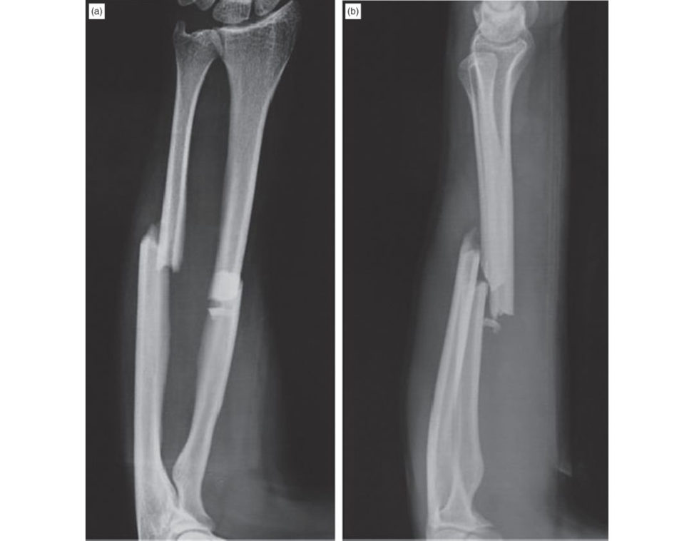
Figure 11.10a and 11.10b Anteroposterior and lateral radiographs, right radius and ulnar.
Evidence base
As stated in the BOAST 10 guidelines there are no RCTs on compartment syndrome and the guideline is based on, predominantly, retrospective studies.
Structured oral examination question 11#
A 49-year-old male is bought into the Emergency room having been blasted off his feet by an exploding air cylinder at work.
Minute 1
How would you initially assess this patient?
I would assess this patient utilizing the principles of A TLS. In this particular case I would be concerned about a deteriorating airway due to possible scorching from the explosion. There would also be concerns about thoracic or abdominal injuries due to rapid acceleration and deceleration, which could impact on breathing and circulation. The cervical spine should be immobilized, and ABC assessed using simple monitoring and examination in the first instance. If the patient is conscious I would like to obtain a history and finally assess the limbs for injuries. If the patient is not in extremis, then I would like to consider get inga trauma series CT and plain radiographs of any obviously injured limbs.
Minute 2
The patient is conscious but muddled. He is maintaining his airway and oxygen saturations. However, the ambulance crew were unable to cannulate him, he is tachycardic at 124 bp mand hypotensive at 90/56. He has a tender abdomen, an open distal tibial fracture with no evidence of major bleeding and a deformed forearm. Pelvic binder is in place. How would you proceed?
This patient is demonstrating signs of shock which is likely to be hypovolaemic. Assuming there are no other external sources of blood loss I would assume this is either intra-abdominal or pelvic in nature. Initial management would involve gaining access to start replacing lost volume. This can be IV or IO and bloods including FBC, U&E, cloting , group, save and cross-match should be sent to the labI would activate the major haemorrhage protocol and start giving fluid resuscitation with crystalloid solution un til blood arrives. I would also want to obtain an ABG to look at pH, base excess and lactate levels as these are good surrogate markers of tissue perfusion. Further to this I would like to give the pa tienta dose of tranexamic acid (1 g IV), a tetanus booster and a dose of IV antibiotics inline with trus t policy for open fractures. I would also check the pelvic binder is positioned correctly.
Minute 3
Do you still want to send this patient for a CT scan?
Yes. Assuming the department is compliant with the Royal College of Radiology guidelines then obtaining a trauma CT should be a quick process and still allow continuing fluid resuscitation. The contrast CT will help identify the source of blood loss as well as any other occult injuries.
Minutes 4–5
CT scan shows evidence of a ruptured spleen and some blood in the pelvis but no active extravasation here. The patient is transferred back to the emergency where departmental radiographs are obtained of the patient limb injuries. The general surgeons are intending to take the patient straight to theatre for a splenectomy. How would you proceed?
This patient has multiple serious injuries. His splenectomy is life-saving and should take priority. From an orthopaedic point of view he has a confirmed open tibial shaft fracture, a closed forearm fracture and a potential pelvic injury which is being stabilized by the binder. Further orthopaedic management will be determined by how the patient has responded to his resuscitation. I would use serial ABG measurements to determine whether I should proceed with early total care or move towards damage control orthopaedic surgery.
What do you mean by damage control orthopaedics?
Damage control orthopaedics is an approach to contain and stabilize an orthopaedic injury to improve apa tien t’s physiology. It is designed to avoid worsening apa tien t’s condition duet o the ‘second-hit’ phenomenon. Delay definitive surgery until a patient’s condition is optimized. It focuses on haemorrhagic control, management of soft -tissue injury and provisional fracture stability.
Anything else?
DCO involves rapid emergency surgery to save life or limb – NOT involving complex reconstructive surgery. Control of bleeding. Decompress cranium, pericardium, thorax, abdomen and limbs. Decontaminate wounds and ruptured viscera. Splint fractures. Cast, traction, pelvic binder , ex-fix. Get back to ITU environment ASAP. Definitive surgery is performed several days later.
Following his splenectomy the patient has a pH of 7.35, base excess of –3 mmol/l and lactate of 2.0 mmol/l.
These values indicate that the patient is currently adequately resuscitated and so I would look to proceed with early total care. I would plan to start with the forearm fracture with the arm extended on an arm table and to be honest with a fracture of this complexity, restoring alignment and performing carpal tunnel decompression with stability through an external fixator would be my plan A. For the tibial fracture, debriding this as a combined case with a consultant plastic surgical colleague to ensure appropriate soft -tissue c over and follow-up will be possible. I would deliver the bone ends through the wound to debride these before reducing the fracture. As a diaphyseal injury I would look to obtain restoration of length, alignment and rotation before stabilizing with a relative stability device. My personal preference here would be an intramedullary nail. If adequate soft -tissue cover was not available, then I would debride as above before using a spanning external fixator to stabilize until the patient was able to better tolerate graft surgery.
Having dealt with the forearm, a repeat AB Gis obtained, this shows a pH of 7.35, base excess of –4.1 mmol/l and lactate of 2.9 mmol/l. Does this change your management?
Yes. The patient while not in extremis, is struggling to cope physiologically, with an early trend for underperfusion showing. As such I would move to a damage control philosophy. This would entail a thorough debridement and then simple external fixation of the tibia. His tibial fracture can be formally stabilized in a planned fashion once his physiology has improved.
What is SIRS?
This is a condition characterized by systemic inflammation, or gan dysfunction and or gan failure.
How is SIRS diagnosed?
1. Heart rate > 90/min. 2. Breathing rate > 20/min, hyperventilation with decrease of arterial CO2 partial pressure (PaCO2) under 32 mmHg. 3. Temperature > 38.8°C or < 36.8°C. 4. Number of leukocytes < 4000/mm3 or > 12,000/mm3. For the definition of SIR S, two or more parameters must be fulfilled. Sepsis is defined as SIRS with detection of bacteraemia or bacterial focus. Do not forget to mention early appropriate care. Accept different patients respond differently to first and second hits. Consider severity of initial injury. Consider response to resuscitation. What further surgery is required? Continued reassessment and ability to change from ET Oto DCO.
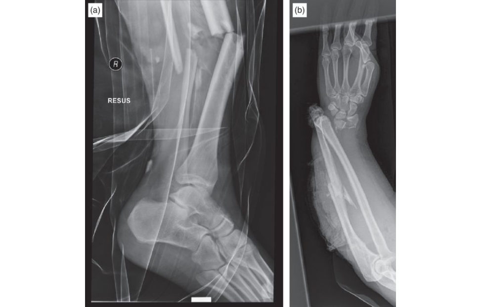
Figure 11.11a and 11.11b Lateral radiograph, comminuted fractured midshaft tibia and fibula, and anteroposterior (AP) radiograph, right radius and ulna.
Evidence base
NICE produced a guideline on major trauma in 2016. There islots of evidence around ETC/DCO and candidates should know some of it. Heather Vallie rhas published extensively, and the key values lifted from her papers are: pH > 7.25, BE ≥ 5.5 mmol/l, lactate > 4.0 mmol/l. It is also worth knowing about the
CRASH-2 trial and use of TXA; make sure you know how TX Aworks as well.
NICE Guideline No. 39 – Major Trauma: Assessment and Initial Management.
Vallie rHA, Moore TA, Como JJ, et al. Complications are reduced with a protocol to standardize timing of fixation based on response to resuscitation . J Orthop Surg Res. 2015;10:155.
Roberts I, Shakur H, Coats T, Hunt B, Balogun E. The CRASH-2 trial: a randomized controlled trial and economic evaluation of the effects of tranexamic acid on death, vascular occlusive events and transfusion requirement in bleeding trauma patients Health Technol Assess. 2013;17(10):1–79.
Structured oral examination question 12#
A 36-year-old lady sustained an open tibial fracture while horse riding. It was treated with wound debridement and closure as well as an intramedullary nail. This is her radiograph at 10 months postop.
Minute 1
How would you assess this patient?
This patient has radiographic evidence of an atrophic non-union. I would start by taking a history. Important factors to drawout include pain, weight-bearing status, postoperative wound issues, completion of antibiotic therapy , comorbidities including diabetes, medication steroids and smoking history. I would also examine the patient looking at the wounds and overall condition of the skin, neurovascular status and limb alignment. Having completed a clinical assessment, I would like to obtain baseline bloodtests including CRPES Rand white cell count to look for any signs of infection.
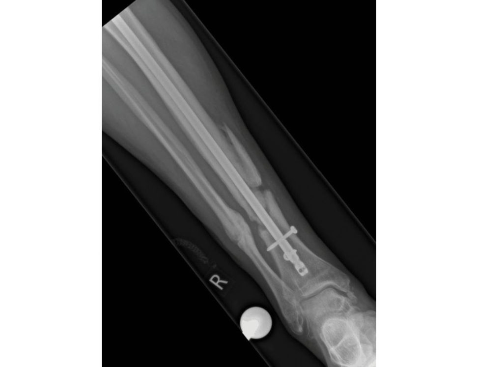
Figure 11.12 Anteroposterior (AP) radiograph, tibial non-union with IM nail in situ.
Minute 2
The patient is a fit and well, non-smoker who had an uneventiul postoperative recovery. She is struggling to weight-bear due to pain at the fracture site. What do you think is the cause of her non-union?
Causes of non-union can be broken down into surgeon factors and patient factors. In this case the patient fracture appears to have been appropriately fixed with good reduction and adequate working length to allow bone healing. There is very litile callus formation, which suggests biomechanical stability itself is not an issue here. There are no negative patient factors. I can only assume, therefore, that this is a problem of biology. This is likely to be due to infection or inadequate blood supply to the fracture site. There are implant factors as well. Distraction a t the fracture site at the time of initial nailing may contribute to the development of a non-union. This is a basic candidate 6 answer, nothing special. On a bad day with more hawkish examiners down to a 5. See Table 11.1 for a more detailed answer. Table 11.1 Causes for non-union.
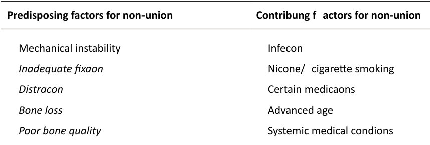
Poor functional le vel
Venous stasis
Burns
Irradiation
Obesity
Alcohol abuse
Metabolic bone disease
Malnutrition
Vitamin deficiencies
Predisposing factors for non-union Contributing factors for non-union
Inadequate vascularity
Severe injury
Excessive soft tissue stripping
Vascular injury
Poor bone contact
Soft tissue interposition
Malposition or malalignment
Bone loss
Distraction
Minutes 3–4
The patient bloodtests come back showing a CRP of 29 (< 4), ESR 40 (< 20) and a WCC of 13.4 (< 10.0). You perform a CT which shows no evidence of fracture healing, but there is a fragment of devascularized bone consistent with a buft erfly fragment. What management options would you give to the patient?
I would counsel the patient that I feel they have an infected non-union and then talk about the broad management strategies of conservative, non-operativ e or operative management. Conservative management is unlikely to lead to resolution of symptoms and will most likely lead to hardware failure, making any future surgery more challenging. Non-operativ e management would involve treatment with suppressive antibiotics to try and allow the fracture to heal despite the infection. This is unlikely to work, as the bone ends are atrophic and therefore the biology of the fracture is unlikely to be favourable for healing even if the infection was suppressed. My management option of choice would be revision surgery. This would involve removal of the current nail, debridement of the fracture site, removal of all dead bone and reaming of the intramedullary canal. I would want to ensure local elution of empirical antibiotics from an antibiotic loaded nail or by using resorbable beads. The choice would be between exchange nailing and ring external fixation Reamed tibial exchange nailing has been used successfully to treat infected tibial non-unions, and is thought to promote healing of a non-union by three different mechanisms.6 Increasing the nail diameter improves mechanical stability and this should stimulate healing. Also, reaming causes a marked increase in periosteal blood flow, which should stimulate the formation of periosteal new bone. Finally, the reaming products are osteoinductiv e. After primary nailing these products are extruded through the fracture site, but at exchange nailing fibrous tissue will t end to confine the reaming to the medullary canal.
Are you sure that you are talking about infected tibial non-unions?
Pardon?
I think you may be talking about exchange tibial nailing for aseptic non-union.
The candidate has mixed up the management of infected and aseptic tibial non-unions. A number of authors report that the treatment of choice for tibial diaphyseal fracture non-union is reamed exchange nailing. It is considered to be a relatively simple procedure which does not expose the fracture site and leads to minimal blood loss, low surgical morbidity and a short hospital stay. However, in the presence of infection it is a more controversial option. The reis currently no consensus in the literature with regard to the use of exchange nailing for tibial diaphyseal fracture non-union in the presence of infection. Simpson et al. from Edinburgh looked at a series of tibial non-unions to identify risk factors for failure of exchange nailing.7 They identified the strongest predictor of failure was infection and suggested that other treatment options such as Iliz arov treatment should be preferred.
I would go for an external ring fixator to stabilize the fracture. I would send reamings and bone samples for culture and sensitivity to allow targeting of suppressive antimicrobial therapy in conjunction with my microbiology colleagues.
What is the role of a circular frame in infected tibial non-unions?
Sorry?
What are the basic requirements for fracture healing?
The examiner has decided to take the viva backwards as he is not sure about the candidate’s level of knowledge. Mechanical stability. Adequate blood supply. Bone vascularity. Bone-to-bone contact. These factors may be negatively influenced by the severity of the injury and suboptimal surgical fixation which predisposes to non-union.
What factors predispose to instability at the fracture site?
1 Mechanical instability, excessive motion at the fracture site
Factors producing mechanical instability include:
Inadequate fixation (implants too small or too few).
Distraction of the fracture surfaces (hardware is as capable of holding bone apart as holding bone together).
Bone loss.
Poor bone quality (i.e. poor purchase).
If an adequate blood supply exists, excessive motion at the fracture site results in abundant callus formation, widening of the fracture line, failure of fibrocartilage to mineralize, and ultimately failure to unite.
2 Inadequate vascularity
Loss of blood supply to the fracture surfaces may arise because of the severity of the injury or because of surgical dissection.
Open fractures and high-energy closed injuries may strip soft tissues, damage the periosteal blood supply, and disrupt the nutrient vessels, impairing the endosteal blood supply.
Injury of certain vessels, such as the posterior tibial artery, may also increase the risk of non-union.
Vascularity may also be compromised by excess stripping of the periosteum as well as damage to bone and the soft tissues during open reduction and hardware insertion.
Whatever the cause, inadequate vascularity results in necrotic bone at the ends of the fracture fragments.
These necrotic surfaces inhibit fracture healing and often result in fracture non-union.
3 Poor bone contact
Poor bone-to-bone contact at the fracture site may result from soft -tissue interposition, malposition or malalignment of the fracture fragments, bone loss, and distraction of the fracture fragments.
Whatever the cause, poor bone-to-bone contact compromises mechanical stability and creates a defect.
The probability of fracture union decreases as defects increase in size. The threshold value for rapid bridging of cortical defects via direct osteonal healing, the so-called osteoblastic jumping distance, is approximately 1 mm.
Larger cortical defects may also heal, but at a much slower rate and bridge via woven bone.
The ‘critical defect’ represents the distance between fracture surfaces that will not be bridged by bone without intervention. The critical defect size depends on a variety of injury-related factors.
4 Infection
Infection in the z one of fracture increases the risk of non-union.
Infection may result in instability at the fracture site as implants loosen in infected bone. Avascular, necrotic bone a t the fracture site (sequestrum), common with infection discourages bony union.
Infection also produces poor bony contact as osteolysis at the fracture site results from ingrowth of infected granulation tissue.
Are there any other investigations you would like to obtain preoperatively?
Yes. I would like to obtain blood cultures to look for evidence of bacteraemia which may help guide antimicrobial therapy. I would also like to obtain an echocardiogram, ideally transoesophageal, to rule out endocarditis as a source for bacterial seeding.
Minute 5
What are the principles of management of an infected non-union?
I would need to consider host factors, the microorganism involved, debridement, antibiotic therapy and reconstruction. Soft -tissue coverage would need to be planned along with bone stabilization and the need for bone gratiing.
What is a biofilm?
A layer-like aggregation of cells and cellular products attached to a solid surface or substratum. An established biofilm structure comprises microbial cells and extracellular polymeric substance sEPS) produced by the microorganisms themselves.
Have you heard of the Masquelet technique?
No, sorry. This is a method to deal with a large amount of bone loss following radical debridement and excision of infected bone down to healthy bleeding bone. Antibiotic-impr egnated cement beads or spacers are used for local antibiotic administration to the soft -tissue bed. In the case of infection following IM nailing the canal should be reamed for debridement and irrigation. A circular external fixator is usually applied to the limb that allows for shortening of any defect if required. The soft - tissue envelope is repaired with vascularized flap transfer if needed. A second stage of bone gratiing is performed around 8 weeks after the initial surgery. An induction membrane should have formed which serves the critical function of preventing gr aft resorption. It act slik e a chamber around the bony defect to contain the bone graft ands timula te bone regeneration. This membrane should be disturbed as liti leas possible a t the time of the second stage.
Structured oral examination question 13#
A 33-year-old barrister slips on some ice and sustains an isolated closed ankle fracture. This is his radiograph in the Emergency Department.
Minute 1
Talk to me about this injury.
These are lateral and oblique radiographs of a left ankle showing a complex fracture– dislocation of the ankle such that the talus is almost completely displaced from the mortice and there is considerable rotation seen. The fibula fracture is suggestive of a Weber B injury. It is unclear if there is any syndesmotic widening on these films. Ideally , I would like to see a mortise view of the ankle. This is a serious injury. My first move would be to fully assess the patient quickly and reduce the dislocation to prevent ongoing swelling and compromise to the skin, nerves and blood vessels. The leg should be placed in a below-knee back-slab followed by check radiographs to confirm satisfactory reduction of the ankle mortice. The patient needs to be admift ed to the ward for elevation and a plan for operative intervention worked out.
OK. He is much more comfortable now and you obtain a good reduction. It is late in the evening and theatres are already busy with emergencies.
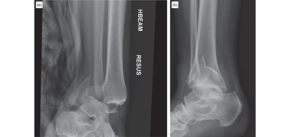
Figure 11.13a and 11.13b Oblique and lateral radiographs, left ankle showing a fracture/dislocation.
Minutes 2–3
Timing is important with these injuries due to soft -tissues welling. I know this may be debatable, but operating through a severely contused and swollen soft -tissue envelope is associated with poor outcomes and as such I would plan to temporize the patient with a spanning external fixator on the theatre list in the morning if soft tissue review is concerning. I would consent the patient for internal fixation and also for external fixation and I would discuss the fact that this is a significant injury with a risk of compromised function in the future.
What is debatable about operating in the presence of severely contused and swollen soft tissues with marked subcutaneous oedema? This practice would increase the risks of wound dehiscence, infection, need for plastic surgery involvement reoperations and adverse outcomes.
Some surgeons believe that soft -tissues welling should not be a contraindication to early operative fixation of ankle fractures. The key point is early fixation within 6 –12 hours of injury only. Late presentation of the patient, no availability of theatre time, the presence of general medical comorbidities and more severe concurrent injuries might point to external fixation being used as a temporary method for fracture stabilization Treatment with a temporary external fixator increases the length of hospital inpatient stay, possibly increases the complication rate and definitely increases the cost of treatment. The paper from Giannoudis from Leeds in the RCSEng bulletin suggested a fast-track method to get ankle fractures into theatre on the day of injury with the development of a streamlined pathway for early surgery.8 The key point is early presentation within a few hours of injury. It is very rare for an ankle to be too swollen to undertake operative fixation on the day of admission, although each patient’s treatment should be decided on an individual basis.9 With anything controversial be very clear about what you are actually saying. Stay safe and avoid ambiguity. Perhaps even better to stay clear of any controversy if you are just averaging a straighfoorward 6.
OK. You go to theatre and find the ankle too swollen to operate on with significant blistering already present and you achieve a good reduction with the external fixator. Please review the image and tellus what you would do now.
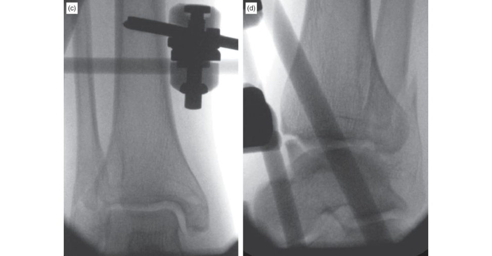
Figure 11.13c and 11.13d Anteroposterior (AP) and lateral radiographs of ankle following external fixator application.
Minute 4
The external fixator has been used in a triangular configuration and the reduction is satisfactory. The tension and pressure are now off the soft -tissue envelope, there is improved articular alignment decreased articular impaction and I can continue the important phase of soft - tissue r est and resuscitation. I will advise the patient that definitive surgery may occur in about 10– 14 days. I would ensure the patient has had aDVT assessment and that the limbis properly elevated. With all complex ankle fractures, when planning reconstruction, 3D cr oss-sectional imaging is useful and so I would order a CT scan. On the available radiographs it is not possible to say if the external fixator has been used in a triangular configuration Try not to make assumptions as some times inferences can be wrong.
What are the concerns with temporary external fixator use?
Pin site infections, superficial wound infections and postoperative loss of reduction. In addition, I would want to inspect the skin daily for blistering and as to when the soft tissues are ready for definitive fixation. Patients should be monitored for compartment syndrome and neuropraxia. The affected limb should be elevated.
What elements in particular are you interested in evaluating here?
Stability in the management of ankle fractures is key. Ligament tension and fracture configuration contribute. The posterior malleolus is important, and I would in particular be interested in the size of the posterior bony chunk, its fracture configuration and its direction. These were classified by Haraguchi et al. (2006) as Types I–III and I find this useful to characterize and treat these fractures.
Here is the axial CT slice – what do you think?
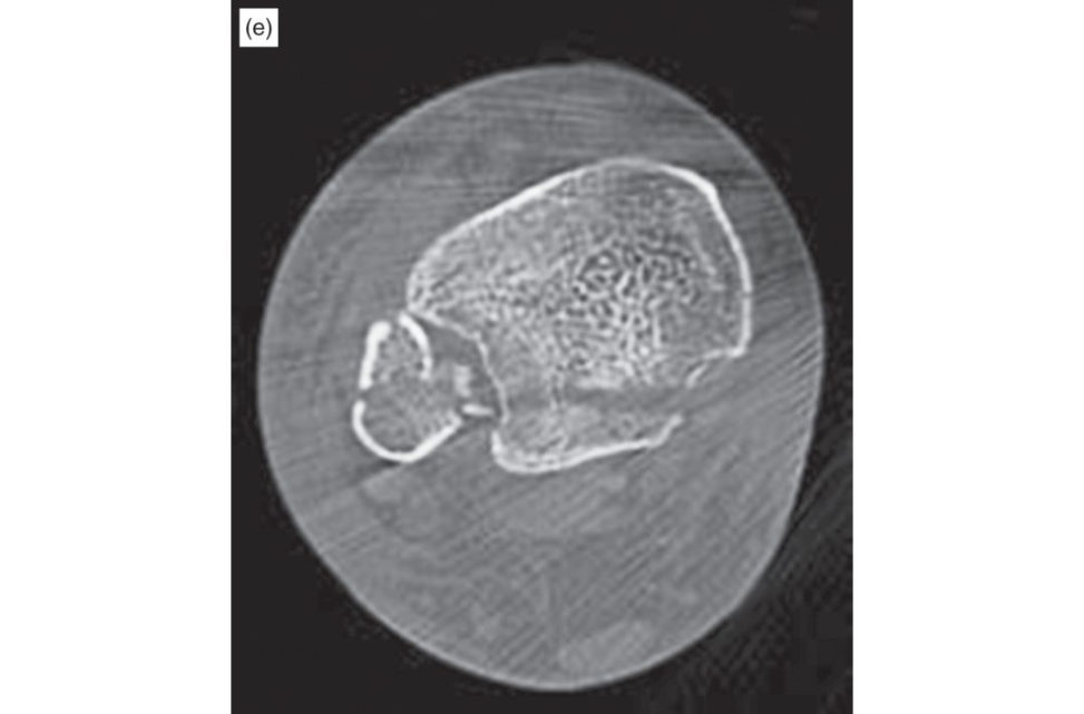
Figure 11.13e Axial CT slices, ankle.
Minute 5
This is a Haraguchi Type I as it is ‘wedge-shaped’ and essentially is a chunk off the posterolateral distal tibia ( Table 11.2). The posterior malleolus plays an integral role in ankle joint stability through its anatomical relationship with the posterior tibiofibular ligament (PTFL), which has been shown through cadaver studies to account for 42% of syndesmotic stability. Ankle fractures involving the posterior malleolus are said to have worse clinical outcomes, possibly related to incongruity and the resultant development of post-traumatic arthrosis. CT is also useful in assessing for syndesmotic injury indicated by widening of the syndesmosis anteriorly or posteriorly on axial scans. A CT would also give information on whether any articular impaction or depression was present.
How would you fix this fracture?
I would fix the lateral malleolar fracture with a lag screw and 3.5 mm one-third tubular plate acting in a neutralizing mode. He is young and has good-quality bone; otherwise, if the patient was elderly and the bone porotic and poor quality I would consider using a locking plate. The posterior fragment can be fixed with lag screws inserted from anterior to posterior.
Anything else to fix the posterior malleolar fragment?
A plate could be used. Contoured one-third tubular plate acting as in bu tiress mode. The plate is not contoured, it will contour itself with placement of the initial screw (just proximal to the posterior fracture line), which then helps with fracture reduction.
How would you put the plate on?
Sorry?
What approach would you use?
Standard lateral approach to the fibula.
Any other approaches you are familiar with?
A posterolateral approach.
Tell me about the posterolateral approach.
Ehh?! [Bell]
Thankyou.
Prone position, skin incision midway between the lateral border of the TA and the fibula. Deep dissection between FHL medially and the peroneal tendons laterally. The sural nerve is at risk and needs to be identified and protected.
The posterior surface of the fibula can be reached by retraction of the peroneal tendons laterally, and the plate should be applied posteriorly. The posterior surface of the tibia can be reached by retraction of the FHL and the deep posterior compartment medially.
Table 11.2 Haraguchi classification of posterior malleolus fracture of ankle
Type I fracture is an isolated PMF
Type II fracture is either a bi- or trimalleolar fracture associated with a Weber B orC fracture pattern. These are further classified depending on the presence or absence of syndesmotic injury
Type III PMF is associated with an ipsilateral tibial diaphyseal fracture. This is further subdivided based on sagift al or coronal plane instability
Evidence base#
Solan MC, Sakellariou A. Posterior malleolus fractures. Bone Joint J. Published Online: 1 Nov 2017.
htip s://doi.org/10.1302/0301-620X.99B11.BJJ-2017–1072
Notes
1. Wasserstein D, Henry P, Paterson JM, Kreder HJ, Jenkinson R. Risk of total knee arthroplasty after operatively treated tibial plateau fracture: a matched-population-based cohort study. J Bone Joint Surg. 2014;96(2):144–150.
2. Griffin D, Parsons N, Shaw E, et al. Operative versus non-operativ e treatment for closed, displaced, intra-articular fractures of the calcaneus: randomised controlled trial. BMJ. 2014;349:g4483.
3. Buckley R, ToughS, McCormack R, Pate G. Operative compared with nonoperative treatment of displaced intra-articular calcaneal fractures: a prospective randomized, controlled multic enter trial. J Bone Joint Surg. 2002;84(10):1733.
4. Baumgaertel F, Buhl M, Rahn BA. Fracture healing in biological plate osteosynthesis. Injury. 1998;29:3–6
5. Sangeorzan BJ, Benirschke SK, Mos caV, Mayo KA, Hansen ST Jr. Displaced intra-articular fractures of the tarsal navicular. J Bone Joint Surg Am. 1989;71:1504–1510.
6. Banaszkiewicz PA, Sabboubeh A, McLeod I, Maffulli N. Femoral exchange nailing for aseptic nonunion: not the end to all problems. Injury. 2003;34(5):349–356.
7. Ts angST, Mills LA, Frantzias J, Baren JP, Keating JF, Simpson AH. Exchange nailing for nonunion of diaphyseal fractures of the tibia: our results and an analysis of the risk factors for failure. Bone Joint J. 2016;98(4):534–541.
8. Kheir E, Charopoulos I, Dimitriou R, Ghoz A, Dahabreh Z, Giannoudis PV. The health economics of ankle fracture fixation . Bull Roy Coll Surg Engl. 2012;94(4):1–5.
9. Pietzik P, Qureshi I, Langdon J, Mol loyS, Solan M. Cost benefit with early operative fixation of unstable ankle fractures. Ann Roy Coll Surg Engl. 2006;88(4):405–407.