Chapter 27 Genetics and cell biology
Introduction#
The average orthopaedic trainee about to sit the FRCS (Tr & Orth) exam requires a basic knowledge of genetics.
This doesn’t need to be encyclopaedic, but candidates will need to have a sound grasp of disease inheritance and genetic disorders. Trainees should be able to draw a family pedigree of single gene inheritance and know the gene mutations of the more common orthopaedic conditions.
By comparison, mention genetic viva questions to any examiner and you get a slightly puzzled look back. Safe to say, it is not a major A-list topic for the vivas and probably doesn’t even make the B-list.
However, the subject does intermift ently appear in the vivas and therefore it is definitely worthwhile knowing how the questions will run in or der to uncomplicate a potentially complicated topic.
Some genetic material will find its way into Section 1 SBA an dEMI papers so common genetic terminology and definitions need to be reasonably well understood.
General information#
Nucleic acid structure (Figure 27.1)
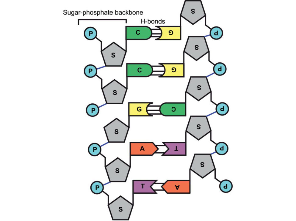
Figure 27.1 Nucleic acid structure.
There are two main types of nucleic acid, DNA (deoxyribonucleic acid) and RNA (ribonucleic acid), which each consist of a sugar-phosphate backbone with projecting nitrogenous bases. The nitrogenous bases are of two types, purines and pyrimidines.
In DNA, there are two purine bases, adenine (A) and guanine (G), and two pyrimidine bases, thymine
(T) and cytosine (C) (Figure 27.2).
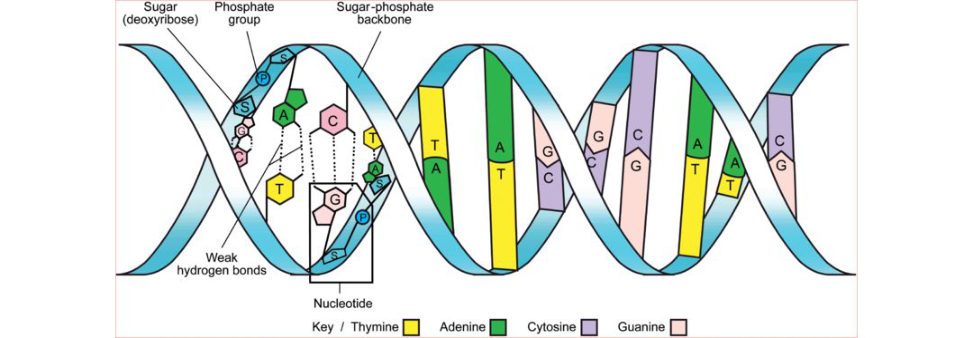
Figure 27.2 DNA structure.
RNA also contains adenine (A), guanine (G) and cytosine (C), but contains uracil (U) instead of thymine (T).
In DNA the sugar is deoxyribose, whereas in RN Aitis ribose. The nitrogenous bases are attached to the 1ʹ (one prime) position of each sugar and the phosphate links 3ʹ and 5ʹ hydroxyl groups.
Each unit of purine or pyrimidine base together with the attached sugar and phosphate group(s) is called a nucleotide.
A molecule of DN Ais composed of two nucleotide chains that are coiled clockwise around oneanother to form a double helix with approximately 10 nucleotides per complete turn of DNA (Figure 27.2).
The two chains run in opposite directions (i.e. 5ʹ to 3ʹ for one and 3ʹ to 5ʹ for the other) and are held together by hydrogen bonds between A in one chain and T in the other or between C and G.
Pedigree charts
Pedigree charts can be complicated to understand if you are unfamiliar with the basics.
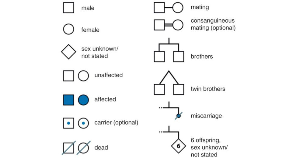
A standard set of symbols are used (Figure 27.14). The father is conventionally placed on the left, and all members of the same generation are placed on the same horizontal level. Roman numerals are used for each generation, starting with the earliest, and Arabic numerals are used to indicate each individual within a generation (numbering from the left).
Single gene disorders are due to mutations in one or both copies or alleles of an autosomal gene or to mutation sin genes on the X orY chromosome (sex-linked inheritance). These disorders show characteristic patterns of inheritance in family pedigrees.
Structured oral examination question 1#
Autosomal dominant (AD) inheritance
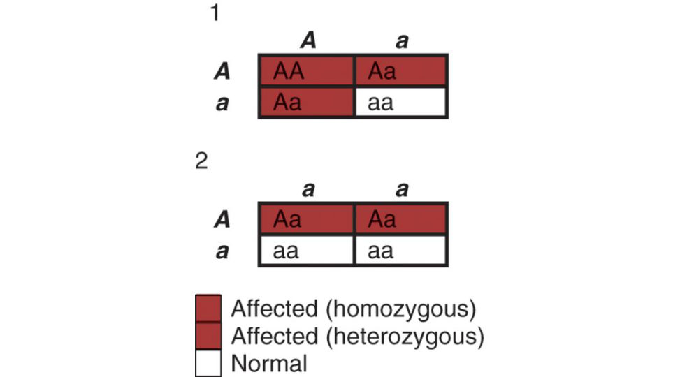
This is much easier to explain if candidates are able to drawout a Punneft square Just get the pen out and practise ( Figure 27.3).
What pattern of inheritance does the pedigree chart show and why (Figure 27.4)?
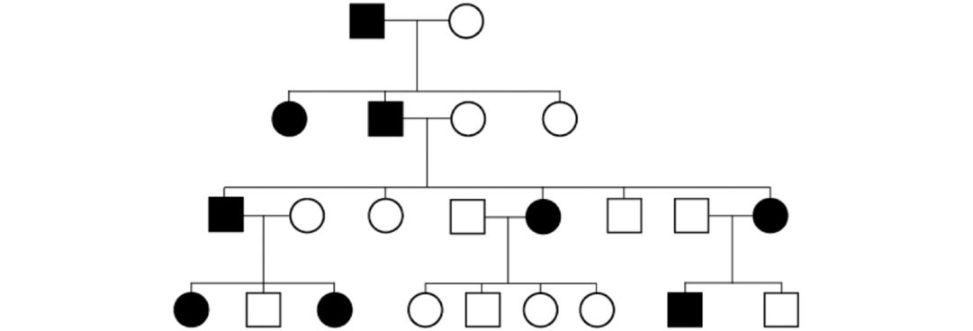
This is autosomal dominant because: There are people with the disease in each generation. Both males and females are affected in approximately equal numbers. All forms of transmission are present (male to female, female to male, female to female and, in particular , male to male, which would not be present if the condition were X- linked). At conception, each child has a 1 in 2 (50%) chance of inheriting the condition. Unaffected persons do not transmit the condition if the condition is fully pene trant (e.g. in achondroplasia). It is equally likely that a child will receive the mutant or normal allele from the affected parent. On average there is a 1 in 2 or 50% chance that each child of a heterozygous parent will inherit the gene mutation. There is usually a variation in time of onset and severity of condition with AD traits. This is likely to be due in part to the effects of other ‘modifier’ genes and also lifestyle/environmental factors.
Choose an autosomal dominant condition and discuss the genetics.
A gift if you have done your homework. The three most obvious disorders to choose are (1) achondroplasia, (2) osteogenesis imperfect aor (3) neurofibromatosis. Plenty to talk about with each condition. It is unwise to choose an obscure disorder that the examiner knows very litile about. The conversation will dry up and the examiners will invariably switchback to more difficult basic science questions. 1 Candidates are throwing away easy scoring opportunities. The other situation is that the clinical condition to discuss has already been decided beforehand.
Figure 27.3 Punneft square demonstrating inheritance of an autosomal dominant trait: (1) two heterozygous parents, (2) one heterozygous and one unaffected parent.
Figure 27.4 Pedigree chart of AD inheritance, one affected heterozygous and one unaffected parent. The disease is passed from father to son – this almost never happens with X-linked traits. The disease occurs in three consecutive generations – this almost never happens with recessive traits.
Achondroplasia
About 80% of people with achondroplasia have the condition as the result of a new mutation; the incidence of the condition increases with increasing paternal age.
Achondroplasia is inherited as an autosomal dominant condition. Each child of someone who has achondroplasia has a 1 in 2 (50%) chance of inheriting the condition.
If both parents have achondroplasia, and a child inherits a copy of the altered gene from both parents, the condition is severe and not compatible with life.
The gene for achondroplasia is on the short arm of human chromosome 4 at locus p16.3.
The condition is the result of a mutation in the gene that codes for fibroblast growth factor receptor
3 (FGFR3), which is a key component of cartilage development.
The main defect isabnormal endochondral bone formation in the cartilaginous proliferative zone of the physis.
Clinical features of achondroplasia may be discussed in more detail and these can easily be mugged up in a standard textbook:2 kyphosis, spinal stenosis, genu varum, trident hand, radial head subluxation, etc.
What is the genetic mutation responsible for this condition, and what effect does this have?
Achondroplasia is caused by a single point mutation in the gene encoding the fibroblast growth factor receptor 3 (FGFR3). The FGFR3 is believed to regulate bone growth by limiting endochondral ossification. T wo mutations in the FGFR3 gene are responsible for 99% of cases of achondroplasia. These mutations both cause substitution of a glycine amino acid by an arginine and lead to prolonged receptor activation after ligand binding and result in excessive growth limitation. Soluble FGFR3 has successfully been used in mice to act as a decoy for FG Fin order to restore normal growth in achondroplasia.
What is the mode of inheritance of this condition?
It is autosomal dominant.
This child’s parents do not have this condition. How is this possible?
A large proportion (80%) of cases occur due to a spontaneous (new) mutation.
Osteogenesis imperfecta
There is a qualitative or quantitative defect of type 1 collagen synthesis. Originally classified by Silence3 into four subgroups, we now know there are many more different subgroups of OI.
The vast majority of cases are autosomal dominant.
Type 1 collagen is the major extracellular protein in bone, skin and tendon.
The structural unit of type 1 collagen is called tropocollagen and is a heterotrimer composed of three polypeptide chains. Two chains are pro-α1(I) and the third chain is pro-α2(I). The three chains form a distinctive unit in which the polypeptide chains wraparound each other for most of their length, forming a tight triple helical braid.
The triple helical structure is not the same as the pro-α helix that is formed by a single polypeptide chain, which is the defining feature of all collagen.
The collagen triple helix forms because both the α1 and α2 chains contain repeat sequences of amino acids (–Gly–X–Y), where Gl yis glycine, X is proline andY is usually hydroxyproline. This arrangement results in a constrained structure that imparts a tight kink in the polypeptide chain with the glycine chain preventing steric hindrance that would otherwise impair wrapping of the helical band.
Only Gly, which has no side chain, can pack into the centre of the triple-helix structure without distortion. A missense mutation 4 leading to the replacement of even one Gl yin the repeating (Gly–X–Y)n sequence by a larger residue may lead to a pathological condition.
The pro-α1(I) chain is encoded from the COL1A1 gene on chromosome 17 and the pro-α2(I) residue is encoded by the COL1A2 gene on chromosome 7.
Mutations in the COL1A1 gene on chromosome 17 or COL1A2 gene on chromosome 7 are responsible fo rOI.
In the type 1 condition the COL1A1 mutant allele results in failure of the production of the pro-α1(I)
chain due to a premature codon stop. There is only 50% production of normal pr o-α1(I) chain.
In more severe forms of the condition there are mutations of either the COL1A1 gene or the COL1A2 gene resulting in a mixture of abnormal and normal collagen chains.
Structured oral examination question 2#
Autosomal recessive (AR) inheritance
Can you please draw a Punneft square demonstrating an autosomal recessive trait (Figure 27.5).
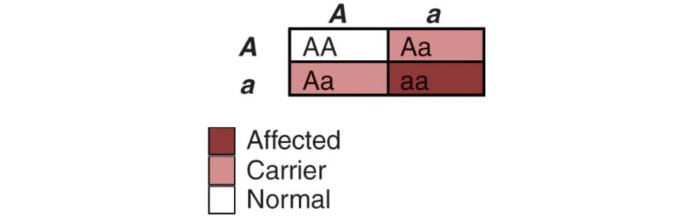
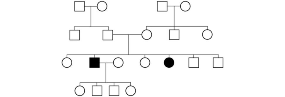
Recessive means two copies of the gene are necessary to have the trait/disease. Parents of affected children are both carriers of the condition. Typically, heterozygotes for the trait/disease are not affected but are gene carriers. Most people do not know they carry a recessive gene for a condition un til the y have a child with the disease. If the parents are both carriers the risk of them having an affected child is 25% (1/4) and the risk of them having a child who is a carrier is 50%. Unaffected adult offspring of carrier parents have a 2/3 risk of carrier status. Alternatively candidates may be shown a pedigree chart of autosomal recessive inheritance and asked to identify the pattern of inheritance (Figure 27.6). 1. Males and females are equally affected. 2. On average, the recurrence risk to the unborn sibling of an affected individual is 1/4. 3. The trait is characteristically found in siblings, not parents of affected or the offspring of affected. 4. Parents of affected children may be related (consanguineous). The rarer the trait in the general population, the more likely a consanguineous relationship is involved. 5. The trait may appear as an isolated (sporadic) event in small sibships. 6. Within the completely unaffected siblings of an affected individual the probability of being a carrier is 2/3.
Can you describe an autosomal recessive disease?
Obvious choices include (1) sickle cell anaemia, (2) mucopolysaccharidoses – all except Type II (Hunter’s syndrome, which is X-linked recessive), (3) Gaucher disease and (4) diastrophic dysplasia.
Figure 27.5 Punneft square demonstrating autosomal recessive inheritance.
Figure 27.6 Pedigree chart of autosomal recessive inheritance.
Sickle cell disease
Sickle cell disease describes a group of disorders caused by a mutation in the beta globin gene (HBB). The altered haemoglobin produced is HbS. SC Ais the commonest of these diseases and is caused by homozygous point mutations in the HBB gene on the short arm of chromosome 11. A single nucleotide mutation (base change) from T to A results in glutamic acid changing to valine at the sixth amino acid in the beta haemoglobin chain. This allows polymerization of the HbS but only underconditions of low oxygen concentration.
Heterozygous carriers of HBB mutations (‘sickle cell trait’) have a selective advantage (heterozygote advantage), due to their resistance to malaria. The malarial parasite causes the defective red blood cells to rupture directly, prior to its reproduction. Hence heterozygotes have increased chances of survival in malaria-prevalent areas. Carriers will only have severe symptoms if they are ever deprived of oxygen (e.g.
at high altitude) or dehydrate, an important factor inconsideration for surgery and when considering using a tourniquet.
Clinical features
SC Ais characterized by episodes of pain owing to vaso-occlusive events, chronic haemolytic anaemia and severe infections from early childhood with splenomegaly.
Any organ may be affected but most commonly bones (ON/osteomyelitis), lungs, liver, kidneys, brain and eyes are involved.
Sickle cell crisis presents with severe abdominal pain, chest or bone pain. Management includes analgesia, fluids, oxygen and in severe cases exchange transfusion.
Structured oral examination question 3#
X-linked inheritance
Can you draw the Punneft square for an X-linked dominant condition ( Figure 27.7 and 27.8)?
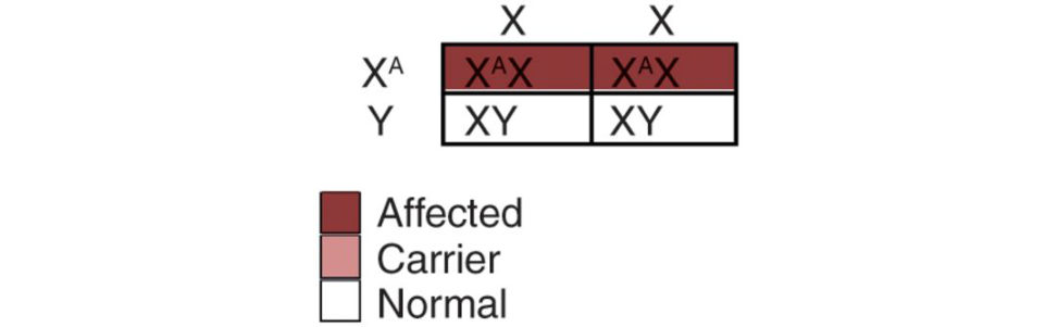
[While drawing the Punneft square.] If the father is affected his sons will be unaffected and all his daughters will be affected (Figure 27.7). If the mother is affected (heterozygous) half of her sons will be affected and half of her daughters will be affected. Other sons and daughters will be unaffected (Figure 27.8).
What is the mode of inheritance here (Figure 27.9)?
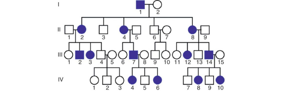
Figure 27.7 Punneft square showing inheritance of an X-linked dominant trait (normal mother and affected father).
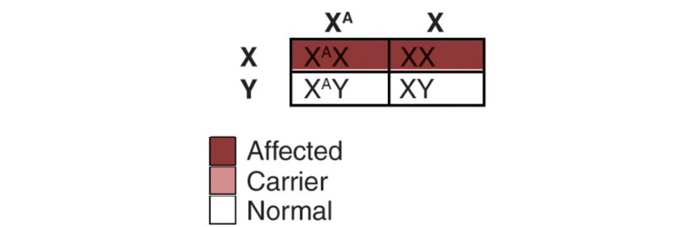
Figure 27.8 Punneft square showing inheritance of an X-linked dominant trait (affected mother and normal father).
Figure 27.9 X-linked dominant inheritance. The key for determining if a dominant trait is X-linked or autosomal is to look at the offspring of the mating of an affected male and a normal female. If the affected male has an affected son, then the disease is not X-linked. All of his daughters must be affected if the disease is X-linked dominant.
This is X-linked dominant inheritance. Hallmarks of X-linked dominant inheritance include: The trait is never passed from father to son. Does not skip a generation. All daughters of an affected male and a normal female are affected. All sons of an affected male and a normal female are normal. Matings of affected females and normal males result in 1/2 of the sons being affected and 1/2 of the daughters being affected. Males may be more severely affected than females. The trait may even be lethal in males during the embryonic or perinatal periods. In the general population females are more likely to be affected than males, even if the disease is not lethal in males.
What is the mode of inheritance here (Figure 27.10)?
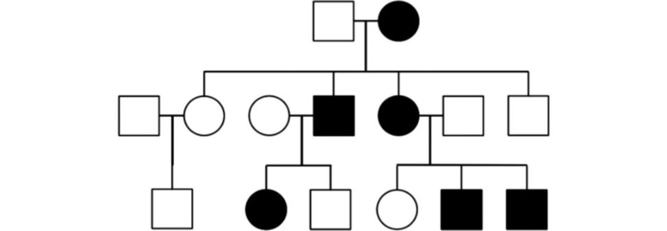
This is the opposite situation to Figure 27.9 where the male parentis affected. This is still X -linked dominant inheritance. An X-linked dominant trait does not skip generations. Affected sons usually have an affected mother, affected daughters usually have either an affected mother or an affected father. Affected fathers will pass the trait on to all their daughters. Affected mothers (if heterozygous) will pass the trait on to half of their sons and half of their daughters.
Do you know of an example of an X-linked dominant inheritance condition?
Hypophosphataemic rickets (vitamin D-resistant rickets).5 The condition is caused by mutations in the PHEX geneP HEX protein regulates fibroblast growth factor 23 protein (FGF23). FGF23 inhibits the renal tubular ability to reabsorb phosphate.
In addition, the absence of PHEX enzymatic activity may cause accumulation ofos teopontin (a mineralization-inhibiting secreted substrate protein found in the extracellular matrix of bone), which contributes to osteomalacia.
Figure 27.10 X-linked dominant inheritance. Parent female is affected. Affected father does not pass disease on to son.
Clinical features
Childhood rickets with growth retardation and poor dental development. Short stature and genu varu min males, genu valgu min females.
In middle age, mineralization of spinal ligaments and thickening of neural arches. Loss of mobility of the spine, shoulders, elbows and hips. Treatment with phosphate supplements and large doses of vitamin
D.
The condition is caused by mutations in the PHEX gene. The change created in the gene is a loss-of-function mutation, resulting in reduced breakdown and circulatory clearance of FGF23. FGF23 acts on the kidney to cause increased phosphate excretion and decreased alpha-1 hydroxylase activity . The gene product is now known to be a zinc-metallopeptidase. Other X-linked dominant conditions: Conradi–Hunermann chondrodysplasia punctata (due to a mutation in the gene encoding EBP). Leri–Weill dyschondrosteosis is very unusual. Because of the gene’s specific location, it is inherited in a manner that is described as pseudo-autosomal (i.e. similar to autosomal dominant inheritance).
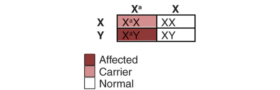
Can you draw the Punneft square for an X-linked recessive condition?
Figure 27.11 and 27.12.
What is the mode of inheritance here (Figure 27.13)?
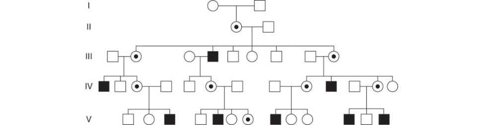
This is X-linked recessive. Hallmarks of X-linked recessive inheritance include: As with any X-linked trait, the disease is never passed from father to son. Males are much more likely to be affected than females. All affected males in a family are related through their mothers. The traitor disease may be passed from an affected grandfather, through his carrier daughters, to half of their sons. For a carrier female, with each pregnancy there is a one in two (50%) chance her sons will inherit the disease allele and a one in two (50%) chance her daughters will be carriers. Affected males transmit the disease allele to all of their daughters who are then carriers, but to none of their sons. Females are affected if they have two copies of the disease allele. All of their sons will be affected, and all of their daughters will be unaffected carriers.
Figure 27.11 Punneft square showing inheritance of an X-linked dominant trait (affected mother and normal father).
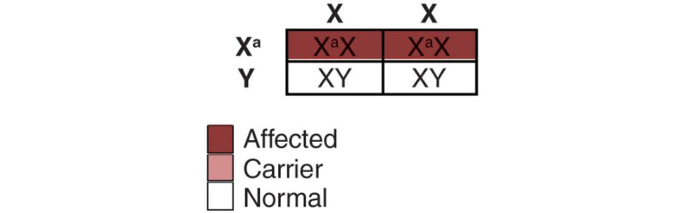
Figure 27.12 Punneft square showing inheritance of an X-linked dominant trait (normal mother and affected father).
Figure 27.13 X-linked recessive inheritance.
Duchenne muscular dystrophy (DMD)
Incidence approximately 1 in 4000 boys.
This is one of the dystrophinopathies caused by a mutation in the dystrophin gene (Xp21). The dystrophin protein provides structural stability to the dystroglycan complex of the muscle cell membrane, and its function is lost as a result of the mutation. Females rarely show musculoskeletal signs of the disease, although there is an increased risk of dilated cardiomyopathy in female carriers. There is a relatively high spontaneous new mutation rate.
Clinical features
Age of onset is usually before 6 years. Progressive proximal myopathy of the lower limbs with noticeable calf pseudohypertrophy.
Compensatory toe walking is an adaptation to knee extensor weakness.
Frequent falls/fatigue.
Speech delay and difficulty with motor skills with learning difficulties in approximately a third of affected boys.
Lumbar lordosis/scoliosis.
Usually wheelchair-bound by 12 years and life expectancy is around 25 years.
Gower’s sign positive the child is unable to jump up quickly from a crossed leg position without bracing their arms against their legs to support the proximally weak muscles.
Other examples of X-linked recessive inheritance:
Becker muscular dystrophy.
Mucopolysaccharidosis Type II (Hunter’s syndrome).
Haemophilia A. Genetic defect in factor VIII.
SED tarda.
Pick one family pedigree and tell me the mode of inheritance (Figure 27.15).
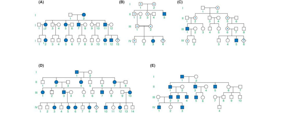
Be able to justify your answer in terms of pedigree features (see above examples).
Figure 27.14 Symbols for constructing a family tree.
Figure 27.15 Family tree for various modes of inheritance. A, autosomal dominant; B, autosomal recessive; C, X-linked recessive; D, X-linked dominant; E, Y-linked inheritance.
Structured oral examination question 4#
Stem cells
Potentially an awkward C-list topic, especially if a candidate hasn’t read through any structured material beforehand.
What are stem cells?
Stem cells represent unspecialized cells that have the ability to differentiate into diverse specialized cell types and self-renew to produce more stem cells.
What two properties must a stem cell demonstrate?6
The two defining features of a stem cell are: Self-renewal: the ability to go through numerous cycles of cell division while remaining undifferentiated. If stem cells could not self-renew, tissues would runout of replacement cells for those that had died. Potency: the capacity to differentiate into specialized cell types. This requires stem cells to be either totipotent or pluripotent – to be able to give rise to any mature cell type.
What are the different types of stem cells that you know?
The two main types of stem cells are: 1. Embryonic stem cells. 2. Adult stem cells. Other types of stem cell include: Foetal stem cells Amniotics tem cells. Induced pluripotent stem cells (IPSCs). Nuclear transplant stem cells (ovasomes). Parthenote stem cells.
What do you mean by pluripotent?
Pluripotent cells have the capacity to differentiate into any cell type in the body. They give rise to most, but not all, of the tissues necessary for foetal development. Totipotent cells have the capacity to form an entire organism as well as the extraembryonic tissue including the placenta Embryonic cells within the first couple of cell divisions after fertilization are the only cells that are totipotent. Multipotent cells can develop into more than one cell type but are more limited incapacity than pluripotent cells. They can form many types of cell in a given lineage, but not cells of other lineages.
Can you think of any uses of stem cells in orthopaedics?
Stem cells have become a focus of regenerative medicine. Adult stem cells, harvested directly from bone marrow, adipose tissue, muscle or blood have the ability to undergo mitosis as well as multipotent differentiation in to a variety of cell lineages. The goal of stem cell therapy is to replace or replenish diseased tissue through the localized differentiation of transplanted stem cells into cells which advance the healing processor directly restore the tissue physically. 1. Articular cartilage damage/degeneration It is hoped that stem cells will create growth of primary hyaline cartilage to restore the normal joint surface. Stem cells assist with growth factor release and alteration of the anatomic microenvironment to facilitate regeneration and repair of the chondral surface. Stem cell application can be combined with microfracture. Due to their role in inhibiting the catabolic activity of matrix metalloproteinases (MMP), mesenchymal stem cells (MSCs) have been shown to have a beneficial effect in OA. 2. Bone fractures and nonunions Stem cells may stimulate bone growth and promote healing of fractures. Traditionally , bone defects have been treated with solid bone graft material placed at the site of the fracture or nonunion. Stem cells and progenitor cells are now placed along with the bone graft to stimulate and speed healing. 3. Ligaments and tendon injury/degeneration Mesenchymal stem cells may also develop into cells that are specific for connective tissue. This would allow faster healing of ligament and tendon injuries, such as quadriceps or Achilles tendon ruptures. 4. Rotator cuff tears Mixed results reported. Some studies have shown increased rates of healing and repair surface integrity. 5. Spinal cord injury7 There has been recent research into cell-based therapies for spinal cord injury. They may be able to limit cell death, stimulate axonal growth, and replace injured cells. 6. Meniscal injury Isolated case reports exist of meniscal regeneration after percutaneous injection of autologous ASCs into an adult human knee.8 It is not clear whether this is a direct action of the mesenchymal-based cells or is rather mediated by secretion of certain stimulating factors on the existing meniscal tissue.
Stem cells have also been added to modify the biomechanical environment of avascular zone meniscal tears at the time of suture repair.
7. Intervertebral disc disease
Various clinical trials have been undertaken using MSCs to biologically repair degenerative disc.
Percutaneous stem cell mediated disc regeneration has the potential to establish itself as a possible treatment option in patients with low back pain hoping to avoid invasive spinal surgery.
8. Spinal fusion
Pseudoarthrosis remains a pressing issue occurring in 13–41.4% of patients undergoing spinal fusion. Risk factors include older age, thoracolumbar kyphosis, smoking, diabetes mellitus, metabolic bone disease and female gender.
MSCs and adipose tissue derived stem cells (ADSCs) have both demonstrated a significant positive effect on spinal fusion in a number of experimental models. Cellular in-vitro expansion is necessary to increase the number of viable pluripotent cells along with the addition of growth factors and/or bone morphogenic proteins (BMPs).
9. Physeal injury/defects
Several animal models have investigated the use of stem cells combined with growth factors to promote the regeneration of damaged regions of the growth plate.
10. Osteonecrosis
Stem cells have angiogenic and osteogenic properties. Core decompression and injection of isolated stem cells have been used in early stages of ON hip.
What are your concerns with the use of stem cells in orthopaedics?
MSCs have been reported to promote tumour growth and metastases. There is very limited clinical experience with pluripotent stem cells (embryonal stem cells and IPSC). Based on their features of self-renewal and high proliferation rate, the risks of tumour formation should be considered high. Ethical issues and controversies regarding the use of embryonic stem cells and embryos exist. Donor site morbidity from stem cell harvesting. Processing time requires separate procedures to harvest and re-implant cells). Retroviruses may be used to generate human IPSCs. These viruses are genetically altered to express the genes that are required for transformation in to an IPSC. Applying this genetic reprogramming, the used viruses can integrate into the cell genome.
Consequently, the cells may contain multiple viral integration sites in their genomes, which could be tumourigenic.
Cost.
Microbial contamination during cell amplification.
Controlling stem cell differentiation in to the desired cell lineage.
Control of their proliferation and differentiation in to complex, viable 3D tissues is challenging.
Lack of adequate vehicles/scaffolds for implantation of s tem cells.
Integration with local tissues.
Immunological rejection and disease transmission (if allogeneic).
Potential modulation of host immune system by implanted stem cells.
Continuous cell amplification of MSCs may lead to chromosomal abnormalities.
Notes
1. This will usually end upmaking the viva more unnecessarily complicated than would have been the case.
2. Ward JG argan A, Smiths onS, Atherton G. Orthopaedic manifestations of achondroplasia. Orthop Trauma. 2013;27(4):229–232.
3. Sillence D. Osteogenesis imperfecta: an expanding panorama of variants. Clin Orthop Rel Res. 1981;159:11–25.
4. A missense mutation is a point mutation in which a single nucleotide change results in a codon that codes for a different amino acid.
5. Best to stick to hypophosphatemic rickets as the rest are just a bit too obscure for your average orthopaedic surgeon.
6. Much better if a candidate volunteers this info rather than being asked.
7. Schroeder GD, Kepler CK, Vaccaro AR. The use of cell transplantation in spinal c ord injuries. J Am Acad Orthop Surg. 2016;24(4):266–275.
8. Pak J, Lee JH, Lee SH. Regenerative repair of damaged meniscus with autologous adipose tissuederived stem cells. BioMe dRes Int. 2014;2014:436029.