Chapter 23 Pain, analgesia and anaesthesia
Introduction#
Pain may be asked as part of a viva topic or candidates may be lucky or unlucky enough to have a full 5-minute viva devoted to the topic. It is an important part of orthopaedic practice which is often neglected, but more important now with the push towards day-case surgery and even day-case arthroplasty. Pain is wellknown to appear as questions in the P art I MCQ/SBA exam.
This viva can be awkward, as bits and pieces such as pain assessment can appear fluffy to orthopods and in real life is best left to the anaesthetists.
Like genetics, ask examiners about pain and one will get a puzzled look back. Suffice to say, it is not an A-list topic and is most likely a C-list category. Again, similar to genetics, the idea is to score a basic 6 for any question asked. Anything else is a bonus.
One word of caution with C list category topics. With the advent of computer generated curriculum sampling C-list topics are quickly becoming the new A list ones.
Section 5 of the Trs+Orth basic science syllabus can be loosely regarded as the pain section. Four topics about which candidates should demonstrate competency are outlined by the JSCFE.
1. Anaesthesia – principles and practice of local and regional anaesthesia and principles of general anaesthesia.
2. Pain management programmes and management of complex regional pain.
3. Pain and pain relief.
4. Behavioural dysfunction and somatization.
If all else fails, try to produce a list of potential pain questions previously asked and work through these in a study group. There is a reasonable chance these questions will be repeated in future diets of exams.
Structured oral examination question 1#
Pain
What is pain?
An unpleasant sensory and emotional experience associated with actual or potential tissue damage. (International Association of the Study of P ain (IASP)). The viva could start off awkwardly with a definition that may catch the unsuspecting candidate off-guard. Go for an uncomplicated, straighfoorward definition that allows you to build on this foundation if you are able to do so.
Your patients have pain. You see many patients with pain. How do you assess pain?
Pain must be assessed using a multidimensional approach, with determination of the following: Chronicity. Severity. Quality. Contributing /associated factors. Location/dis tribution. Aetiology of pain, if identifiable. Mechanism of injury, if applicable. Barriers to pain assessment. SOCRATES pain mnemonic: Site – where is the pain? Onset – when did it start? How long ago? Character – description: aching , stabbing, burning? Radiation – where does it go? Associations – impact on QOL: social, emotional family, financial. Time course – does the pain follow a pattern? Exacerbating /relieving factors. Severity score – how bad is the pain? Score 7/8 candidate Pain scales can be useful.
1. Single dimensional scale
Measures a single dimension of pain, usually pain intensity. Useful in acute pain where the aetiology is clear.
2. Multidimensional sc ale
These measure the intensity, nature and location of pain, and in some cases, the impact that pain is having on apa tien t’s activity or mood Useful incomplex or persistent acute or chronic pain.
Visual analogue pain scales are easy for patients to use. Can be either continuous or discrete.
What is the difference between acute and chronic pain?
With acute pain there is pain of recent onset and probably limited duration. It usually has an identifiable temporal and causal relationship to injury or disease. Chronic pain persists beyond the time of healing of an injury and frequently there may not be any clear identifiable cause.
Anything newly introduced for acute postoperative pain relief in orthopaedics?
I am not sure what you mean.
For lower limb arthroplasty surgery?
Adductor canal block. For many years a femoral nerve block (FNB) was used as the main peripheral nerve block for postop analgesia following TKA. One major issue with FN Bis quadriceps weakness, which can significantly affect early physiotherapy input and thus interfere with rapid recovery programmes following TKA. Adductor canal block is a pure sensory nerve block for postop analgesia. The saphenous nerve (sensory nerve) and part of the obturator nerve travelling through the adductor canal of thigh are targeted with local anaesthetics injected into the canal to provide adequate analgesia by blocking these nerves. The block is performed under ultrasound guidance. This block seems to work well without causing quadriceps weakness. The candidate should have expanded on their answer if able to do so.
That’s for knee replacements. Anything else?
Stop before you block.
OK, tell me about this.
This is a national patient safety initiative aimed at reducing the incidence of inadvertent wrong-sided nerve block during regional anaesthesia. It reduces the chance of a never event occurring.
What is a never event?
The National Patient Safety Agency (NPSA) describes a ‘never event’ as a serious, largely preventable patient safety incident that should not occur if the available preventative measures have been implemented.
Have you heard of the sufentanil sublingual tablet system?
No. The sufentanil sublingual tablet system is pre-programmed to dispense a single tablet, when the unique radiofrequency adhesive tag wrapped around the patient thumb is activate dIt provides an alternative option to IV morphine PCA for some people with moderate to severe acute postoperative pain. Its use is restricted to acute moderate to severe postoperative pain, in the hospital seting and for a maximum duration of 72 hours. It is a user-friendly device that is especially useful inpatients for whom improved mobility is an advantage.
What about day-case lower limb joint replacement? CANDIDATE 1 : In my hospital we don’t have the set up for this.
This is a gift question if you have done a bit of homework. CANDIDATE 2: Enhanced recovery is multidisciplinary standardized peri operative care aimed at early mobility, discharge and return to normal life with both reduced morbidity and potentially mortality. The strategy has fourstrands: 1. Improving preoperative care. 2. Reducing the physical stress of the operation. 3. Decreasing postoperative discomfort. 4. Improving postoperative mobility. There is no single protocol for all hospitals and each centre must develop its own ERAS (enhanced recovery after surgery) programme based on its own strengths and limitations. Patient selection is important. Ideally, patients should be fully optimized with well-controlled systemic disease and be well motivated to complete the programme. Preoperative anaesthetic preparation includes correction of anaemia, optimization of hypertension and diabetic control. Anaesthetic technique is tailored to facilitate enhanced and early mobility. The use of a widespread local anaesthetic cocktail infiltration may significantly reduce postop pain. In my hospital we use ropivacaine, ketorolac (NSAID) 30 mg and morphine 5 mg. Ketorolac is sometimes omift ed if apa tien t has chronic renal disease and the latest evidence has questioned the effectiveness of morphine. Despite initial concerns over toxicity, this technique has been shown to be safe and aid postoperative recovery. Adrenaline may be added to prolong the duration of action.
You have mentioned ERAS, but whatabout day-case surgery? CANDIDATE 2: This would involve preoperative patient education and motivation for the programme. Anaesthesia should be standardized and would typically involve a low-dose spinal and sedation or light GA, local anaesthetic infiltration, IV paracetamol and use of tranexamic acid. Peri operative measures would include adequate postop analgesia, physiotherapy, a blood transfusion protocol, standardized discharge medications and arrangements for a nurse specialist to check on the patient to make sure they are safe when discharged. Anything else?
Sorry. The patient should be young with no significant comorbidities and live within an acceptable distance from the hospital. Discharge hurdles would include physiotherapy (mobility, stairs, hip precautions radiographs and dry wound.
Structured oral examination question 2#
Managing neuropathic pain
What do we mean by neuropathic pain?
Neuropathic pain arises from damage, or pathological change, in the peripheral or central nervous system. Neuropathic pain is defined by the IA SPas ‘pain initiate dor caused by a primary lesion or dysfunction in the nervous system’.
How is neuropathic pain different to nociceptive pain?
Nociceptive pain is caused by actual tissue damage whereas neuropathic pain is produced either by damage to or pathological change in the peripheral or central nervous system, the system that normally signals pain.
And?
The mechanisms of neuropathic pain differ significantly from nociceptive pain. For example, there is: A lower threshold for activation of injured primary afferents causing ectopic discharges from the injured nerve or the dorsal root ganglion. There is downregulation of dorsal horn opioid receptors and reduced opioid sensitivity . Windup occurs, that is increased activity of glutamate in the dorsal horn which increases the response to C fibre stimulation.
Can you give examples of neuropathic pain?
Common neuropathic conditions affecting the peripheral nervous system include peripheral diabetic neuropathic pain (PDNP), postherpetic neuralgia (PH NAIDS polyneuropathy, cervical or lumbar radiculopathy, mechanical compression such as entrapment syndromes (e.g. carpal tunnel syndrome), phantom limb pain after amputation trigeminal neuralgia and traumatic nerve injury. Central causes for neuropathic pain include spinal cord injury (SCI), multiple sclerosis (MS) and stroke leading to central post stroke pain (CPSP).
How does neuropathic pain present?
Patients usually complain of dysaesthesias (unpleasant and strange sensations in the skin (tingling , pins and needles)), deep-seated gnawing pain, and abnormal thermal sensations (burning , on fireLess commonly, paroxysmal pains such as shooting , stabbing, or electric shocks. Patients may also complain that the painful area is abnormally sensitive to any innocuous mechanical or thermal stimulus; such as clothes brushing against the area being intensely painful.
How do we treat nerve pain?
Nerve pain can be difficult to treat, as standard treatment with conventional analgesics does not typically provide effective relief of pain. I would use a step-wise ladder: 1. Non-opioid analgesic/basic analgesia paracetamol 1 g QDS. Unlike most other types of pain, neuropathic pain doesn’t always respond well to these common painkillers. Higher doses may be better at managing the pain, but are also more likely to cause side effects. 2. Tricyclic antidepressant (TCA). Amitriptyline. TCAs block thereup take of noradrenaline and serotonin. The pain-relieving effect of TCAs is independent of their antidepressant effect. The most common adverse events include sedation, anticholinergic side effects (namely dry mouth, constipation and urinary retention) and orthostatic hypotension. TC Ascan cause or exacerbate cognitive impairment and gait disturbances in elderly patients and may predispose them to falls. They are associated with cardiac toxicity and must be avoided in the elderly and in those with cardiac pathology. They offer moderate relief of neuropathic pain. 3. Anticonvulsant gabapentin. If T CA is contraindicated or there is lancinating pain (electric shock or stabbing). Proposed mechanism of action is the interaction with the v oltage-gated calcium channel alpha-2-delta subunit. 4. Trama dolCan be used to treat resistant neuropathic pain. Side effects include nausea and vomiting , dizziness and constipation. 5. Secondary pain care referral. Indicated if pain persists or remains uncontrolled. I would also need to assess apa tien t’s perception of pain, coping strategies, mood changes, disturbed sleep and anxiety. Treating an y associated anxiety or depression may reduce the need for analgesics. Pain is a subjective internal experience, and reliable assessment of pain relies heavily on the patient’s self-report. Psychosocial factors play critical roles in the development of persistent pain and associated experiences of functional disability and emotional distress.
Structured oral examination question 3#
WHO pain ladder
Your clinic patient has osteoarthritis of the knee Referred in for a knee replacement, but symptoms aren’t severe enough yet for surgery. How would you control his knee pain?
I would prescribe him morphine.
Are you sure?
Yes. Morphine is a good choice for pain control.
What is the WHO pain ladder? CANDIDATE 1: I have heard of it, but I am not sure. CANDIDATE 2: The WHO pain ladder was originally introduced as a framework for treating cancer pain in 1986 with modifications in 1997. Treatment of pain should begin with a non-opioid medication. If the pain is not properly controlled, one should then introduce a weak opioid. If the use of this medication is insufficient to treat the pain, one can begin a more powerful opioid. One should never use two products belonging to the same category simultaneously. The analgesic ladder also includes the possibility of adding adjuvant treatments for neuropathic pain or for symptoms associated with cancer. The WHO guidelines can be used for all patients with either acute or chronic pain who require analgesia. Although there has been a number of criticisms due in part to omissions, developments of new techniques and medications, the WHO treatment guidelines are still considered a valid tool to use. What are the five recommendations of the WHO ladder? CANDIDATE 2: 1. Analgesics should be administered orally, wherever possible. 2. Analgesics should be given at regular definite intervals. 3. Analgesics should be prescribed according to pain intensity. 4. The dosing of medication should bea dap ted to the individual. The correct dosage is one that will allow adequate pain relief. 5. The patient should be given all the necessary information about when and how to administer the medication.
Structured oral examination question 4#
Complex regional pain syndrome
You meet apa tien t in clinic complaining of severe pain in a wrist preventing them from using it normally after a fracture. It is unrelenting pain keeping them awake at night, associated with swelling, temperature and skin changes, and light touch provokes severe pain. What are your thoughts?
This is a giveaway diagnosis of CRPS. These features are very suggestive of complex regional pain syndrome. I would like to take a fuller history and examine the patient to confirm my initial provisional diagnosis.
The patient is a female, 43 years old, right-hand dominant secretary who sustained a straighfoorward undisplaced fractured radius 10 weeks ago after a fall. Managed conservatively in a Colles’ cast.
Examination wise I would examine to see if the wrist was red or swollen. I would assess if they hold their limb protectively and if wrist movements were severely restricted and painful. I would order AP and lateral radiographs of the wrist and look to see if diffuse patchy osteopenia was present.
What is complex regional pain syndrome?
Complex regional pain syndrome is a syndrome associated with severe pain in a distal limb with associated peripheral sensory, vasomotor, sudomotor/oedema and motor/trophic changes.
How is it diagnosed? CANDIDATE 1: Diagnosis is based on clinical history and examinations. Investigations can be used as adjuncts.
This is a score 5/6. CANDIDATE 2: Diagnosis is based on clinical history and examinations. Investigations can be used as adjuncts. However, it is very much a diagnosis of exclusion. It is important to exclude other causes of pain such as fracture malunion or non-union, post-traumatic arthritis, infection , peripheral vascular disease in any patient who develops a red, hot and swollen or cold and poorly perfused limb after a fracture or surgery.
Any other differentials you need to consider?
Diabetic polyneuropathy may also present with pain, skin colour changes and motor deficit.
Do you know any criteria?
No, sorry. The Budapest Criteria (specificity 0.69) allow a clinical diagnosis to be made on the basis of a combination of symptoms and signs seen in four clinical categories.
The patient has continuing pain that is disproportionate to any inciting e vent.
The patient has at least one sign in two or more categories below.
The patient reports at least one symptom in three or more categories below.
No other diagnosis can better explain the signs or symptoms.
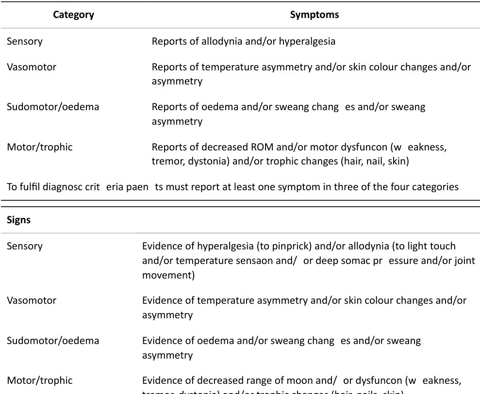
Must display ≤ 1 sign at time of evaluation in ≥ 2 of the categories.
What is the natural history of CRPS?
After a year, around half of patients affected willfully recover while the other half may complain of some residual stiffness and pain. A very small number of patients will continue to have significant disabling symptoms which can become chronic in nature.
What is the pathophysiology of CRPS?
It is thought that CRPS develops when persistent noxious stimuli from an injured body region leads to peripheral and central sensitization, whereby primary afferent nociceptive mechanisms demonstrate abnormally heightened sensation, including spontaneous pain and hyperalgesia.
What do you mean by a noxious stimulus?
This is a stimulus that is damaging to normal tissues.
What is a nociceptor?
This is a receptor preferentially sensitive to a noxious stimulus ort o a stimulus which would become noxious if prolonged.
How are nociceptors activated?
Peripheral activation of nociceptor sis modulated by a number of chemical substances, which are produced and released when there is cellular damage (e.g. potassium, serotonin, bradykinin, histamine prostaglandins, leukotrienes and substance P). These substances influence the degree of nerve activity and intensity of the pain sensation.
What are the current approaches to management? CANDIDATE 1: A recent paper demonstrating le vel I evidence looked at the use of antioxidant vitamin C. A dose of 500 mg a day for 50 days was shown to reduce symptoms of CRPS.
Jumping straight in and quoting a paper isnt the best viva tactic approach to use. CANDIDATE 2: There are several management options which would include: Physiotherapy. Pharmacological management. Nerve stimulation. Regional nerve blocks. Chemical sympathectomy. Surgical sympathectomy.
What pharmacological management? CANDIDATE 2 : Drugs used include calcitonin, bisphosphonates, steroids and gabapentin. Oral and intravenous biphosphonates, but not calcitonin, have been shown to decrease pain and swelling and increase range of motion in patients with CRPS. A short course of oral steroids may be beneficial but limited evidence exists for the use of gabapentin. What about physiotherapy? CANDIDATE 2 : Although we generally refer patients with CRPS to physiotherapy the evidence for its effectivenes sis unclear. It may improve ROM of the affected limb but does not affect pain. What about regional nerve blocks?
CANDIDATE 2 : IV regional sympathetic block typically using guanethidine may help with pain control, but the evidence for improved final outcome is poor.
How do we minimize the risk of CRPS occurring?
Patient risk factors would include complex pain issues pre-surgery, such as if the patient was on lots of pain medication such asg abapentin, codeine or morphine-based tablets before surgery. Surgical factors that would concern me would be if the patient required complex elbow or hand surgery that would need prolonged tourniquet use. Both patient and surgical risk factors for CRPS would mean being proactive preoperatively perhaps starting them on vitamin C prophylactically and thinking about regional or epidural anaesthesia rather than a general anaesthetic. I would avoid tight casts, painful manipulations of fractures and unphysiological manipulated wrist positions incast. Mirror physiotherapy postop seems to be a promising new development and TENS may have a role. I would make sure the patient had good pain control postoperatively.
Structured oral examination question 5#
Intravenous regional anaesthesia (Bier’s block)
What are the indications for use of a Bier’s block?
Mainly for manipulation of wrist fractures in A&E. Other indications might include suturing of multiple forearm lacerations foreign body removal from forearm wounds, excision of wrist ganglia and palmar fasciotomy.
What are the contraindications to Bier’s block?
This mainly relates to tourniquet use. Absolute contraindications include: Allergy to local anaesthetic. Methaemoglobinaemia. Severe hypertension (SBP > 200 mmHg). Compartment syndrome. Uncooperative or confused patient. No IV access on affected hand and other limb. Relative contraindications include: Coagulopathy. Cardiac conduction abnormalities. Peripheral vascular disease. Sickle cell disease or trait. Epilepsy. Local inflammation/in fection. Children (< 10 years). Pregnancy. Lymphoedema.
Anything else?
I can’t think of anything. Morbid obesity (as the cuff is unreliable on obese arms), scleroderma and Raynaud’s phenomenon. Paget’s disease (local anaesthetic may spread to the systemic circulation via venous channels in bone).
What local anaesthetic is used?
Recommended agents are either prilocaine or lignocaine. 0.5% Prilocaine. Dose is 3 mg/kg. A 70 kg patient gets 210 mg (42 ml) 0.5% prilocaine.
What about cuff deflation?
With single cuff use the cuff should not be letdown until a t least 30 minutes have elapsed. Deflate the cuff briefly (for 5–10 s) then reinflate. If there are no signs of toxicity after 45 s, then deflate again (for 5–10 s). This is repeated once more prior to being permanently removed. This allows some of the local anaesthetic in to the systemic circulation a t short intervals to avoid local anaesthetic toxicity from large amounts of local anaesthetic being released all at once.
Why all this protocol?
This is because systemic toxic doses of local anaesthetic may be released. After 20 minutes, 30% of the injected drug is fixed within the tissues and is unavailable for immediate release into the systemic circulation.
What are the dangers of a Bier’s block?
Major complications include nerve injuries, anaphylaxis, methaemaglobinaemia seizures, arrhythmias, cardiac arrest and death. Compartment syndrome, but it is difficult to separate out the influence of the distal radius fracture itself as the causative factor. Tourniquet pain, rash and thrombophlebitis.
How do you recognize prilocaine toxicity?
Symptoms include dizziness, restlessness, anxiety, perioral tingling , metallic taste, altered mental status, muscle twitching and seizures.
What is a double tourniquet?
The double tourniquet is used to increase safety and to reduce tourniquet pain in the awake patient.
Yes, but what do we mean by the double tourniquet method?
This allows better analgesia of the area under the tourniquet. Step 1: Exsanguinate limb. Step 2: INFLATE proximal/top cuff of tourniquet to 100 mmHg above the patient systolic blood pressure.
Step 3: After 10 min INFLATE boft om cuff of the tourniquet. Then ONLY AFTER THE BOTTOM CUFF IS
FULLY INFLATED, DEFLATE THE TOP CUFF. The result is that the BOTTOM tourniquet is over a now anaesthetized area of arm.
Step 4: After 30 min from injection deflate theb oft om/distal cuff of the tourniquet.
It is important that there is always at least one tourniquet cuff inflated for the full 30 min from injection of priloc aine.
There has been a prolonged period of icy weather at your local hospital and the trauma list is overloaded with cases. The orthopaedics service manager has called you into their office and asked if you would agree to the temporary introduction of Biers block use in casualty for distal radius fractures. This is to free up more trauma list time. What are your thoughts?
Most wrist fractures managed on a trauma list usually require either ORIF or K-wire fixation rather than just manipulation. I would worry about the risk of fracture re-displacement with just simple manipulation and application of a moulded c ast. If there is a regular list of Bier’s block cases taking place in casualty and working well then it may simply be a case of reviewing practice and auditing procedures to ensure optimal patient selection, patient care and clinical governance. My own hospital does not have a Bier’s block list for Colles’ fractures. This type of arrangement has generally fallen out of favour, so reintroducing this technique into casualty would need careful planning. I would need to know more about the business plan submift ed. My initial thoughts are those of concern about safety. There is a move by anaesthetists to do a brachial plexus block (axillary approach) instead of a Bier’s block for procedures in the forearm. Thus, doing Bier’s block may fail with non-anaesthetis ts such as A&E staff. This is a soft viva question. If you go in the wrong direction with this type of question you will struggle to score points. Unfortunately, there is a bit of guesswork required as to what to say. Consent, ethics and duty of candour are all creeping into the vivas. The candidate’s answer has sat on the fence alit ile.
What do you want to know?
We would need to decide if the service was A&E-led, orthopaedic-led or if a combined approach was taken. We would have to ensure staff had the necessary training. Protocols and procedures would need to be put in place. We would need to have guidelines in place and a business case setout. The procedure should only be used for fairly straighfoorward extra-articular distal radius fractures and certainly not in young patients with high-energy displaced intra-articular fractures.
There would need to be an agreed protocol in place for timely patient follow-up, as these fractures may re-displace and require ORIF. Cases that clearly need ORIF should be identified and we should avoid Bier’s block use.
It seems like a lot of extra work for a temporary fix of lack of trauma theatre time. My own preference would be to free up some theatre time from elsewhere. An additional trauma list or perhaps cancelling some elective cases would be much better allround.
Again, I must say I do not think it is a good idea. Tourniquets can fail and release toxic doses of local anaesthetic in to the systemic circulation, the reis a risk of methaemoglobinaemia occurring and there are better methods to anaesthetize a limb.
The candidate has decided not to support the introduction of a Biers block list. It can be difficult to decide which way to go with these types of questions, especially if you are not very familiar with the procedure.
No free theatre time is a vailable.
I can only say this would not be a quick-fix solution and needs careful planning and thought and I would beworried this wasn’t the correct approach. This is a soft question dealing with the way the NHS is set up. It is perhaps more suited for a consultant interview, as this type of question is difficult for examiners to differentiate between the different scoring marks.
Structured oral examination question 6#
Pain
Three mechanisms need to be mentioned: 1. Segmental inhibition system (gate theory). 2. Opioid system. 3. Descending inhibitory system.
What is the gate theory of pain (segmental inhibition system)?
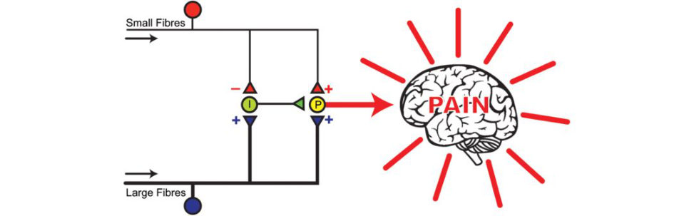
This is made unnecessarily complicated in many textbooks. The theory is easier to explain by drawing a diagram (Figure 23.1). Pain sensation is regulated in the spinal cord. Synapses between nociceptor fibres (Aδ/Type C) and dorsal root ganglia can be diminished or blocked by an inhibitory neuron (substantia g elatinosa) within the spinal c ord. The inhibitory neuron is activated by Aβ/Type A fibres (light touch, large diameter). Therefore stimulation of ligh t-touch fibres can block the pain transmission at the nociceptor– dorsal root ganglia synapse. Without any stimulation, both large and small nerve fibres are quiet and the inhibitory interneuron (I) (substantia g elatinosa) blocks the signal in the projection neuron (P) that connects to the brain. The ‘gate is closed’ and therefore there is no pain. With non-painful stimulation large nerve fibres are activated primarily. This activates the projection neuron (P), but it also activates the inhibitory interneuron (I), which then blocks the signal in the projection neuron (P) that connects to the brain. The ‘gate is closed’ and therefore there is no pain. With pain stimulation, small nerve fibres become active They activate the projection neurons (P) and block the inhibitory interneuron (I). Because activity of the inhibitory interneuron is blocked, it cannot block the output of the projection neuron that connects with the brain. The ‘gate is open’, therefore, there is pain!
How does this explain the action of transcutaneous electrical nerve stimulation (TENS) on pain?
The development ofTENS was the result of stimulating large A fibres that stimulated the inhibitory interneuron (substantia g elatinosa) that blocked the central pain stimulators and thus closed the gate.
Any other mechanisms of pain?
The opioid system.
OK, what is this?
Opioid derivatives are powerful analgesics (morphine, diamorphine, codeine). Opioid receptors are present in the spinal cord, periaqueductal grey matter and the ventral medulla. There are three types: μ (mu), δ (delta), κ (kappa). Enkephalins, endorphins and dynorphin are naturally occurring peptide ligands that bind to opioid receptors. The peptides modulate nociceptive input in two ways: 1. Block neurotransmift er release by inhibiting calcium influx in the presynaptic terminal. 2. Open potassium channels, which hyperpolarize neurons inhibiting excitatory action potentials. Systemically administered opioid analgesics can bind to the opioid receptors and modulate pain transmission.
Anything else?
Descending inhibitory system (adrenergic and serotoninergic): From the periaqueductal grey matter and the rostral medulla descending nerve fibres can modulate the ascent of nociceptor information a t the dorsal root ganglia. Noradrenaline and serotonin are the main neurotransmift ers in this pathway.
Figure 23.1 Gate theory of pain. I, ‘Inhibitory Interneuron’ (substantia g elatinosa); P , ‘Projection Neuron’; –, inhibition (blocking); +, excitation (activation ).
Structured oral examination question 7#
Pain pathways
What about primary afferent fibres?
There are three main types of afferent fibre (Table 23.1). Sensory (Aβ) fibres are highly myelinated and of large diameter, therefore allowing rapid signal conduction. The y have a low activation threshold and usually respond to light touch. Under pathological conditions the y may become hyperexcitable, leading to stimuli that would usually elicit sensations of tactile t ouch causing pain. Alpha delta fibres (Aδ) are lightly myelinated and smaller diameter, and hence conduct more slowly than Aβ fibres. They respond to mechanical and thermal stimuli. The y carry rapid, sharp, shallow pain that is specific to one area. These fast pain pathways composed of Aδ fibres are also responsible for the initial reflex withdrawal response to acute pain. High activation threshold. Group C nerve fibres are unmyelinated and are also the smallest type of primary afferent fibre. Hence, they demonstrate the slowest conduction. C fibres are polymodal because they can respond to various stimuli (chemical, mechanical and thermal). C-fibre activation leads to slow, burning pain spread out over an unspecific area (second pain). High activation threshold.
What about pain afferent fibres?
There are two main types of nociceptive nerve fibres: Aδ and C fibres. Aδ fibres transmit rapid, sharp, localized pain. C fibres transmit slow, diffuse, dull pain. The difference in speeds at which the two types of pain fibres (Aδ and C) conduct nerve impulses explains why, when you are injured, you first feel a sharp, acute, specific pain, which gives way a few seconds later to a more diffuse, dull pain.
What about the lateral spinothalamic pathway (Figure 23.2)?

The lateral spinothalamic tract conveys pain, temperature and crude touch to the somatosensory region of the thalamus. GABA, glycine, serotonin, norepinephrine, dopamine and acetylcholine have an inhibitory effect on spinothalamic tract neurons, whereas glutamate has an excitatory role.
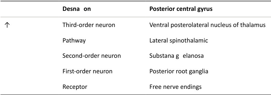
The first-order neuron delivers sensations to the CNS: the cell body is in the dorsal root ganglion.
The second-order neuron: an interneuron with the cell body in the spinal cord or brain.
The third-order neuron: transmits information from the thalamus to the cerebral cortex (Figures 23.3 and 23.4).
Table 23.1 Primary nerve fibre afferents.
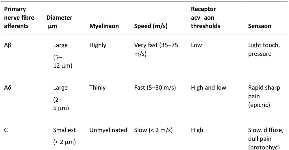
Figure 23.2 Lateral spinothalamic pathway.
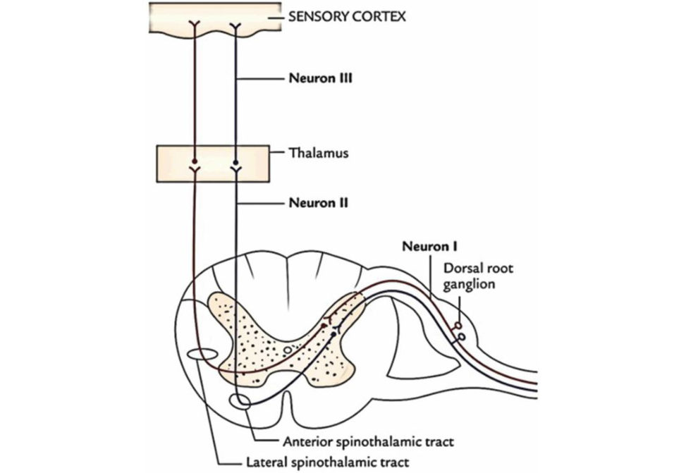
Figure 23.3 Spinothalamic tract.
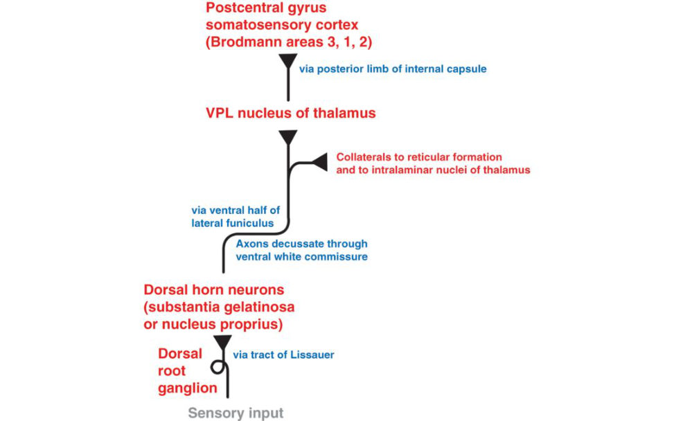
Figure 23.4 Spinothalamic tract. Candidate drawing.
First-order neuron
Aδ and C primary afferent nerve fibres have cell bodies in the dorsal root ganglia and terminate in the dorsal horn of the spinal cord. Once they have entered the spinal cord the nerveroots may bifurcate into ascending and descending branches, which can enter the dorsal horn one or two segments higher or lower than the segment of origin.
The dorsal horn of the spinal cordis the site where the primary afferent fibres synapse with second-order neurons. It is also where complex interactions occur between excitatory and inhibitory interneurons and where descending inhibitory tracts from higher centres exert their effect.
The dorsal horn is divided into six laminae (called Rexed laminae) responsible for processing sensory information and has an important role in the modulation of pain signals ( Figure 23.5).
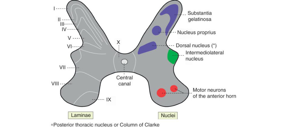
Figure 23.5 Rexed laminae.
Lamina II is known as the substantia gelatinosa and this extends from the trigeminal nucleus in the medulla to the filum terminal eat the caudal end of the spinal cord. It contains inhibitory and excitatory interneurons.
C fibres terminate in lamina II and Aδ fibres terminate in laminae I and V.
Aβ fibres (light touch and vibration) enter the cord medial to the dorsal horn and pass without synapse to the dorsal columns. They give off collateral branches to the dorsal horn which terminate in several laminae (III–V). They also synapse directly with terminals of unmyelinated C fibres in lamina II.
Laminae II and V are important areas for the modulation and localization of pain.
Second-order neurons
Second-order neurons ascend to higher centres via the contralateral spinothalamic and spinoreticular tracts, which are located in the anterolateral white matter of the spinal cord. Some of these neurons branch out to the periaqueductal gray or the reticular formation.
Third-order neurons
The tertiary neurons in the ventral posterolateral nucleus of the thalamus.
Axons from these thalamic neurons follow the thalamic radiations to terminate in the primary somatosensory cortex (Brodmann’s areas 3, 1, 2 of the postcentral gyrus).
What do you mean by nociceptors?
Nociceptors are sensory receptors for pain. Nociceptive fibres do not have specialized structures (such as Pacinian or Messner corpuscles) at their endings. Instead, they have what are known as free (naked) nerve endings that form dense networks with multiple branches. The peripheral activation of nociceptor sis modulated by a number of chemical substances, which are produced and released when there is cellular damage (e.g. potassium, serotonin, bradykinin, histamine prostaglandins, leukotrienes and substance P). These substances influence the degree of nerve activity and intensity of the pain sensation.
Structured oral examination question 8#
Local anaesthetic
Local anaesthetics are important in orthopaedics. What do you know about them?
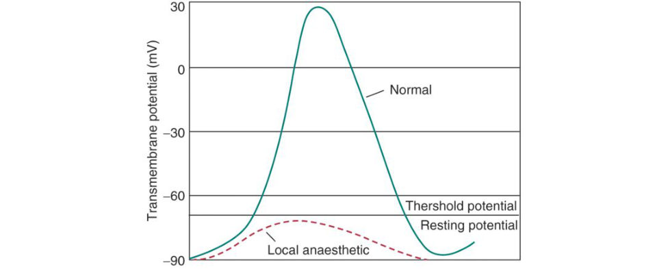
A general startup question that is a gift. All local anaesthetics produce their effects by blocking the transmembrane pore of sodium-gated voltage channels. This prevents depolarization of the nerve cell and propagation of the action potential down the nerve (Figure 23.6). The duration of action of the drug is dependent on protein binding and its clearance from the injection site. Protein binding for the longer-acting agents, i.e. bupivacaine and ropivicaine, is 95%; this is compared to 65% for lidocaine, a shorter-acting drug. The clearance from the injection site is dependent on local blood flow. Short-acting agents, i.e. lignocaine, cause vasodilation, thus potentiating clearance. Vasopressors, i.e. adrenaline, can be added in order to prolong the duration of action. Interestingly , local blood flow has litile influence on the longer-acting agents due to their high percentage of protein binding; therefore, the addition of vasopressors does not prolong their duration of action. Local anaesthetics block conduction in the following order: small myelinated axons, unmyelinated axons and large myelinated axons. As such, nociceptive transmission in Aδ and C fibres is blocked first and large-diameter myelinated motor fibres last.
Tell me more about how they work.
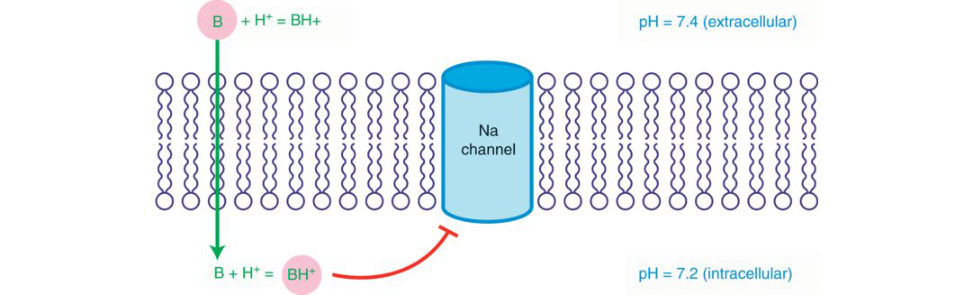
Local anaesthetics act by inhibiting sodium through sodium-specific ion channels in the neuronal cell. Entry of Na+ is essential for the generation of an action potential . The y interact with a receptor within the voltage-sensitiv e Na+ channel and raise the threshold of opening the channel (Figure 23.7). The rate and rise of an action potential and the maximum depolarization decreases leading to a slowing of conduction. Finally , local depolarization f ails to reach threshold potential resulting in a conduction block. Local anaesthetic binds more readily to sodium channels in an activated state and slows its reversion to the restings tate. The refractory period is increased. This could easily be asked in the basic science nerve section on action potentials as a sc ore 7/8 question.
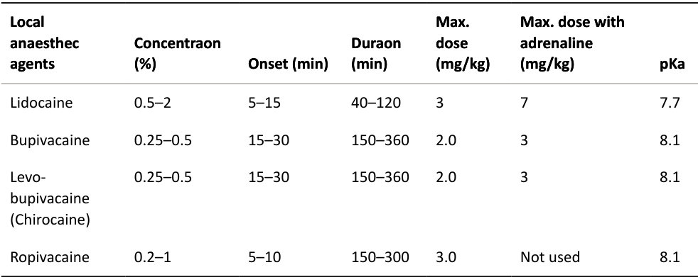
What are the commonly used local anaesthetics and their classes ( Table 23.2)?
Local anaesthetics can besplit depending on their chemical composition in to two classes. 1. Amides. Longer-acting. Metabolized by liver enzymes and excreted in urine.
2. Esters. Short-acting , metabolized in the plasma and tissue fluids by a cholinesterase enzyme and excreted in urine.
Amides are more commonly used, i.e. lidocaine, prilocaine, bupivacaine, levo-bupivacaine, ropivacaine.
Esters, e.g. cocaine, chlorprocaine and benzocaine, are now rarely used.
All the amides have an ‘I’ before the ‘caine’.
What determines the onset and potency of different local anaesthetics?
The activity of local anaesthetics is strongly pH-dependent. All local anaesthetics are weak bases which exist in two forms: ionized (BH+) and unionized (B). It is the unionized form that crosses the nerve lipid and once through the ionized form affects sodium depolarization. Thea mount that is ionized is determined by the equation
Figure 23.6 Local anaesthetics slow the rate of depolarization of the nerve action potential such that the threshold potential is not reached. As a result, an action potential cannot be propagated in the presence of local anaesthetic, and conduction blockade results.
Figure 23.7 Mode of action of local anaesthetics: Na-channel blocker.
Table 23.2 Most commonly used local anaesthetics with their maximum doses and duration of onset and action. A 2% solution of lignocaine contains 20 mg in 1ml.
pKa – pH = log [BH+]/[B].
How may the pKa of a local anaesthetic influence its speed of onset?
Onset is determined by the pKa. The pKa of a local anaesthetic determines the amount which exists in an ionized format any given pH. At physiological pH (7.4) all local anaesthetics are more ionized than unionized (as all the pKa values are greater than 7.4). However, the proportions vary between the drugs: lignocaine has a pKa of 7.9 and is approximately 25% unionized at p. 7.4. Bupivacaine has a pKa of 8.1 and hence less of the drug is unionized at p. 7.4 (about 15%). As the drug must enter the cell in order to have its effect, it must pass through the lipid cell membrane. Unionized drug will do this more readily than ionized drug. Therefore, the drug which is more unionized at physiological pH will reach its target site more quickly than the drug which isless so. This explains why lignocaine has a faster onset of action than bupiv acaine.
What about potency?
The aromatic ring structure and hydrocarbon chain length of a particular local anaesthetic determine the lipid-solubility of the drug and hence its potency. A more lipid-soluble drug penetrates the cell membrane more easily to exert its effect. The more potent the drug, the smaller the amount required to produce a given effect. Thus bupivacaine – which is highly lipid-soluble – is approximately four times more potent than lignocaine. Potency ∝ effect/dose Potency increases with increased lipid-solubility, which is related to hydrocarbon chain length.
What about duration of action?
Duration of action increases with increased protein binding, which is determined by the length of the intermediate chain which joins onto the aromatic and amine group. Lignocaine: 65% protein-bound.
Bupivacaine: 95% protein-bound.
As such, bupivacaine will have a longer duration of action than lignocaine .
Lipid-solubility is the second leading determining factor, greater percentage protein-bound and increased lipid solubility = longer duration of action.
Why do we avoid local anaesthetics use with abscess surgery?
There are two reasons. 1. Infection in a tissue decreases its pH, which increases the pKa–pH difference, which leads to more ionized form which does not cross the lipid nerve membrane. 2. Infection is often associated with localized increased blood supply and hence more anaesthetic may be removed from the area before it can affect the neuron.
You have infiltrated the skin and local tissues for a carpal tunnel release and used the maximum amount of local anaesthetic permift ed, but the local anaesthetic doesnt seem to be working. What are your thoughts?
Pathological reasons may include infection previous trauma or surgery or inflammation. Psychological reasons include fear and anxiety. Poor technique with the local anaesthetic injected away from the proposed site of surgery. I wouldn’t inject more than the recommended dose of local and would consider using a GA instead assuming the patient was fully fasted and there was an anaesthetist available to do this in a safe and timely manner .
Why may levobupivacaine or bupivacaine be preferred for use as a local anaesthetic compared to lignocaine?
They are associated with less vasodilation and have a longer duration of action, which is good to cover the operation duration and for postoperative analgesia.
What are the complications from local anaesthetics?
Complications are frequently associated with errors of dose or intravenous administration. Complications are related to membrane destabilization of cells. Neurological – perioral and glossitic paraesthesia, dizziness, drowsiness, tinnitus seizures. Cardiovascular – bradycardia, hypotension, cardiac arrhythmias, i.e. ventricular fibrillation and asystole. Hypersensitivity and allergy – these are rare and can vary from a mild skin irritation or r ash to anaphylactic shock. The main signs and symptoms of anaphylactic shock are chest discomfort, urticaria stomach pain and dyspnoea. An anaphylactic reaction can rapidly lead to a life- threatening condition duet o airway passage obstruction in association with laryngeal oedema and needs immediate treatment.
Pain at injection – many factors. Low pH and cold solution may irritate the tissue. F ast and high injection pressure may cause rapid swelling of tissues and pain. This can be avoided by a slower injection. A void aggressive, rough insertion of a needle.
Neurological effects are usually witnessed first followed by cardiovascular collapse. Management is supportive. A definitive airway should be established, ensuring adequate oxygenation and ventilation. Benz odiazepines and phenytoin can be used to control seizures. Cardiac monitoring is mandatory, as arrhythmias require urgent treatment.
The addition of adrenaline to local anaesthetics should not be used on tissues with end arteries , i.e. digit and penis, as the induced vasoconstriction may result in tissue ischaemia. Caution should be used when infiltrating around skin flaps for the same reason. Earlobes and nose should also be avoided
Structured oral examination question 9#
General anaesthesia
Anaesthesia gas machine laminated photo is shown to the candidate (Figure 23.8).
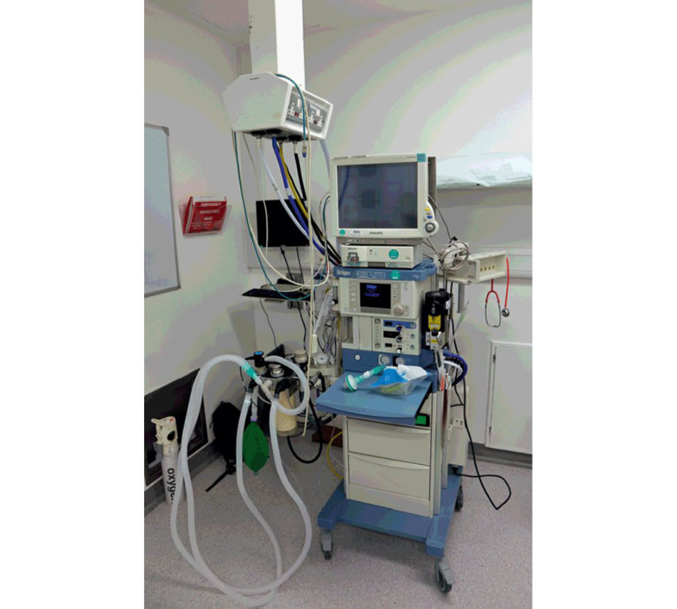
Figure 23.8 Anaesthesia gas machine.
What are the principles of general anaesthesia?
General anaesthesia encompasses the triad of analgesia, amnesia and muscle relaxation. Rather than using a large dose of a single drug to establish the clinical triad, a combination of drugs is used in smaller doses, thus avoiding dose-related adverse effects, and is termed ‘balance anaesthesia’.
What else?
General anaesthetic agents act in the CNS to bring about a reversible loss of consciousness, amnesia (memory loss), analgesia, loss of motor reflexes and skeletal muscle relaxation.
What drugs are used?
The three main classes of drugs used for GAs are intravenous induction agents, inhalation induction agents and muscle relaxants. Drugs used as intravenous agents include thiopentone, propofol and ketamine (Table 23.3).
Do you know which intravenous agents are most useful incertain situations ( Table 23.4)?
Propofol is mainly used in day case anaesthesia. It would be difficult to do day-case surgery without propofol. Thiopentone has fallen out of favour because it can cause significant bronchospasm and severe extravasation injuries. It has a longer postoperative recovery than propofol. Ketamine is the most cardiovascular-stable induction agent. It is therefore good to use in the trauma situation if there has been a large amount of haemorrhage occurring. Etomidate is a short-acting intravenous anaesthetic agent historically used for short procedures, such as reduction of dislocated joints.
You mentioned halothane as an inhalation agent, but what is the problem with using it?
It can cause hepatis and as such is rarely used these days. Around 1 in 50,000 can develop a severe life-threatening hepatis.
Why do we use muscle relaxants as part of a GA?
Neuromuscular blocking agents is another way to refer to muscle relaxants. Muscle relaxants are used to facilitate tracheal intubation and provide optima loper ating conditions. Muscle relaxants target the neuromuscular junction (NMJ).
How do muscle relaxants workTable 23.5)?
There are two types of muscle relaxants. 1. Non-depolarizing. 2. Depolarizing. Non-depolarizing agents competiv ely blockthe binding of acetylcholine (ACh) to nicotinic receptors at the post-synaptic membrane. Thisleaves fewer receptors available for ACH to bind to and initiate channel opening. No endplate potential is generated with paralysis of theaffected skeletal muscles. Depolarizing agents act by depolarizing thepost-synaptic plasma membrane of the musclefibre similar to Ach, but as these agents aremore resistant to breakdown they persistentlydepolarize the muscle fibre. As such there aretwo phases to action, the first being an initial depolarization phase where the agent causesmuscle fasciculations while the muscle fibres are depolarizing. The second phase involvesa desensitizing phase in which the muscle is no longer responsive to neuromuscular transmift er released by the motor neurons.
How are muscle relaxants reversed?
At the end of the procedure muscle relaxants can be rapidly ‘reversed’ with the use of neostigmine, which has a plasma halflife of 60 minutes, longer than all the commonly used muscle relaxants. Neostigmine is an antagonist to acetylcholine esterase (AChe) (the enzyme that breaks down ACh) resulting in a flux of A Ch at the NMJ. Neostigmine is rarely used on its own to reverse as it can cause significant bradycardia, being often given with glycopyrrolate (with a similar but milder action to atropine). Adjuncts to anaesthesia include anft-eme tics, which aim to reduce the incidence of postoperative nausea and vomiting. Local anaesthetic and peripheral blocks used in conjunction with general anaesthesia have led to improved postoperative pain relief and aided postoperative physiotherapy. Table 23.3 Intravenous induction agents.
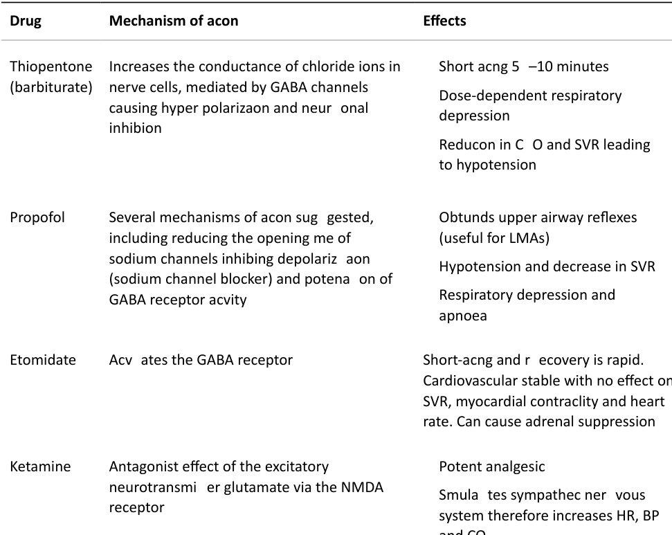
BP, blood pressure; CO, cardiac output; HR, heart rate; LMA, laryngeal mask airway; NMDA, N-methyl-Daspartate receptor; SVR, systemic vascular resistance.
Table 23.4 Inhalation agents.
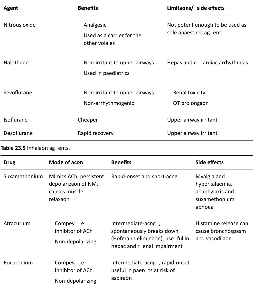
ACh, acetylcholine; NMJ, neuromuscular junction.
Analgesia
Anaesthetists use analgesics which are different to those used in non-anaesthetiz ed patients These often have a very quick onset of action – alfentanil, fentanyl and remifentanil.
Remifentanil has a very short half-life, which is the same even after many hours of receiving the infusion.
In the doses anaesthetists give, these can have a potent respiratory depressant effect (as do most other anaesthetic agents).
Have you heard of total intravenous anaesthesia (TIVA)?
No, sorry. TIVA has been used for many years as an alternative to inhalational anaesthesia. Some anaesthetists choose to use this as their standard. Others use it for the following reasons: Reduced postoperative nausea and vomiting. Rapid recovery from anaesthesia. Some evidence that it may be better to reduce cancer recurrence (disputed). When inhalational agents are absolutely contraindicated – malignant hyperthermia. When you wish to avoid muscle relaxant drugs due to certain neurological diseases. When you wish to avoid muscle relaxants for endotracheal intubation. It is possible to intubate without using neuromuscular blocking agents. This can be done with infusions of propofol and remifentanil, or with boluses of propofol and alfentanil/fentanyl.
What do you understand by the term regional anaesthesia? CANDIDATE 1: With regional anaesthesia you are rendering a specific area of the body, e.g. foot, arm, insensate to the stimulus of surgery or other instrumentation. What types of regional anaesthesia do you know? CANDIDATE 1: Radial nerve block, brachial plexus block, epidural and spinal anaesthesia.
Scoring 5/6. CANDIDATE 2: Regional anaesthesia involves infiltration of local anaesthetic to block sensory and motor nerves. Regional anaesthesia makes the operative site insensate and provides peri- and postoperative pain relief. The duration of mot or and sensory blockade depends on the type and concentration of local anaesthetic agents and whether any additive agents are used.
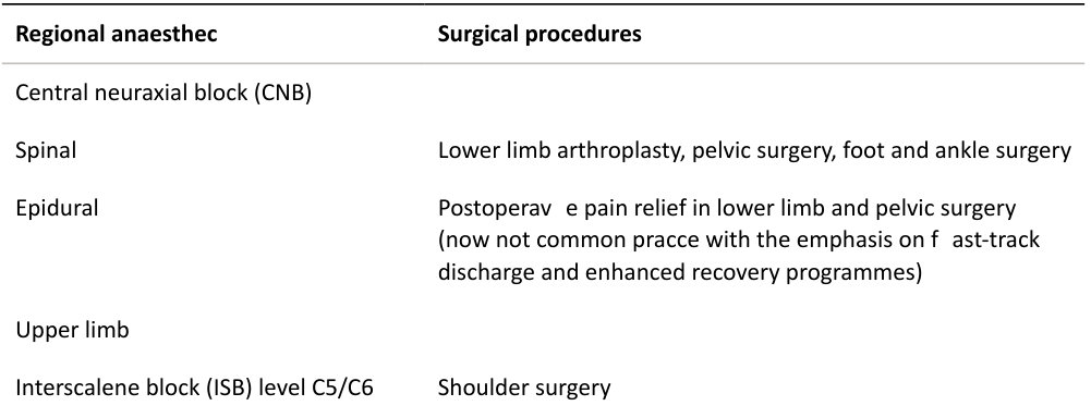
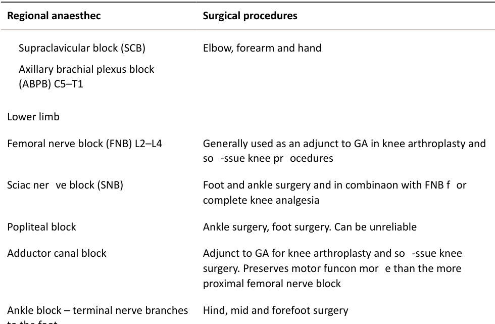
What types of regional anaesthetic are used in orthopaedic surgery (Table 23.6)?
A peripheral nerve block involves injecting local anaesthetic near the course of a named nerve. The advantages are that a relatively small dose of local anaesthetic can cover a large area with rapid onset of action Disadvantages include that it is technically challenging. To increase the accuracy and safety, nerves or plexuses are located using ultrasound guidance and peripheral nerve stimulation. A plexus block involves injection of local anaesthetic adjacent to a plexus. The advantages are a large area of anaesthesia can be produced, but the procedure is technically complex with the potential for damage of the plexus. A spinal anaesthetic involves an injection of local anaesthetic in to the CS Fand provides profound anaesthesia of the lower abdomen and extremities. It is technically easy (LP technique) with a high success rate and rapid onset of action Disadvantages include a ‘high spinal’, hypotension due to sympathetic block and postdural headache. An epidural involves injection of a local anaesthetic in to the epidural space at any level of the spinal cordItis used for anaesthesia/analgesia of the thorax, abdomen and lower extremities. Advantages include controlled onset of blockade, long duration of action with catheter use and postoperative analgesia. Disadvantages include postdural puncture headache if the epidural needle is accidentally advanced through the dura, toxicity of doses intended for the epidural space injected into the subarachnoid space as well as higher failure rates than for performing spinal anaesthesia. Complications of an y regional anaesthetic include bleeding , infection nerve damage and local anaesthetic systemic toxicity (LAST). Good score 6, possibly 7. Knows the general principles of regional anaesthesia, able to discuss uses and aware of advantages/disadvantages. Table 23.6 Types of regional anaesthetic used in orthopaedic surgery.