Chapter 18 Children’s orthopaedics
Introduction#
Feedback from candidates who did the FRCS exam showed that the pediatric viva section has certainly changed since the introduction of the first edition of this book. The paediatric viva section seems to contain the followings three areas:
1. One of the big paediatric topics such as DDH, septic hip, SUFE, clubfoot or knee deformities, during which in-depth knowledge is expected from candidates.
2. A common significant paediatric trauma such as elbow/supracondylar fracture, forearm fractures, femur fractures, NAI or paediatric ankle fractures. Again, the candidate is expected to have a solid knowledge about these subjects.
3. The last area is about common conditions that could face orthopaedic surgeons in any subspecialty such as bone cyst, multiple hereditary exostosis, tarsal coalition, pes cavus, osteochondritis dissec ans.
In this secft on, we follow the exam format with a simple question around a clinical picture, X-ray or a video clip, followed by increasingly difficult questions to explore candidate depth and breadth of knowledge. Some of the questions are made deliberately difficult and beyond average candidate level, some are easy and the majority are average. We also support some of the answers with clinical photographs to create mental images to aid recall information during the exam. The online version of this book will have more cases, videos and discussion. This section complements the first edition of this book, the postgraduate paediatric orthopaedic book (the green book) [1] and the third edition of the parent book. Candidates are strongly encouraged to read all of them to have a better overview of the paediatric section of the exam.
Candidate 1#
This is a clinical photograph of a child (Figure 18.1) who tripped and fell, hurting his knee. He was seen in the A&E department and referred to your fracture clinic. Describe what you see. How would you approach him?
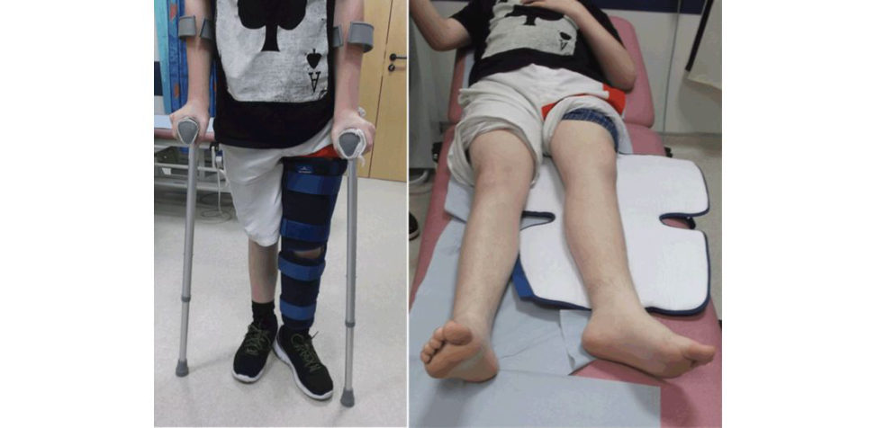
My approach is to take a detailed history, perform a thorough examination and or der the appropriate investigations guided by my examination and provisional diagnosis. The left image shows a child standing with two crutches, wearing a knee splint on the left lo wer limb. The left leg is externally rotated and may be short. My first impressionis that this child may have a slipped upper femoral epiphysis (SUFE).
How can you confirm your diagnosis?
History, examination and radiological tests to confirm my diagnosis. History of previous pain in the hip before the fall is an important clue. He stands with crutches (if he has a slip, it is probably a stable type). Hip examination may reveal limited internal rotation or even obligatory external rotation on flexing the hip (Drehmann sign). I also request pelvis X-ray (AP and cross-table lateral views of both hips). I do not prefer frog lateral as it may worsen the severity in unstable slips; however, it is reasonable to request in a stable slip.
This is his pelvis X-ray (Figure 18.2). What can you see?
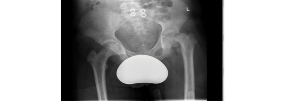
This is a plain X-ray of the pelvis (AP view only) showing both hips. The most obvious abnormality is the slipped upper femoral (capital) epiphysis on the left side. The head remained in the socket while the neck moves anteriorly and superiorly. Trethowan’s sign is positive; a line (often referred to as Klein’s line) drawn on the superior border of the femoral neck on the AP view should pass through the femoral head. In SUFE, the line passes over the head rather than through the head. There are remodelling changes of the neck with sclerotic, smooth superior part of the neck and callus formation on the inferior border. This indicates the slip is not acute and has been subclinical for a while.
What other radiological signs might you see in SUFE?
Several radiological signs are described to aid diagnosing SUFE (particularly subtle ones). These are not present in every case of SUFE, such as Trethowan’s sign that I just mentioned; widening and irregularity of the growth plate (early sign); decreased epiphyseal height as the head slipped posteriorly behind the neck; remodelling changes of the neck and increased distance between the teardrop and the femoral neck metaphysis. Capener’s sign: normally, on the AP pelvis the posterior acetabular margin cuts across the medial corner of the upper femoral metaphysis. In SUFE, the entire metaphysis is lateral to the posterior acetabular margin. Steel’s blanch sign, which is a crescent shape dense area in the metaphysis due to superimposition of the neck and the head.
What if this child’s X-ray was normal?
Normal X-ray does not exclude SUFE (it may be in the preslip stage); therefore, I would request an MRI scan but also, I would consider other possible diagnoses.
Can you grade the severity?
I measure the severity using either Wilson grading on the AP views or the Southwick angle on the lateral views. I consider that this is a severe slip as the head almost slipped by more than two- thirds of the physis width.
Can you draw for me how these two classifications measure the severity of the slip?
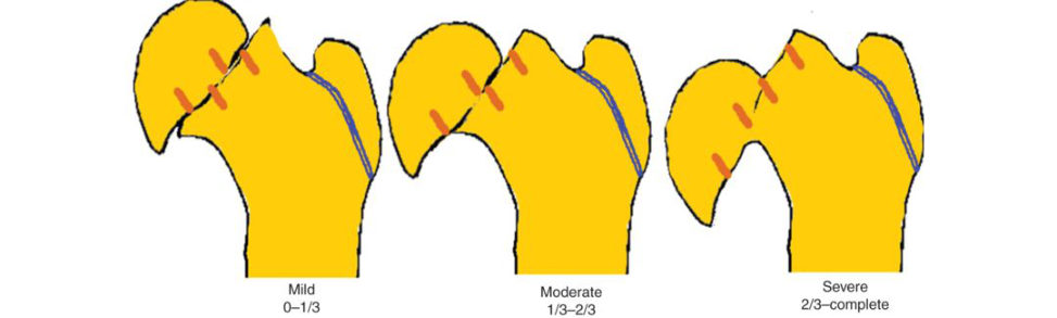
Radiological grading of the severity of the slip has been based on either the degree of displacement of the head relative to the neck (Wilson), or by degree of the angulation of the head relative to the shaft (South wick). Wilson recognized three grades on the AP view: mild slip (grade I) is one where the displacement of the head as a proportion of neck physis) width isless than a third, moderate slip (grade II), displacement is between a third and a half of the neck width and severe slip (grade III) has displacement of greater than half of the neck width (Figure 18.3). Southwick graded the severity on the frog lateral view by measuring the Southwick angle, which is the difference between the lateral epiphyseal shaft angle of the slipped and the non-slipped sides (Figure 18.4). Mild slip (grade I) has an angle difference of less than 30°, moderate slip (grade II) has an angle difference of between 30° and 50° and severe slip has a difference of over 50°. If both sides slipped, Southwick angle is calculated by subtracting 12° from the corresponding lateral epiphyseal shaft angle.
You mentioned that the slip is stable. Why?
Randall Loder [2] classified SUFE into two types: I. Stable slip: child is able to weight bear. II. Unstable slip: child is notable to weight bear on the affected side even with crutches. This classification has been shown to be of prognostic value. The risk of AVN is high in an unstable SUFE and low in a stable one. In Loder’s original paper the AVN rate was 47% in unstable SUFE and 0% instable ones. Similar findings were shown in other centres [3,4]. There has been some confusion about the exact meaning of ‘able to weight bear’ in Loder’s original paper. ‘Ambulation ’ may be a better term to describe slip stability. So, to me the slip is unstable if the child cannot weight bear and ambulate even with crutches. This child is standing and even if he does not put weight on the affected limb, there must be enough stability to allow him to stand. That is why I said it is a stable slip.
Figure 18.1 Twelve-year-old boy who tripped and fell.
Figure 18.2 Pelvis X-ray of 12-year-old child with knee pain shown in Figure 18.1.
Figure 18.3 SUFE radiological grading.
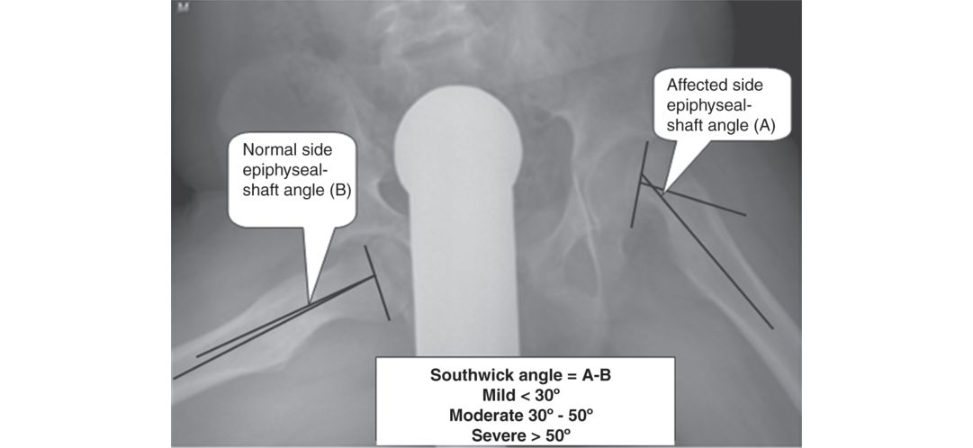
Figure 18.4 Southwick angle.
How would you treat this child?
This child has grade III stable slip. My options are: 1. Pinning in situ to stabilize the slip and prevent further progression until physis closure. I anticipate that he would have impingement symptoms that may require future surgery if remodelling is not enough, which is the case in most cases with such severity. 2. Primary open reduction and internal fixation Several techniques have been described and currently the Ganz surgical dislocation is the preferred option. It is technically demanding and better performed in specialized centres that do it on a regular basis.
Do you think you can pin this SUFE?
Yes, although it will not be easy and the worse the deformity is the more difficult the pinning in situ will be.
Take me though how you would pin it.
Before I perform any operation, I will make sure that my patient is as fit as can be for surgery. I review their health records investigations and obtain an informed consent for surgery. I will make sure that all the required equipment and implants are available. The operation is done undergeneral anaesthetic (GA Intravenous antibiotic is given at induction. The patient is positioned supine on a fracture table (without traction). The other limb can be placed in abduction or flexed and abducted on stirrup to allow for imaging. Optimum visualization of the femoral head before the procedure is essential. (In bilateral stable slip, a radiolucent table is preferred over the fracture table because it reduces the chance of worsening the contralateral slip by overenthusiastic positioning. This also reduces the time for re-positioning and r e-draping the contralateral side. The stability is usually adequate to obtain a lateral view of the femoral neck by gentle flexion of the hip.)
The trajectory of my screw is identified and marked using a free guide wire placed on the skin overlying the proximal part of the femoral neck and head, crossing the physis in a perpendicular fashion in the AP and lateral views (Figure 18.5).
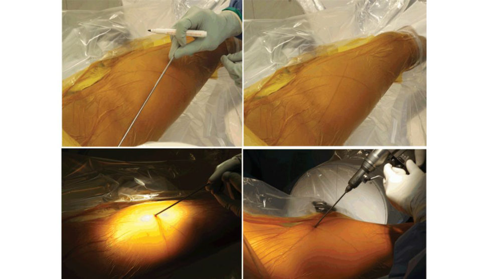
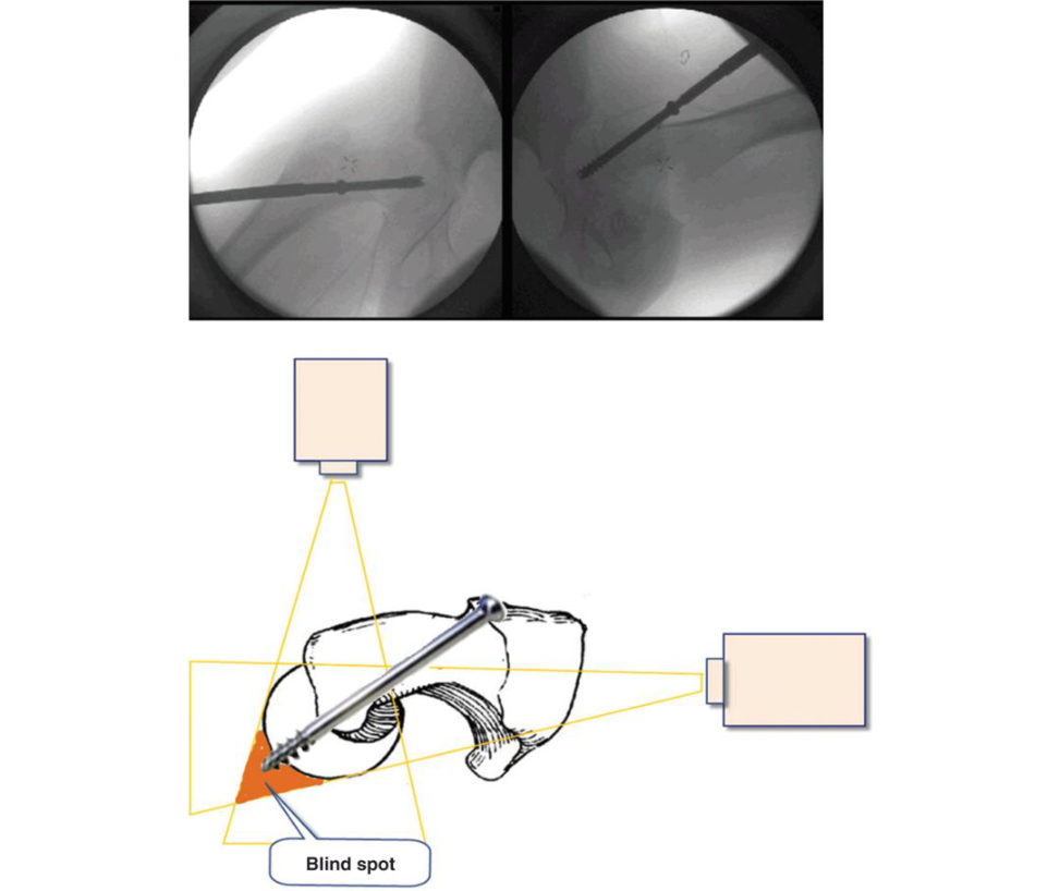
The guide pin is advanced freehand where the lines intersect through the soft tissues to engage the anterolateral femoral cortex. The position and angulations of the guide pin are adjusted under fluoroscopic guidance, to obtain the proper alignment before the guide pin is advanced into the bone. The entry point is usually quite anterior. It is essential to screen the hip to ensure there is no protrusion of the guide pin in the joint; particularly in the blind spot ( Figure 18.6). For unstable slips, a second guide wire is useful to provide some rotational stability and can be used for the insertion of a second cannulated screw if desired.
After the appropriate screw length has been determined, the femoral neck and epiphysis is drilled using the cannulated instruments while periodically checking that the guide wire position is not advancing into the hip.
I prefer to use a 7.3- or 6.5-mm fully threaded, reversed cuting cannulated screw. The screw position should be carefully checked (using the withdrawal technique) to ensure there is no protrusion. If available, 3D C-arm is valuable for this purpose. Several other methods have been proposed for the same reason, but none is without limitations.
The patient is allowed touch weight-bearing with the use of crutches and gradually advances to full weight-bearing as tolerated. Follow-up is until ph yseal closure.
Would you pin the other side?
I would consider pinning the other side in the following situations: 1. Age of the child (< 10 years is associated with a higher risk of bilaterality). 2. Slips associated with renal osteodystrophy and endocrine disorders (a high incidence of bilaterality approaching 95%). 3. Poor compliance of the child and family. 4. The nature of the current slip (a very bad slip occurring over a very short period of time may justify pinning the other side). The quoted risk of contralateral slip varies from 18% to 60%. Prophylactic PIS is not devoid of risk and should be weighed against the benefit. The proponents and opponents have some evidence to support their views [5]. Some radiological markers have been proposed to aid decision-making about pinning the other asymptomatic side in patients with SUFE, such as the posterior sloping angle and the modified Oxford bone age [6,7]. Both of these markers are not perfect and do not have 100% positive or negative predictive value. I would have a low threshold to prophylactically pin the other side if the posterior sloping angle is more than 14° as research has shown that the risk of contralateral slip would be around 83%.
Can you draw the posterior slip angle?
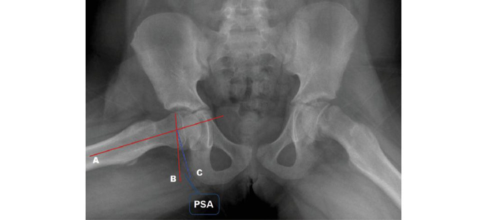
The posterior sloping angle (PS Ais measured by a line (A) from the centre of the femoral shaft through the centre of the metaphysis; a second line (B) is drawn from one edge of the physis to the other, which represents the angle of the physis. Where lines A and B intersect, a third line (C) is drawn perpendicular to line A. The PS Ais the angle formed bylines B and C posteriorly, as illustrated in Figure 18.7. Authors’ note: 1 SU FEis a very important topic for the exam. Excellent knowledge is expected. The above scenario is typical; however, the examiners may wish to explore the following (this is a good sign indicating that you have done well to reach these areas): 1. When pinning in situ: a. Radiolucent table vs. fracture table. b. Single or double screws for unstable severe slips: double screws offer 66% stiffer construct than single screws, but the risk of intra-articular penetration increases from 4% to 20% with double screws. c. Fully threaded vs. partially threaded. d. Would you remove screws routinely? 2. Management of grade III slips; open reduction with or without surgical dislocation: both can be used as longas there is no undue pressure on the retanicular blood vessles. Parsch [8] reported 4.7% AVN with open reduction without surgical dislocation while Ganz [ 9] reported no AVN with surgical dislocation. In a systematic review and meta-analysis we compared all treatment options and risk of AVN, and surgical dislocation has 3% A VN rate but better patient satisfaction rates whereas PIS has 1.5% AVN rate and lower satisfaction rates [3,10]. 3. If you do well, you may be asked about the technique of surgical dislocation or the Par sch technique. 4. Treatment of residual deformity (corrective osteotomies, arthroscopic osteochondroplasty). 5. AVN (and its medical and surgical treatments).
This is a clinical photograph of the elbow of a 5-year-old child who fell off a monkey bar (Figure 18.8). How would you manage this child?
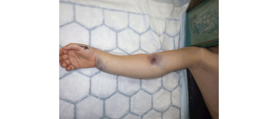
The photograph shows wrist and elbow swelling, deformity and bruises. There may be skin puckering in front of the elbow. This child most likely has wrist and supracondylar fractures of the humerus (SCH#). I always follow the ATLS protocols in assessing and managing traumatic injuries in children. Primary survey, then secondary survey and followed by AMPLE history. AMPLE stands for Allergies, Medication, P ast medical history, Last food and drinks and Events around the accident.
You perform primary and secondary surveys and find that this is an isolated upper limb injury.
Then I perform a thorough assessment of that limb. Optimum analgesia is extremely helpful to calm the child and makes my assessment pleasant. I look for obvious swelling, bruises, wounds, colour of the hands and fingers, etc. in that limbI feel for skin temperature, pulses and tenderness. I assess the movement of the joints that are not involved to ensure that there are no unexpected problems. I would not move the elbow joint at this stage as it is expected to be painful. I assess the neurovascular status of the limb using special tests to test for nerve injuries.
You find the hand warm and relatively well perfused but there was no radial pulse.
What about the neurological status?
The child was not cooperative and you could not assess him optimally despite your best effort.
This is not an uncommon scenario. I would document this accurately (i.e. warm hand with no radial pulse) and I could not assess his neurological status (because of the pain and the lack of cooperation). I would like to confirm the diagnosis by requesting an X -ray.
This was his X-ray.
The radiograph confirms my thoughts that this child has supracondylar humeral and distal radius fractures. Both are displaced. The supracondylar humerus fracture is an extension type (Gartland type III) injury with the distal fragment displaced posteriorly and rotated externally. It is not very clear from the X-ray, but there may be some comminution of the lateral column. This is a limb-threatening injury and requires an immediate intervention. I would like to take the patient to theatre to reduce and stabilize the fracture. A vascular surgeon or a plastics surgeon (with microvascular skills) must be consulted before taking patients to theatre in case hand perfusion becomes worse after the reduction.
How did Gartland classify these fractures?
Gartland classified this fracture into three types: Type I (undisplaced), type II angulated and type III complete displacement. The classification has undergone several modifications as more knowledge developed.
How do you fix this fracture?
Preoperative planning and thoughtiul preparation are key to successful outcome. I obtain an informed consent for surgery. I inform theatre about the child and the urgency of surgery. I liaise with my anaesthetist and vascular colleagues. The fracture pattern is extension type (Gartland type III), there may be some comminution. The reis significant elbow swelling and there may be tethering. There is a wrist fracture distally which complicates things further. The risk of compartment syndrome is high. I still think closed reduction and percutaneous pinning (using two or three 2-mm K-wires) will be successful; however, an open reduction may become necessary. After the WHO check, the child will have a GA and IV antibiotic. I position the child supine on the operating t able and his hand is stretched on a hand table. The child’s head should be placed in a position closet o the hand table, so it will not falloff the main table. The C-arm should come parallel to the main table to allow viewing the AP and lateral of the elbow without changing the elbow position. I will gently check how easily I can reduce both fractures. Reduction is usually achieved by a gentle traction inline with the humerus, with the elbow in slight flexion. Traction in full extension may cause tethering of neurovascular structures over the proximal fragment. I will use the mid-forearm part for applying the traction to avoid traction damage to the distal neurovascular structures crossing the wrist fracture. If the proximal fragment has pierced through the brachialis muscle, a ‘milking’ manoeuvre over the brachialis can help untethering of the muscle off the bone. Traction usually takes between 1 and 3 minutes to allow for the muscle to relax and the soft tissue to stretch. Successful traction should allow the visibility of the fracture without overlapping of the proximal and the distal fragments on the AP screening. Then medial or lateral translation is corrected by pushing the distal fragment medially or laterally. An important precaution here is not to cause varus or valgus deformities. It should be pure translation. I then flex the elbow while keeping traction. This makes reduction more stable because the main pull vector of the triceps muscle becomes the fracture compressor (or even flexor) rather than the extensor. In words, the triceps muscle force changes from a deforming force into a stabilizing force. Successful completion of this step is marked by the fact that the child’s fingertips can touch the shoulder easily. Fluoroscopic assessment to confirm reduction using AP and two oblique views at 90° (lateral and medial oblique views). A lateral view can betaken either by carefully rotating the elbow if the fracture is stable enough or by rotating the X -ray machine if not. After flexing the elbow, there will be overlapping of the proximal ulna and radius over the fracture site Jone’s view) but it is still possible to assess the continuity of the medial and lateral columns and Baumann’s angle (see Figure 18.10). Once the fracture is reduced, it needs to beheld in the reduced position untils tabilized. There are two methods that I use depending on the fracture stability and the experience of my assistant (Figure 18.11). If the fracture is stable and I do not have an experienced assistant, I would tape the limb (hand to shoulder as in Figure 18.11) to keep it reduced; otherwise, I will rely on my assistant to hold it reduced during the procedure. In this child, I cannot use the taping method as there is a wrist fracture.
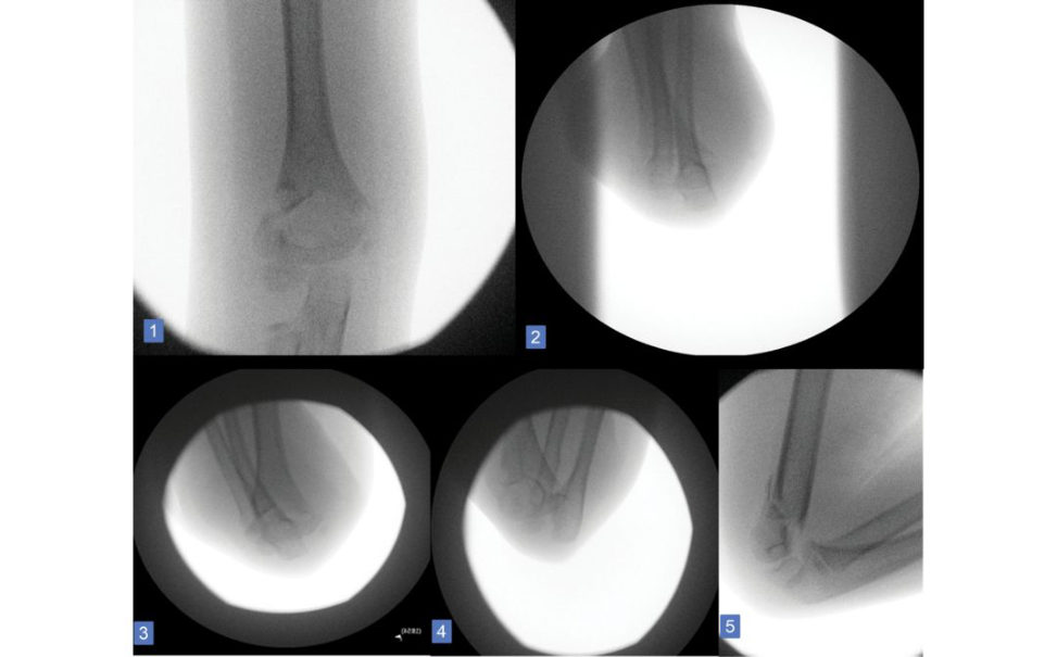
Then I prepare the skin using antiseptic solution and dr ape the upper limbI use two smooth K-wires (size 2 mm unless the child is very small, when I use a smaller size). I pass the first wire through the capitellum in a superomedial direction through the olecranon fossa and the medial cortex
(holding up to six cortices – this is not always possible depending on the site and direction of the fracture). I pass the second wire through the distal fragment with maximal spread at the fracture site from the first wire. The wires should not be crossed inside the bone (or even worse, at the fracture site). I check the wires on the AP and lateral views to ensure optimum positioning. Then I t est the fracture stability; if it is stable, I will do the wrist, but if not, I may supplement with a third wire or I may use the medial wire. The latter requires a small incision to visualize the ulnar nerve.
This is what has been done. Any thoughts?
The images showed a good reduction of the humeral fracture. There may be a slight lateral translation on the AP view. This needs careful follow-up. The positions of the wires are perfect. The medial wire is passing through the capitellum, olecranon fossa and as low as possible through the medial cortex. This gives good purchase to the bones as it passes through six cortices. The lateral wire is going through the lateral column and is not crossing with the medial wire. On the lateral they are both in good position. The same can be said about the wrist.
Was the second wire in the wrist necessary?
I think so, although it is not always possible to correctly judge the stability of a fracture from a static 2D picture Moreover, the risk of compartment syndrome is high in this patient and it is important to obtain a stable fixation in an ticipa tion of c ast splinting or even removal if compartment syndrome happens or if the vascular surgeon decides to explore and repair a blood vessel in this case. So, I think the surgeon was right to use two wires in the wrist.
Fair enough. You stabilized the fracture but the radial pulse did not return. What would you do?
BOAST guidelines suggest that a well-perfused limb does not require brachial artery exploration whether or not the radial pulse is present. However, if the upper limb remains ischaemic (pale, cold, delayed capillary refill and pulseless limb) after fracture reduction, a surgeon competent to perform small vessel vascular repair should explore the brachial artery and that is why I informed them before taking the child to surgery [11].
Authors’ note: 2
Fractures around the elbow are commonly featured in the exam as they have diagnostic, therapeutic and prognostic challenges which are ideal to explore high-level thinking of candidates.
Try to master them. Always practise with your colleagues and seniors the five ‘whys’ questions until you become confident to tackle any potential questions in these areas.
Candidates must also know the following topics inequivalent detail:
1. Gunstock deformity.
2. Lateral condyle fracture.
3. Medial condyle fracture.
This young boy presented with the above toe deformity. What is your thought?
The pictures show a typical overlapping fitih toe. It is a congenital deformity of the fitih toe which overlaps the fourth. Children and parents seek advice for either cosmetic reasons or problems with footwear. The toe is usually adducted and externally rotated and the MTPJ is dorsiflexed. The nail is sometimes smaller than the contralateral normal toe. I usually advise non-operativ e treatment in the form of passive stretching, neighbour taping and shoe modifications. If this f ails I would offer a modified Butler’s procedure if they cannot cope with the symptoms.
How do you perform Butler’s procedure?
The essence of the surgery is to lengthen all the tight structures at the back and pull the toe into the correct place using the dermodesis principle. I perform a double racket incision then release the extensor digitorum longus and release the dorsal capsule. Then I close the skin (Y inV at the plantar aspect and V in Y at the dorsal aspect) (see Figure 18.14).
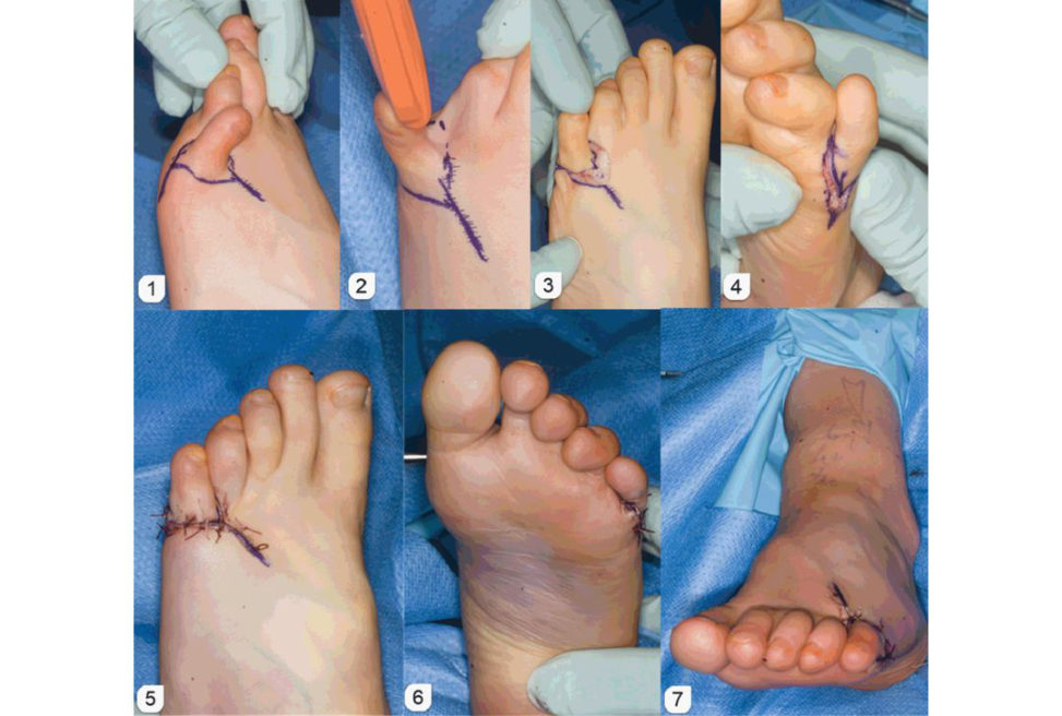
Figure 18.5 Pinning in situ.
Figure 18.6 Blind spot.
Figure 18.7 Posterior sloping angle.
Figure 18.8 A clinical photograph of a child who fell off a monkey bar.
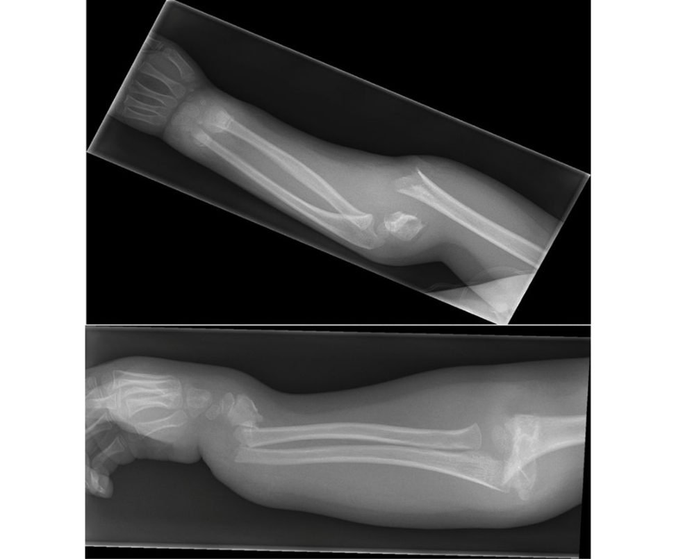
Figure 18.9 Plain radiograph of the child who fell off a monkey bar.
Figure 18.10 Reducing supracondylar humeral fracture.
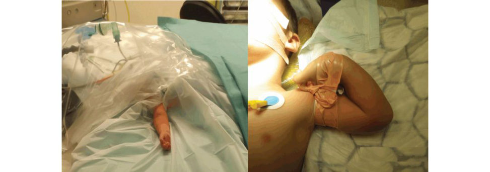
Figure 18.11 Supracondylar methods of fixation.
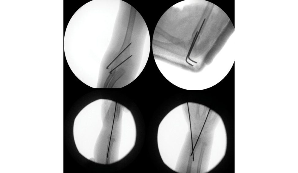
Figure 18.12 Intraoperative fluoroscopy images.
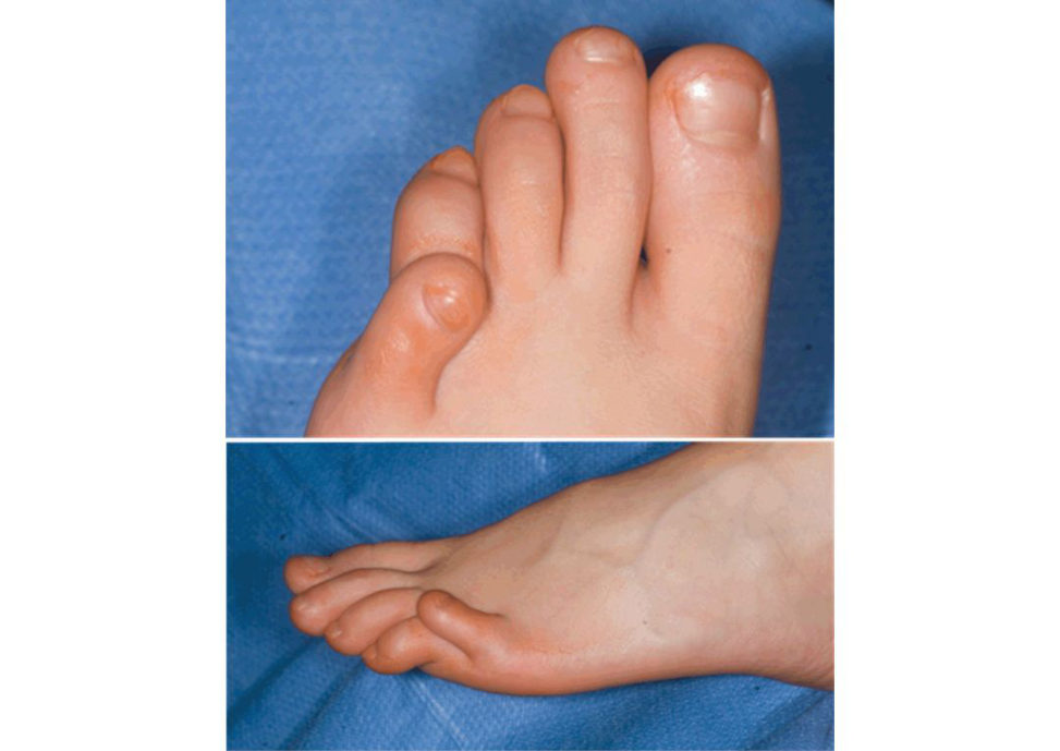
Figure 18.13 A child with the liti let oe deformity.
Figure 18.14 Butler’s procedure.
Table 18.1 Modified Gartland classification
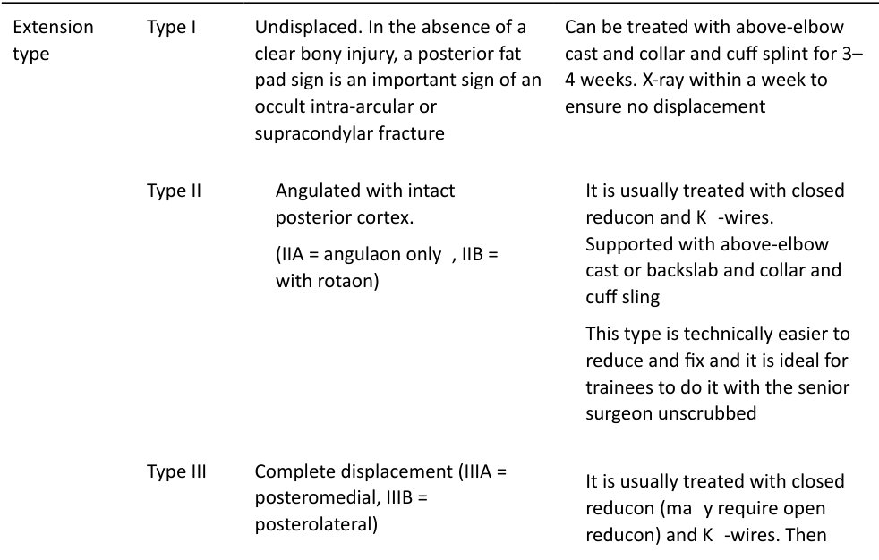
above-elbow back slab and collar and cuff sling
This type can be technically demanding and some experience is required
As in type III, but even with three wires this type can still be unstable
Flexion type As the name implies, the distal fragment flexes. This indicates that the thin posterior periosteum has been disrupted rendering the fracture very unstable.
1. Low and high SCH fractures (below and above the olecranon fossa), flexion type and medial comminutions are radiological signs that predict technically difficult surgery – be prepared.
2. Collar and cuff is part of the stabilization and should beworn 24/7. It is not for comfort as in forearm or wrist fractures.
Candidate 2#
This is a clinical photograph of newly born foot (Figure 18.15). What can you see?
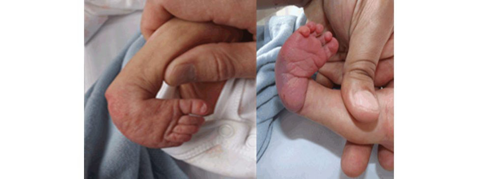
This photograph shows typical features of a clubfoot (congenital talipes equinovarus (CTEV)) deformity. This is often summarized as (CAVE) deformity: Cavus (high arched foot due to tight intrinsic foot muscles, FHL, FDL); Adductus of forefoot (tight tibialis posterior); Varus (tight tendoachillis, tibialis posterior, tibialis anterior); Equinus (tight tendoachillis). It looks severe. There are two classifications in use to assess the severity: Pirani score and Dimeglio scoring system. The former is more popular int heUK.
How does Pirani score clubfeet?
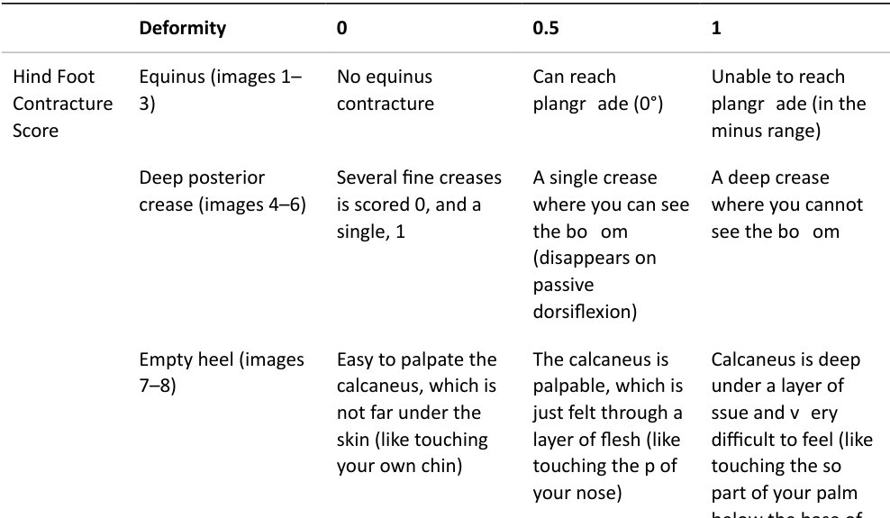
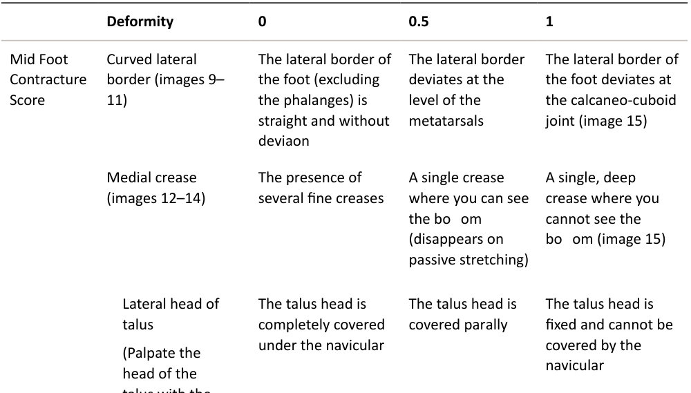
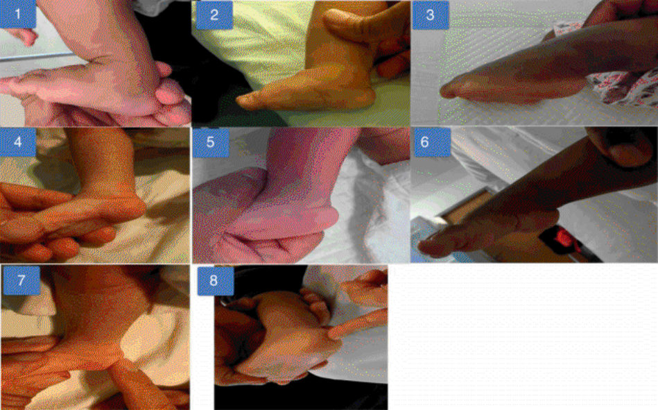
The Pirani score is simple and reproducible. It uses six clinical signs to quantify the severity of each hind foot and mid foot deformity. Each component is scored as 0 (normal), 0.5 (mildly abnormal) or 1 (severely abnormal) (Table 18.2). The six clinical signs are divided equally between the hind foot and midfoot as follows:
Hind Foot Contracture Score (HFCS) 0–3
1. Equinus.
2. Deep posterior crease.
3. Empty heel.
Midfoot Contracture Score (MFCS) 0–3
4. Curved lateral border.
5. Medial crease.
6. Lateral head of talus.
Figure 18.15 A newly born child with a foot deformity.
Table 18.2 Pirani score for clubfeet.
(Palpate the head of the talus with the foot uncorrected) (image 15)
Deformity 0 0.5 1
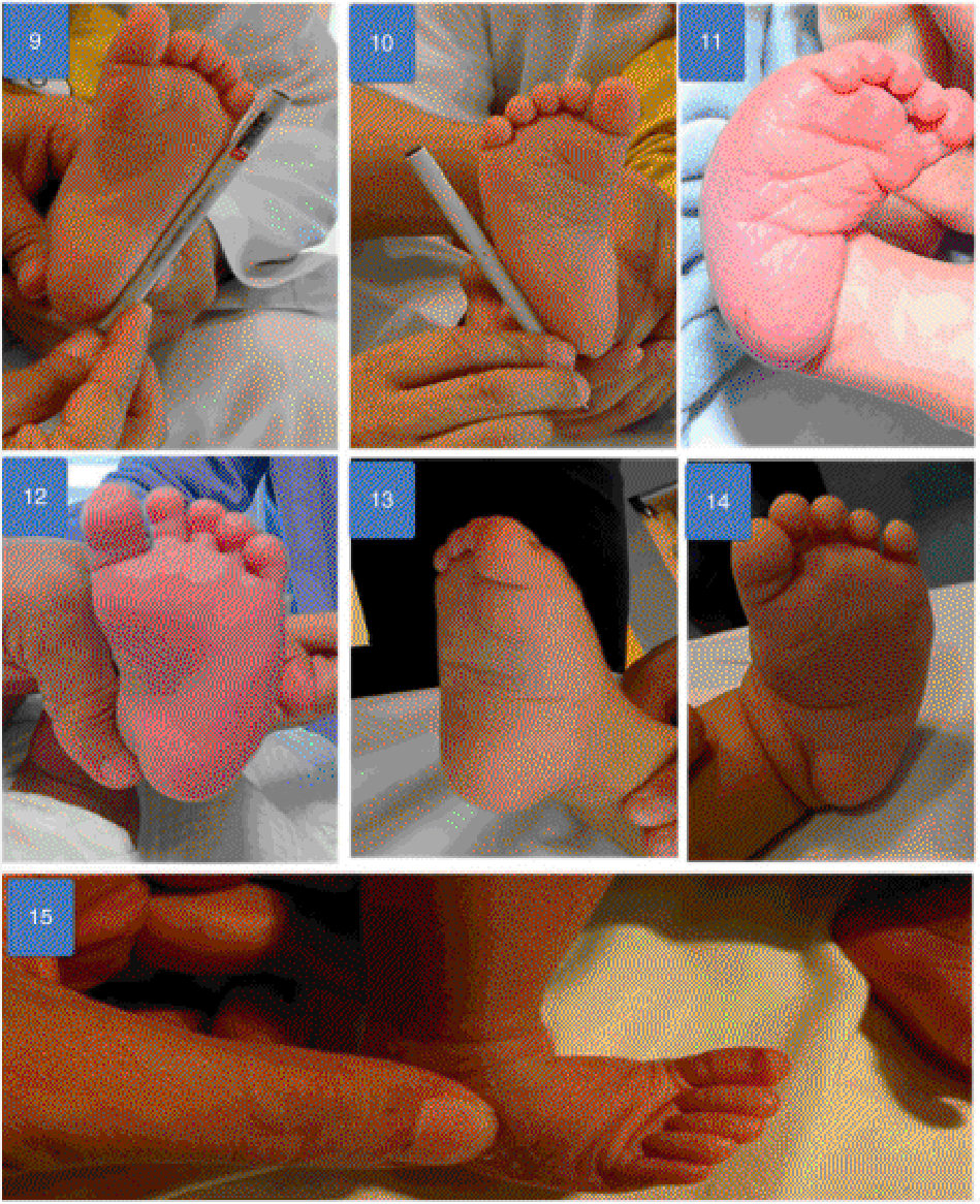
Pictures courtesy of Dr Sattar Alshryda.
Total Score (TS) 0–6
What causes clubfeet?
The cause in the majority of cases is unknown (idiopathic). A few theories have been postulated to explain the aetiology . 1. The neuropathic theory [12]: biopsies were taken from the posteromedial and peroneal muscle groups in 60 patients mostly under the age of 5 years. Evidence of neurogenic disease was seen in most instances and was more obvious in the older patients. 2. The myopathic theory [13]: a histochemical analysis was made of 103 muscle biopsies taken from 62 patients with idiopathic clubfeet. Authors noticed the muscles in patients aged under 6 months contained 61% Type 1 fibres in the affected legs compared to 44.3% in normal legs. 3. Genetic theory: it is common incertain races such as Polynesian and rare in the Japanese race. There is a 10% risk if a first-degree relative is affected; combination of en vironmental/genetic [ 14]. Twenty-five percent have a family history. Recent link to PITX1, transcription factor critical for limb development. 4. Arrested development of the growing limb bud. 5. Congenital constriction annular band. 6. Viral infection. 7. Mechanical moulding theory. 8. Multifactorial. a. Common in Polynesian race and rare in the Japanese race. b. Not more common in consanguinity. c. 10% risk if a first-degree relative is affected: combination of en vironmental/genetic [ 14]. d. 25% have a family history. Several associated conditions have been identified and these need to be excluded. These include the following: 1. Neurological causes: spina bifida (myelomeningocele), polio, CP. 2. Sacral agenesis. 3. Foetal alcohol syndrome. 4. Congenital myopathy. 5. Down’s syndrome (may include vertical talus). 6. Arthrogryposis.
7. Hand anomalies Streeter dysplasia/constriction band syndrome).
8. Diastrophic dwarfism.
9. Prune belly syndrome.
10. Opitz syndrome.
11. Larsen syndrome.
12. Anterior tibial artery hypoplasia or absence is common.
13. Tibial hemimelia.
How would you manage this child?
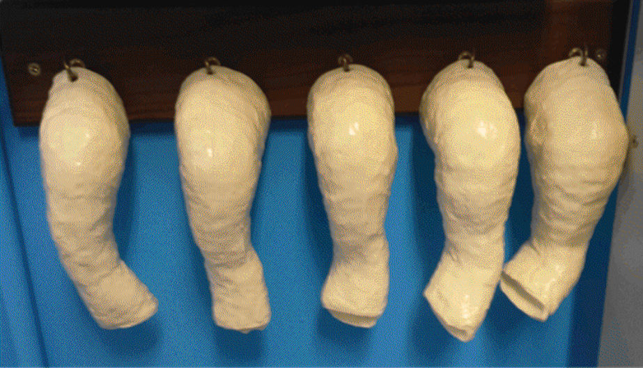
Most clubfeet are diagnosed prenatally in the 20-week scan and management and counselling starts before birth. Prenatal counselling is usually focused on the accuracy of diagnosis (65–90%) [15], potential associated conditions (see above) and treatments. Having established the diagnosis of idiopathic clubfeet, I recommend the Ponseft serial casting [16,17] (Figure 18.16). The treatment should be started as early as possible; the severity of the deformity is quantified using the Pir ani score, then serial casting weekly for an average of 4–6 cast changes. This usually corrects all deformities (CA V) with the exception of equinus, which requires a tendoachillis tenotomy in more than 90% of the cases. Sequence of deformity corrections follows the deformity pattern which is (CAVE): 1. Cavus (high medial arch) is due to the pronation of the forefoot in relation to the hindfoot. This is corrected by positioning the forefoot in a proper alignment with the hindfoot. The cavus is supple in newborns and requires only elevating the first ray of the forefoot to achieve a normal longitudinal arch of the foot. The foot usually looks worse in the first cast as the forefoot is facing inward. 2. Adduction of the forefeet and varus of the heel are corrected concomitantly using the talus head (not the cuboid) as a fulcrum. This is achieved while keeping the foot in equinus. 3. Equinus of the heel corrections tarts in the serial casting stage, but often requires tendoachillis tenotomy for full correction in 90% of patients. The timing to start correction of the equinus is critical. Premature correction could lead to midfoot break and rocker boft om foot. Three signs indicate the right timet o start equinus correction: i. The ability to palpate the anterior process of the calcaneus as it abducts out from beneath the talus. ii. Forefoot abduction of approximately 60° in relationship to the frontal plane of the tibia. iii. Neutral or slight valgus heel.
Tendoachillis tenotomy is performed when the residual equinus is about 0–5°. It is performed under local anaesthetic, then the final c ast applied for another 3 weeks.
Successful correction is followed by a regime of using Denis Browne (DB) splint (Figure 18.17) on a full-time basis for 3 months, after which the splint will be used at nap and nightime un til the age of
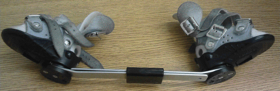
4–5 years. The splint consists of open-toe straight shoes attached to a bar. The bar should be of sufficient length so that the heels of the shoes are at shoulder width. This can be adjusted using the sliding clamp in the middle. The bar should be bent 5–10° to hold the feet in dorsiflexion. For unilateral cases, the brace is set at 60–70°of external rotation on the clubfoot side and 30–40° of external rotation on the normal side. In bilateral cases, it is set at 70° of external rotation on each side.
This is another child who was referred to you with a clubfoot (Figure 18.18). Have a look at the picture and tellus what your thoughts are.
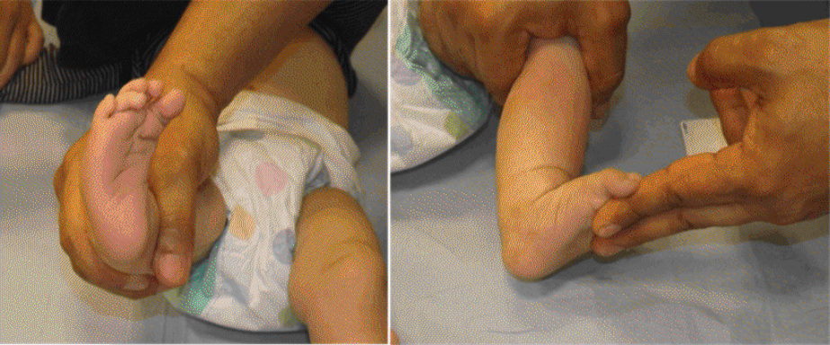
This clinical photograph of the right foot shows a marked foot deformity with forefoot adducted; however, there is no convincing equinus or varus deformity. My thoughts are that the child has a metatarsus adductus and not a clubfoot. I need to examine the child to confirm my thoughts.
Tell me more about metatarsus adductus (MA)?
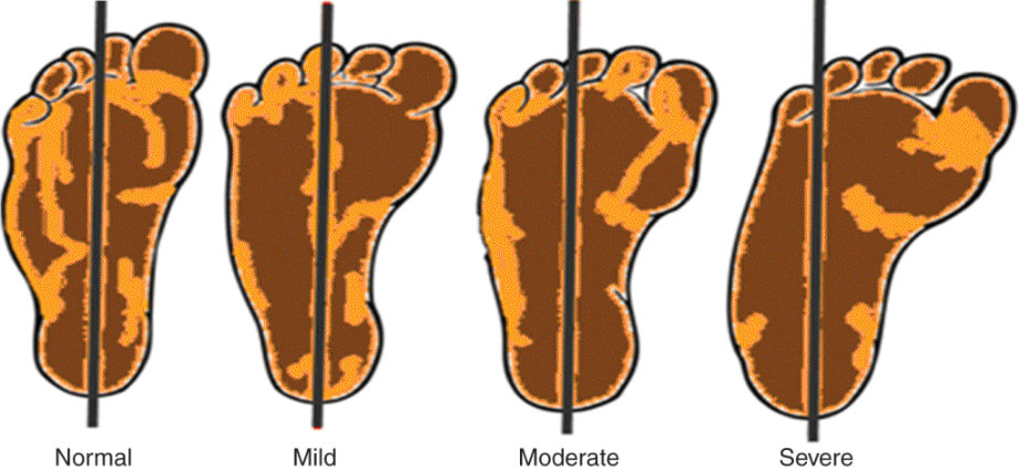
As the name implies, the metatarsus (forefoot) is adducted in relation to the hind foot. It is as common as clubfeet (1 in 1000 births), no sex predominance and bilateral in approximately 50% of cases. It is widely considered as a foetal packaging disorder (that is why it is common in late pregnancies, first pregnancies, twin pregnancies and oligohydramnios). For the same reasons, MA is linked to DDH (15–20%), torticollis and plagiocephaly. It can bepart of a more complex foot deformity such as clubfoot and skew foot. Bleck classified the severity using the heel bisector line into mild, moderate and severe (Figure 18.19). Normally, the heel bisector line passes through the second and third toe web space. In a mild MA, the heel bisector line passes through the third toe; in a moderate MA the line passes through the third and fourth toe web space and in a severe MA, it passes through the fourth and fitih toe web space.
How would you treat metatarsus adductus?
Isolated MA is a benign condition that resolves by the age of 5 years (or even earlier). I would advise serial stretching by parents if the MA is flexible and can be actively or passively corrected to midline, whereas a rigid deformity with medial crease requires serial casting. Surgery is rarely indicated (and often unnecessary) before the age of 5. However, persistent and symptomatic MA after the age of 5 may warrant lateral column shortening if the foot is long or medial column lengthening if the foot is short. Lateral column shortening is done with cuboid closing wedge osteotomy. Medial column lengthening includes a cuneiform opening wedge osteotomy with medial capsular release and abductor hallucis longus recession.
How do you know whether the foot is long or short?
By comparing it to the standard growth charts.
Authors’ note: 3
Clubfoot is another A-list topic in the paediatric section. It should be an area where you get a full mark. The above scenario is a typical example for a straighfoorward pass performance. The candidate answered all the questions in a comprehensive and correct matter and clearly talked more than the examiner. He did not need prompting and supplemented some evidence.
Our advice is that you should aim for more than a pass by considering the following:
1. The Ponseft vs. the French method.
2. Complications of treatments.
3. Relapse and its treatment.
4. Potential operative intervention:
a. Posteromedial soft -tissue release and tendon lengthening.
b. Medial column lengthening or lateral column-shortening osteotomies.
c. Talectomy (in severe, rigid recurrent clubfoot in children with arthrogryposis).
d. Gradual correction using a circular frame.
Figure 18.16 The classic shapes of casts in Ponseft weekly serial casting.
Figure 18.17 Denis Browne boots.
Figure 18.19 Bleck classification of metatarsus adductus severity.
Figure 18.18 A child with right foot deformity.
Clubfoot, metatarsus adductus and congenital vertical talus are completely different conditions and clear understanding of these three conditions and their treatment is essential.
Make sure that you do not confuse them in the exam (and real life!).
The below is a photograph of a child with a thumb deformity (Figure 18.20). What is the diagnosis?
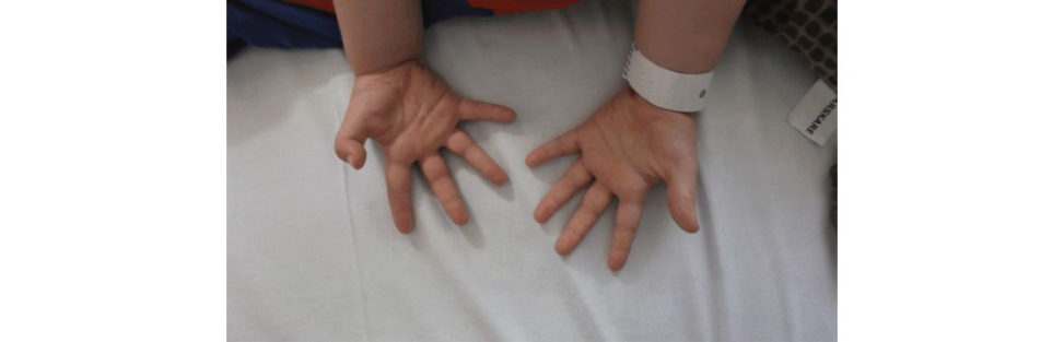
The right thumb IPJ looks in a fixed flexion deformity; the likely diagnosis is a congenital trigger thumb; however, without a proper examination, I cannot be certain. I need to see that the IPJ can be flexed further, I would like to feel the mobile lump at the base of the thumb. Sometimes, the deformity can be corrected with a palpable (and even visible) clunk.
You are correct. This is a trigger thumb. What would you tell the parents?
I would tell them about the condition and the known reason behind it. I will explain the management options including conservative (observation exercises, splinting) and surgical (open A1 pulley release or percutaneous A1 pulley release). In my practice, if conservative treatment fails, I would offer the child open A1 pulley release. It is a small operation, with low comorbidity and a high success rate. Unfortunately, the natural history of congenital trigger thumb is not very well studied, and the evidence accumulated is predominantly level III and IV. Many of the study interpretations are vulnerable tobias [18].
Figure 18.20 A child with a thumb deformity.
Candidate 3#
You have been called to see an 8-year-old boy who presented to the A&E with limping on the right leg. How would you approach this child?
Limping is one of the commonest reasons why parents seek medical advice. The list of causes for a limping child is long and ranges from trivial conditions that require just reassurance such as minor trauma to the most serious conditions that require admission investigations and urgent surgery, such as infections and tumours. A thorough history and examination are essential to narrow the list to a few working diagnoses. It is essential not to miss important diagnoses even if they are not common (such as infections or tumours), as delay may jeopardize the outcome.
What do you want to know in the history?
I would like to know whether there is a history of trauma (trauma or SCFE), temperature (infection), upper respiratory tract infection or earache (transient synovitis constitutional symptoms and weight loss (tumour or infection). I would like to know about the onset of symptoms, precipitating and relieving factors, impact on his daily activities.
There was no history of trauma, no temperature and no recent URTI or contact with an unwell child. He has never had this problem before and there are no swollen joints.
I will proceed with my examination, I would start with general signs and walking, then I examine his lower limbs, back and upper limbs. I will look, feel, move his joints and perform special tests relevant to particular joints and what I am looking for.
You examined him and he was walking with obvious limp, he was holding his leg in a position of rest and there was a reluctance to move the right hip.
This is important information to know as I can now focus on hip causes of limping and I can investigate accordingly. I would like to obtain a pelvic X-ray and request some bloodtests (FBC, ESR, CRP).
Your junior has already requested these as well as rheumatoid factor (RF) and serum uric acid (UA).
That is OK. I would not recommend checking RF and UA at this stage. Juvenile rheumatoid arthritis and g out are rare in children and these are not at the top of my list at this stage unless there are clear signs to indicate the contrary, such as gouty tophi or the patient being on cytotoxic medicines. My investigations are guided by what I want to rule in and rule out at this stage of presentation. When I want to find the diagnosis ASAP, I do not want to miss or delay important diagnosis such as infection Therefore, I requested the above bloodtests to estimate the probability of infection in this child using K ocher’s criteria.
What are Kocher’s criteria?
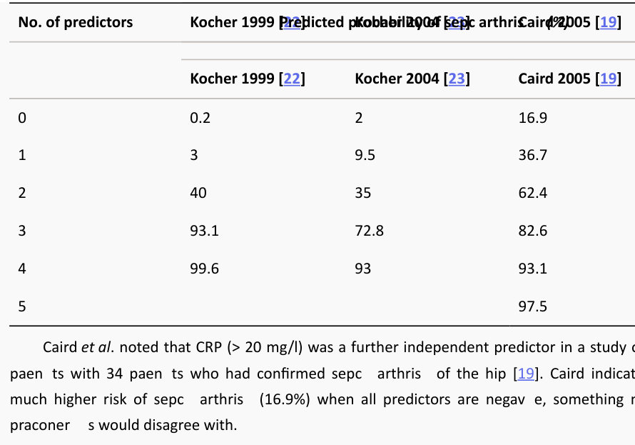
These are criteria to help differentiate between transient synovitis and septic arthritis, as both have a similar early presentation. The criteria include: 1. Fever (> 38.5°C). 2. Inability to weight-bear. 3. ESR > 40. 4. WBC > 12,000/mm3. InKo cher’s original paper, the predicted probability of septic arthritis with one positive predictor was 3%, two predictors 40%, three predictors 93% and four predictors 99.6%.
What about the CRP that you requested?
CR Pis more sensitive to infection thanE SR (although it was not mentioned in the K ocher criteria). It usually rises within 6 hours after an insult (whether infection or injury Caird and colleagues [19] noted that CRP (> 20 mg/l) was a further independent predictor of septic arthritis. However, the fact that CRP can be elevated after injury (including surgery) precludes its diagnostic value. Procalcitonin (PCT) is another infection marker that has become increasingly popular. PCT has even greater specificity thanES Rand CR Pin identifying patients with infection. It is s till expensive and not widely available [20,21].
Authors’ note: 4
The FRCS exam is about safe and high-standard practice. This should be based on evidence. There is a myth that candidates should not quote published evidence. You can pass the exam without quoting a paper; however, quoting papers and published evidence will enhance your performance. It is even more impressive if you know the evidence, its strength and weakness and how you would utilize this evidence in your practice knowing its limitations. The above candidate scored high by quoting a well-recognized paper and showed that he kept updated with recent advances in the field (the PCT value in diagnosing septic arthritis). He could have emphasized the limitation of the evidence that he quoted, for example by saying, ‘It is important to appreciate that even when all Kocher’s criteria are negative the risk of infection is still present ranging from
0.3% to 17%’.
As this area is extremely important, we summarize the evidence below.
In a study of 282 patients, Kocher and colleagues [22] identified the above four independent predictors to differentiate between transient synovitis and septic arthritis: They put the patients into three groups: confirmed septic arthritis, presumed septic arthritis and transient synovitis.
The diagnosis of true septic arthritis which occurred in 38 patients was assigned when a patient had one of the following:
1. A positive finding on culture of joint fluid.
2. WCC ≥ 50,000 cells per cubic millimeter with positive findings on blood culture.
The diagnosis of presumed septic arthritis (44 patients was assigned when apa tien t had
WCC ≥ 50,000 cell per cubic millimeter in the joint fluid with negative findings on culture of joint aspirate and blood.
The diagnosis of transient synovitis (86 patients was assigned when the patient had WCC
< 50,000 cells per cubic millimetre in the joint with negative findings on culture, resolution of symptoms without antimicrobial therapy, and no further development of a disease process as documented in the medical record.
The group with septic arthritis (88 patients included both groups with confirmed and presumed septic arthritis. Predicted probability of septic arthritis with one predictor was 3%, two predictors 40%, three predictors 93% and four predictors 99.6%.
Kocher validated his criteria prospectively in another study of 154 patients [23]: 24 had true septic arthritis, 27 were presumed to have septic arthritis, 103 had transient synovitis. He had slightly different findings (see Table 18.3).
Table 18.3 Predictors for septic arthritis.
No. of predictors Predicted probability of septic arthritis (%)
The CRP did not perform as well as PCT in a systematic review and meta-analysis by Zhao
[21]. The review included 10 studies (838 patients and found that the overall sensitivity of serum
PCT levels for the diagnosis of septic arthritis was 0.54 (95% CI, 0.41–0.66), and the specificity was
0.95 (95% CI, 0.87–0.98). The sensitivity and specificity of CRP were 0.45 (95% CI, 0.35–0.55) and
0.079 (95% CI, 0.0.021–0.25), respectively.
The bloodtests that you requested came all within normal. This is the X-ray that you requested.
This AP pelvis radiograph (unlabelled) shows obvious abnormality of the right femoral head, the epiphysis is small, denser (whiter in colour) and there is some fragmentation Several possibilities can cause such a picture. On the top of my list isL egg Calves Perthes disease (LCPD), but there are others such as: 1. Infections (normal blood does not exclude septic arthritis or osteomyelitis ). 2. Multiple epiphyseal dysplasia (MED: as the name implies, there will be involvement of the other side and other joints). 3. Spondyloepiphyseal dysplasia (similar to the MED but with spinal involvement). 4. Sickle cell disease (history and family history). 5. Eosinophilic granuloma (other lesions of the skull, radiological feature, biopsy). 6. Gaucher’s disease.
7. Hypothyroidism.
8. Meyer’s dysplasia.
How do you confirm your diagnosis?
The history, examination and initial investigations indicate that the likely diagnosis isL CPD. However, if I am still in doubt, other investigations can rule out other possibilities. Joint aspiration and MRI sc an could largely exclude joint and bone infection Thyroid function t est to rule out hypothyroidism, sickling testor HB electrophoresis to rule out sickle cell diseases, etc. Every test that I may use to confirm my top diagnosis (LCPD) has some limitation and the eventual diagnosis will have some uncertainty.
You did all these tests and you are convinced that this child has LCPD. How would you treat him?
My first line of treatment for all children with LCPD is to treat their symptoms and educate them and their parents about the disease itself. Rest, analgesia, anft-in flammatory, temporary non- weight-bearing with crutches and I may consider admiting for a short period of gentle traction if his hip is very stiff. Physiotherapy plays an important role in improving range of motion Several types of braces have been advocated, but their values have been contested and compliance is a real issue in this age group.
How would you educate them?
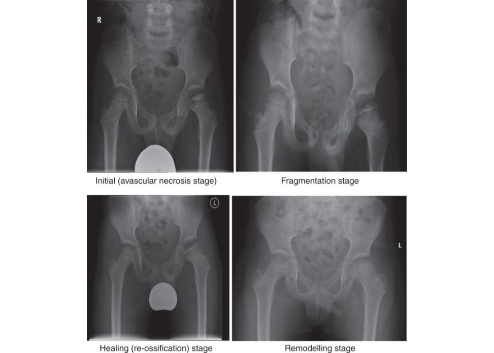
I would explain the condition to them verbally and I would also provide them with aw rift en leaflet about the condition. I would explain that LCPD is a unique disease that is not fully understood. The disease is caused by interruption of blood supply to the femoral head. We do not know why this happens. There are a few theories suggested to explain the reasons, but none has been proven. The blood supply is restored spontaneously over a period of 2–4 years. During this period the femoral head passes through distinctive stages (Figure 18.22): 1. Initial (also called necrotic or a vascular necrosis stage). 2. Fragmentation (or resorption stage). 3. Healing (re-ossification or reconstitution stage). 4. Remodelling stage. Each stage lasts 6 months on average (range 3–18 months). There is overlap between these stages. Some areas of the femoral head may start healing while other parts are still fragmenting. The childs symptoms tend to get worse as the disease progresses into the fragmentation stage regardless of the types of treatment that the child gets. Several radiological classifications have been advised to grade the severity of the disease. The most popular is the lateral pillar classification (by Dr Herring). The classification depicts that the femoral head is made of three equal pillars (medial, middle and lateral). The severity is divided into four groups based on the height of the lateral pillar or third on the AP film at the beginning of the fragmentation stage (Figure 18.23). The groups are:
Group A: Normal height of the lateral third of the head is maintained.
Group B: More than 50% of the original lateral pillar height is maintained.
Group C: Less than 50% of the original lateral pillar height is maintained.
Group B/C: Less than 50% of the original lateral pillar height is maintained but the lateral pillar is higher than the central segment.
I would also explain that when LCPD is fully healed, there are other classification systems that can help us predict how well the child’s hip would do in the future. I personally prefer the modified
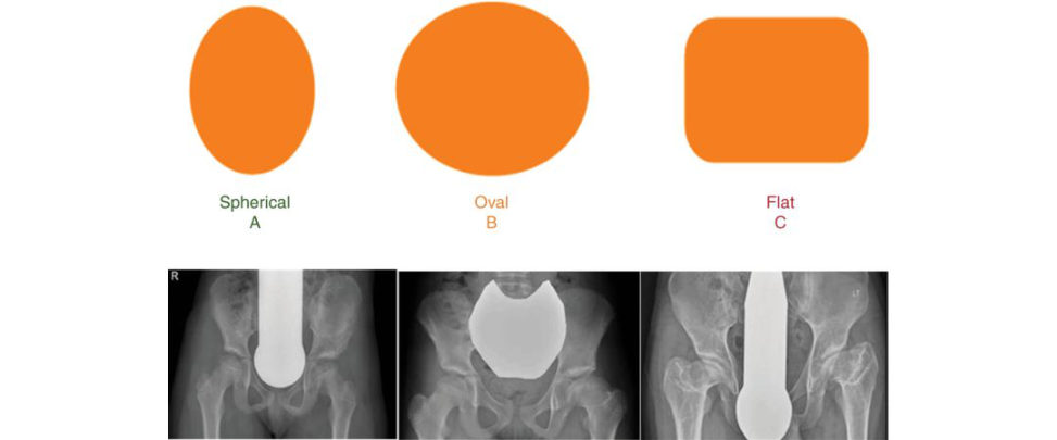
Stulberg classification ( Figure 18.24). It consists of three groups: Group A hips have a spherical femoral head, group B have an ovoid (or mushroom-shaped) femoral head and group C have a flat femoral head. Group A usually do well. Group C do not usually do well, and they would require total hip replacement in their fitiies. Group B usually run a course between groups A and C.
There are some clinical and radiological factors that help predict which hip would do well and which does not. The following are not good signs (often referred to as the FOOBS):
1. Females.
2. Older children (older than 6 years).
3. Overweight.
4. Bilateral hip involvement.
5. Stiffness of the involved hip.
So which Herring group would you think this child belongs to?
I would classify this as group B. The lateral pillar is more than 50% (compared to the other side).
Would you consider surgery?
Yes, I would consider containment surgery (femoral varus derotation osteotomy, Salter osteotomy or both). My preferred option is femoral varus derotation osteotomy. The evidence shows that containment surgery is beneficial inpatients with lateral pillar B and B/C stages who are 8 years or older [24,25]. It increases the number of patients with Stulberg A by 30%.
Have you done or seen femoral varus osteotomy for LCPD? Can you take me through it?
Yes. Preoperative planning is essential. It is important to ensure that there is a good hip abduction to compensate for the varisation of the femur. We can also estimate how much varus is required to achieve good containment by obtaining a plain radiograph, in the neutral position and abduction (in neutral rotation). This step can be done in theatre using fluoroscopy.
The patient is positioned supine on theo per ating t able and surgery is performed under GA. The lower limbs are prepared and draped free. Adductors release may be required if there is significant tightness.
I use the lateral subvastus approach. The periosteum is incised and elevated with a Cobb elevator. I prefer the paediatric proximal locking plate, I use the size which is appropriate for the patient weight
(3.5 mm for those under 35 kg and 5 mm for those above). I set my guiding jig based on the plate angle and the desired amount of varisation.
Jig angle = plate angle + the desired amount of varisation
So, if I want a varisation of 30°, I will use 110° plate and I set my jig to 140°.
I fix the plate proximally then I remove it (so that it will be ready to be re-fixed again after the femoral osteotomy). It is important to mark rotation before performing the femoral osteotomy.
Several techniques have been used. I use two K-wires; one above and one below the osteotomy, using the hockey stick (a small curved spanner on the paediatric locking plate that looks like a hockey stick) to identify the level of the femoral osteotomy.
I perform femoral osteotomy using an oscillating saw. Gentle soft -tissue releases around the osteotomy site to aid varisation. I fix the plate proximally then distally. Depending on the femoral head damage, I may add some internal rotation or extension for better containment. I washout the wound and close it in layers.
Would you do the same if this child has lateral pillar type C?
The evidence shows that surgery does not improve the outcome in this group. However, I may consider other options such as r est, traction, adductor release and Petri if there are significant stiffness and lateral extrusion.
Would you do the same if this child has lateral pillar type A?
For the best result, surgery should be performed in the earliest stage of the disease; maybe even before being able to classify the severity. There is uncertainty whether type A is a genuine type or will progress to type B or even C. If all children older than 8 years were to be offered surgical treatment, the group A and C hips would not likely benefit. These groups combined represent only 13% of hips presenting a t age older than 8, and this approach may be justified [ 26].
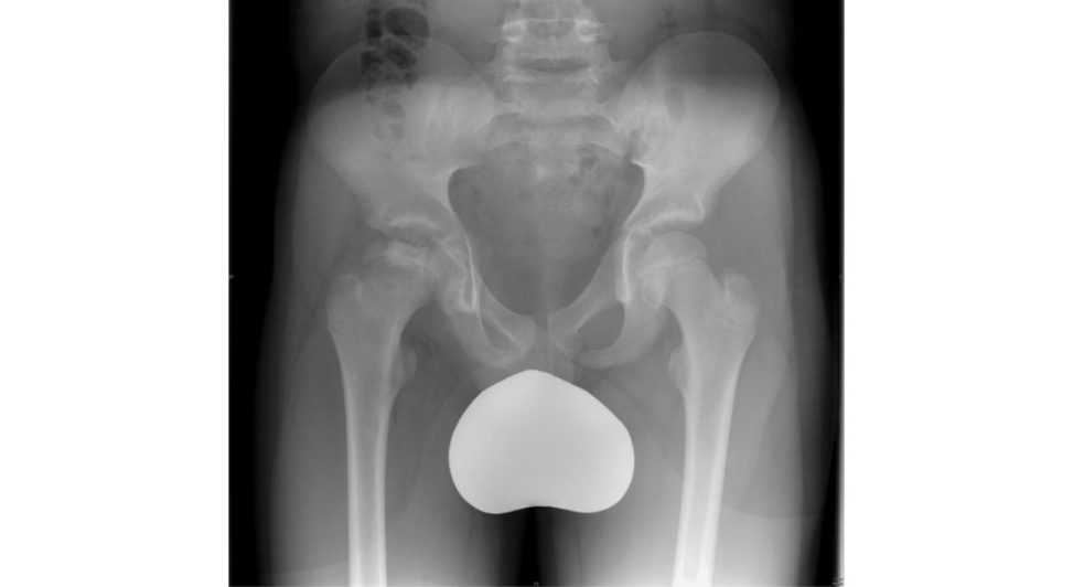
Figure 18.21 Pelvis X-ray of a child with right hip pain and limping.
Figure 18.22 LCPD stages.
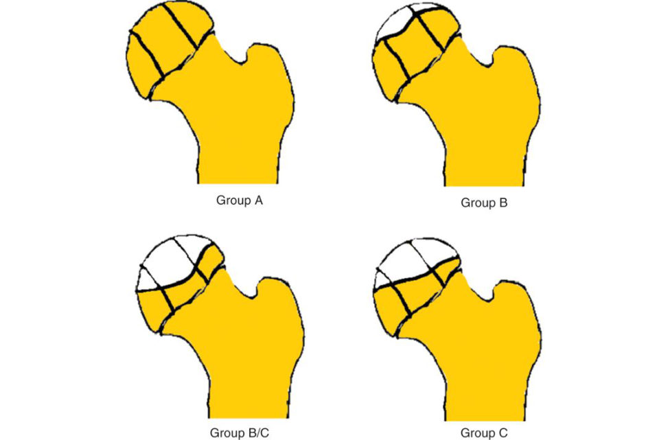
Figure 18.23 Herring classification.
Figure 18.24 Modified Stulberg classification.
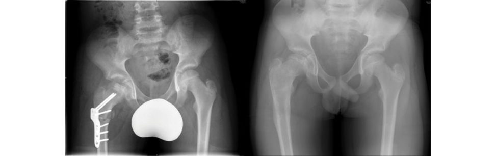
Figure 18.25 Varus osteotomy of patient with LCPD and a few years later.
Authors’ note: 5
LCPD is another A-list topic. The ability to discuss the findings and differential diagnosis is a must topass the exam. Treatment is very controversial and there is a paucity of evidence to support treatment. There are only two level II evidence for intervention and the y are worth reading:
Herring et al. [24] reported on the results of the Legg Perthes Study Group. Thirty-nine surgeons from 28 centres took part in a prospective study. Each surgeon agreed to apply a single treatment method to each patient who met the study criteria. All patients were between 6 and
12 years of age at the onset of the disease, and none had had prior treatment. The treatment groups were no treatment, range of motion treatment in which the patient did exercises once a day, Atlanta brace treatment, femoral varus osteotomy and Salter osteotomy.
The study showed that age, lateral pillar grading and treatment methods were significantly related to outcome.
In group B hips with an age at onset of more than 8 years, 73% of the operated hips had a
Stulberg I or II result compared with 44% of the non-operated hips (P = 0.02). In the group B hips with onset at 8 years or younger, there was no advantage demonstrated for the surgical group.
The group C hips were not shown to benefit from surgical or non-surgical treatments.
Wiig [25] reported on a nationwide prospective study. Twenty-eight hospitals in Norway were instructed to report all new cases of LCPD over a period of 5 years.
A total number of 368 with unilateral disease were included in the study. For patients over
6 years of age at diagnosis with more than 50% necrosis of the femoral head (152 patients the surgeons at the different hospitals had chosen one of three methods of treatment: physiotherapy
(55 patients the Scotish Rite abduction orthosis (26) and proximal femoral varus osteotomy (71).
The study showed that the strongest predictor of poor outcome was femoral head involvement of more than 50% (modified Catterall classification) followed by age at diagnosis, then lateral pillar grades. In children over 6 years at diagnosis with more than 50% of femoral head necrosis, proximal femoral varus osteotomy gave a significantly better outcome than orthosis or physiotherapy. There was no difference in outcome after any of the treatments in children under
6 years.
It is worth knowing more details about Stulberg classification and its modification. The original classification is based on sphericity of the head and congruency of the hip joint.
A modified version of the Stulberg classification is becoming more popular. It consists of three groups: group A hips (Stulberg I and II) have a spherical femoral head, group B (Stulberg III)
have an ovoid femoral head and group C (Stulberg IV and V) have a flat femoral head.
Other small studies showed the benefit of shelf acetabuloplasty as a salvage operation for extruded head [27,28]; valgus osteotomy in hinged abduction [29]; and trochanteric growth arrestor advancement when there is overgrowth.
Soft -tissue release and articulated hip distractor intreating late-onset LCPD (even severe ones) have shown promising results [30,31]. Segev and colleagues studied 16 children with late-onset and type CL CPD. Fourteen patients had a saddle-shaped subluxating femoral head with hinge abduction. They found that soft -tissue release and articulated hip distraction produced good results, including the disappearance of the saddle-shaped femoral head in 10 of the 14 patients.
Table 18.4 Stulberg grading.
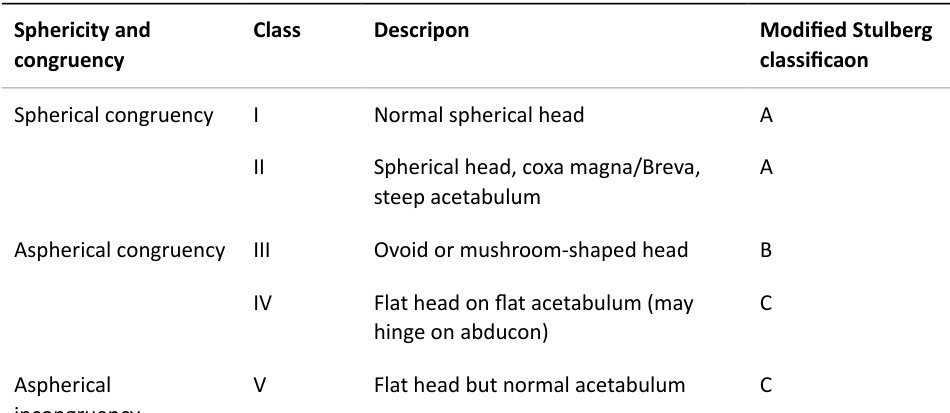
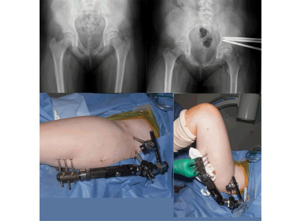
Figure 18.26 Articulated hip distractor in LCPD.
Candidate 4#
This is a clinical photograph of a 2-year, 8-months-old girl who was brought to you by her parents because they are concerned about the bent legs that she has (Figure 18.27). What are your thoughts?
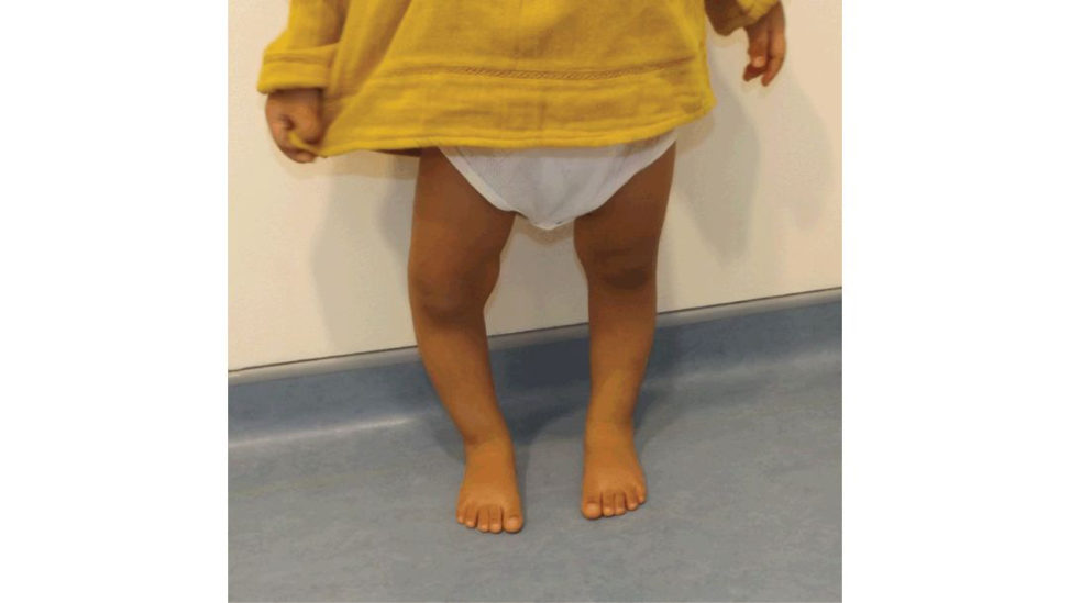
This is a clinical photograph of a standing child. There is an obvious asymmetry of leg alignment, with the right leg more varus than the left, which looks almost straight. I anticipate a child of this age has almost straight knees (or slight valgus) as depicted by the Salenius curve.
Tell me more about the Salenius curve.
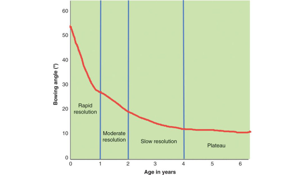
Salenius and Vankka reported on the development of the tibiofemoral angles in 979 children from Finland. They showed that the tibiofemoral angle (coronal alignment for the knee) follows a distinct pattern. Newborn babies were born with an average knee varus of 10–15°. As children started standing and walking, the knee became straight at around 18 months of age. This was followed by a progression into valgus of an average of 10° around the age of 4. Thereafter, knee valgus gradually reduced to the adult valgus of about 6° over the next several years (Figure 18.28). The standard deviation (SD) of the abo ve-quoted angles was 8° (more in boys (10°) and less in girls (7°)).
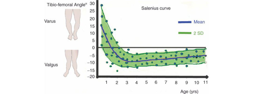
How would you approach this patient?
I would like to take a detailed history and perform a thorough examination. I want to know when they noticed the deformity, is it geting worse or better, are there any symptoms, did they have any treatment? I want to know whether there is any history of trauma or previous infection ( growth arrest). Any family history of similar conditions, joint diseases, congenital or genetic disorders. I would perform a general examination looking for any signs to explain the deformity. Overweight children from Afro-Caribbean backgrounds (Blount’s disease), other limb deformities ( growth arrest, congenital disorders), widened metaphysis and rachitic rosary (Rickets). Is there a lateral knee thrustor rotational lo wer limb deformity on walking? Then I would examine the lower limbs (looking, feeling, moving and special tests). I will look for any obvious abnormalities, f eel for swellings, tenderness or abnormal sounds (clicking, clunking or crepitus), I will check joint movements (active and passive) then I will do relevant special tests. The cover-up testis a useful screening test to assess this child’s lower limb alignment. A positive test (bowing in the upper tibia) or a neutral test (a straight thigh–upper leg axis) is an indication for radiographic evaluation. A negative test (slight valgus at the upper tibia) indicates physiologic bowing. A positive cover-up test has a high sensitivity; a negative cover-up test has a high specificity and negative predictive value [32].
The below clinical photograph (Figure 18.29) shows the cover-up test. What do you think?
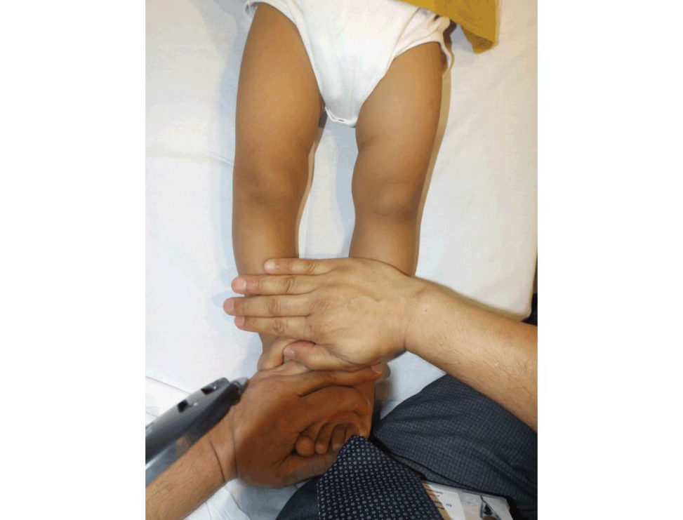
I think the child has a positive cover-up teston the right and neutral on the left and I would request a longlegs alignment view.
This is the X-ray that you have requested (Figure 18.30). Tell me what you can see.
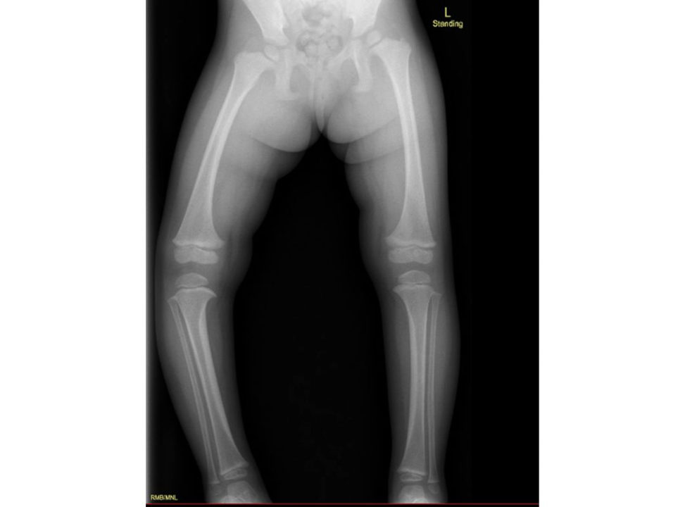
The X-ray shows most of the lower limbs. Ideally, I would like to see the top of the iliac crest and the heels. I also noted the X-rays are not fully labelled (names, dates, time, etc.) so I have to bear this in my mind when I make my decision. It confirms my clinical impression that there is an asymmetry of the lower limb alignment. There are features that are suggestive of Blount’s disease (stage I) with a varus deformity of the knee and medial beaking of the metaphysis. I would like to draw and measure some angles and distances to confirm my suspicions and predict the probability that the deformity may progress.
OK; go ahead. Show us what angles you measure and how.
I usually measure the tibiofemoral angle (TFA), mechanical axis deviation (MAD), metaphyseal–diaphyseal angle of Levine and Drennan (MDA)[33], epiphyseal–metaphyseal angle (EMA), femoral–metaphyseal–diaphyseal angle (FMDA) of O’Neill and MacEwen [34] and femoral– tibial ratio of McCarth y (FTR) [35] and tabulate them comparing right and left.
Figure 18.27 A child with bent legs.
Figure 18.28 Salenius curve of tibiofemoral angle.
Figure 18.29 Cover-up test (positive on the right and neutral on the left – both are indication for X-ray).
Figure 18.30 A longlegs alignment view in a child.
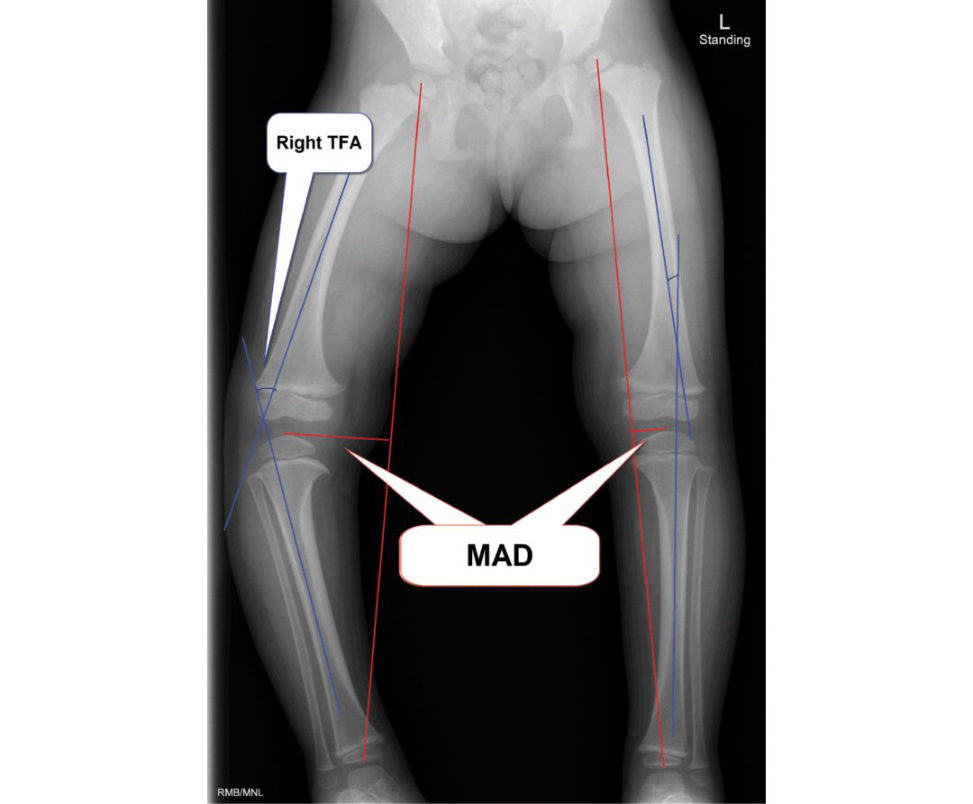
Figure 18.31 Tibiofemoral angle and mechanical axis deviation. The TF A is the angle the femoral and tibial mid-diaph yseal. MAD is the distance between the mechanical axis and the centre of the knee.
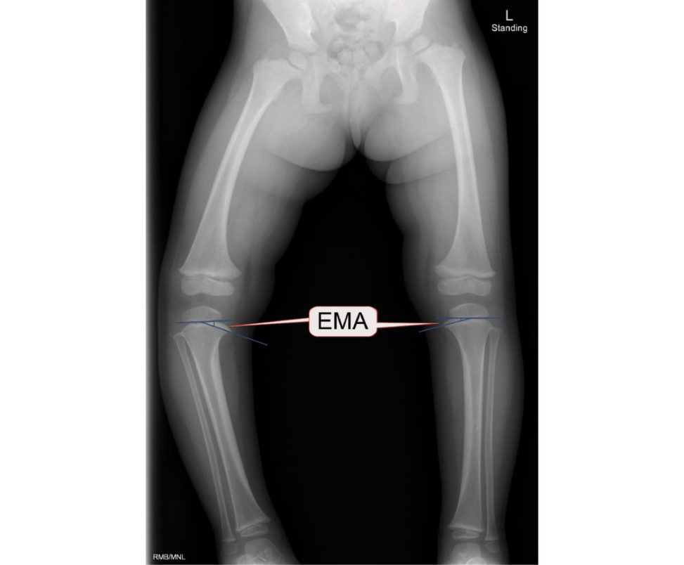
Figure 18.32 Epiphyseal–metaphyseal angle (EMA). The EM Ais the angle formed by the epiphyseal line (a line through the proximal tibial physis parallel to the base of the epiphyseal ossification centre) and a line connecting the midpoint of the base of the epiphyseal ossification centre with the most distal point on the medial beak of the proximal tibial physis.
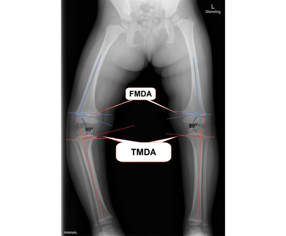
Figure 18.33 Metaphyseal–diaphyseal angles. Th eMDA is the angle formed by a line connecting the most distal point on the medial and lateral beaks of the metaphysis (distal femur in FMDA and proximal tibia in the TMD A) and a line perpendicular to the anatomic axis (or lateral cortex) of the bone. The femoral-tibial ratio (FTR) is defined as the FMDA divided by the TMDA.
Table 18.5 Angle measurements around the knee.
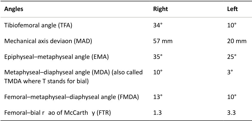
The TFAs show bilateral varus with the right side worse. The right side is probably more than 2 SD of the average predicted by the Salenius curve; however, the left side is still within the expected range. This is also reflected by the MAD, which is more than the normal value (0 ± 3 mm from the centre of the knee).
MAD is valuable to monitor progression of the disease.
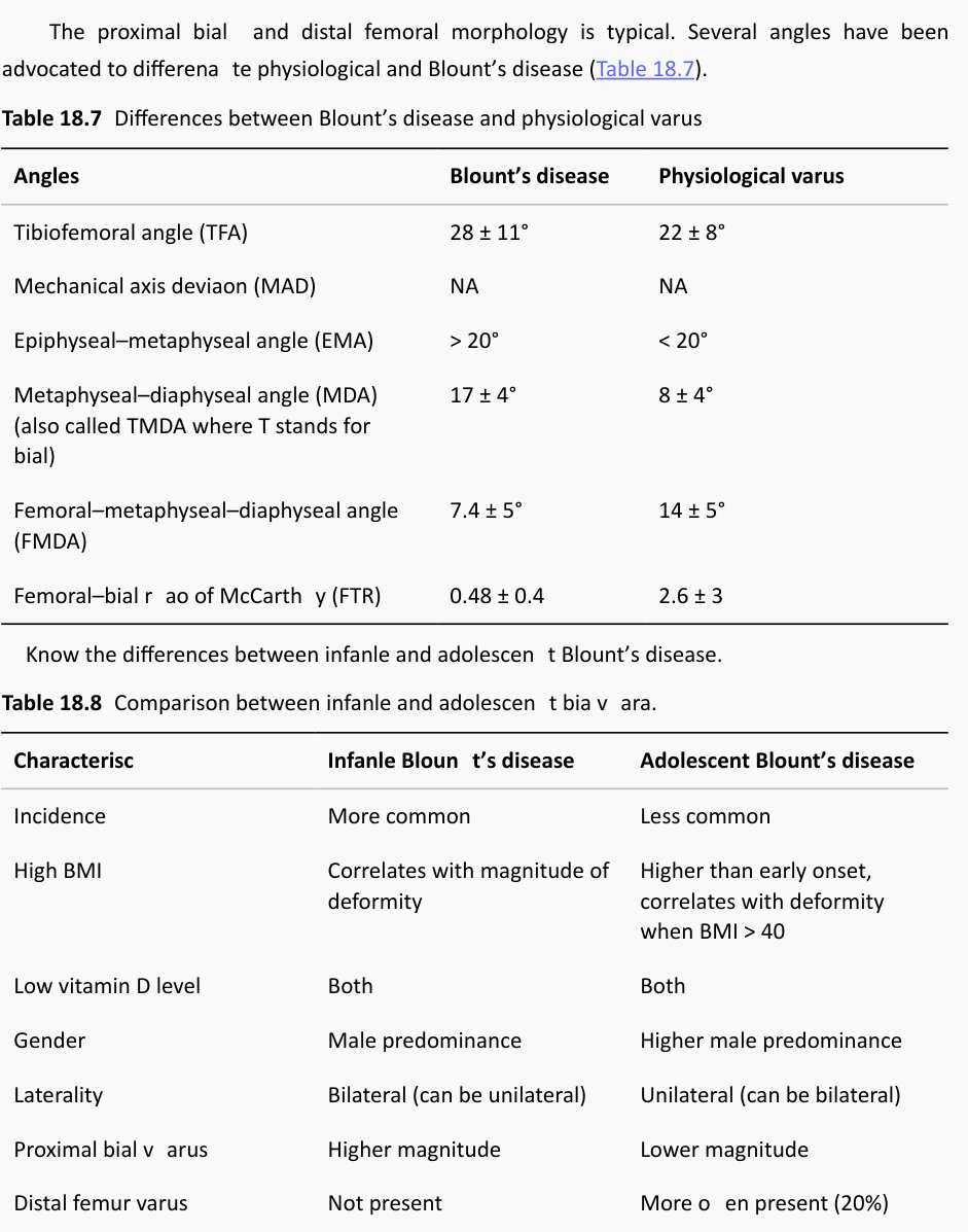
The other angles help to differentiate between physiological and pathological genu varu mAs with any test, they are not 100% sensitive or specific and clinical judgement is often required when the values fall in the grey zone. In general, when the EMA < 20°, TMDA < 10°, TFA < 10° and the FTR > 1.4 Blount’s disease is very unlikely, whereas if EMA > 20°, TMDA ≥ 16° and FTR < 0.7 Blount’s disease is likely. The grey zone is when the values are between this range.
So, what is Blount’s disease?
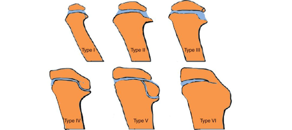
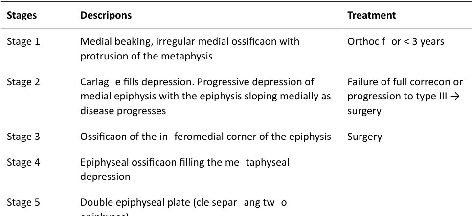
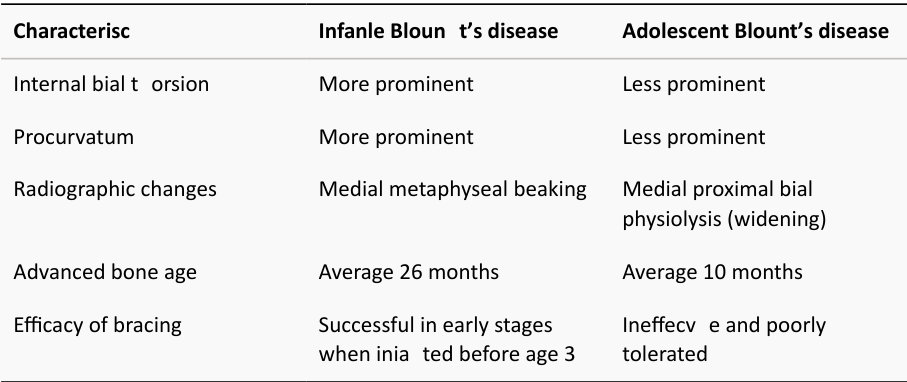
Blount’s disease is an uncommon paediatric knee condition in which the reis a growth disorder of the medial aspect of the proximal tibial physis. This is probably caused by a combination of excessive compressive forces on the proximal medial physis of the tibia. There are two recognized types: 1. Infantile (0 –4). 2. Adolescent (> 10 years). In the infantile tibia v ara, patients generally start to walk early (9–10 months; it is more prevalent in females, blacks and those with marked obesity. It is bilateral in approximately 80% of cases and associated with a prominent metaphyseal beak, internal tibial torsion and LLD. The deformity is usually painless. In the adolescent type, patients complain of pain at the medial aspect of the knee. These patients are normally similar to the SUFE type of patient with more incidences in overweight Afro-Caribbean males and unilateral involvement in 80% of cases. Langenskiold recognized six radiological stages of the disease (Figure 18.34). Spontaneous resolution according to Langenskiold is common for stages 1–2, possible for stages 3–4, while stages 5 and 6 are associated with recurrence and permanent deformity even after mechanical realignment.
So, what do you advise parents?
My advice is that bowed legs are common (in fact, the norm) before the age of 2 years. I get concerned when I see it after the age of 2 years or if there is asymmetry, which is the case with this child. This may resolve spontaneously; however, there are several predictors against spontaneous resolution. My options are either start treatment now or observe it for another 4–6 months. If things improve, observation will continue; otherwise, I will treat with a brace.
Do you think the brace will help?
There is evidence that braces can improve deformity in younger thanage 3 or prior to Langenskiold stage II. An elastic Bloun t brace provides valgus force in conjunction with a medial upright, with drop locks to increase corrective force during weight-bearing [36]. Bracing schedules vary from full-time to day or night only, with frequent adjustment of the medial upright every couple of months to provide a continuous valgus force. Compliance is an issue. The success of the true bracing effect is confounded by the benign natural history of physiologic bowed legs treated as Blount’s disease. One study limited topa tien ts with Drennan’s angles over 16° showed 86% success. Bracing failure was more likely with ligamentous instability, bodyweight exceeding the 90th percentile, or late initiation [ 37]. Another study demonstrated 70% success in Langenskiold stage II disease, although this was mainly in unilateral disease. Seventy percent of patients with bilateral involvement required surgical management [38].
Figure 18.34 Langenskiold classification of Bloun t’s disease.
Table 18.6 Langenskiold classification of Bloun t’s disease.
Stages Descriptions Treatment
Stage 6 Medial physeal closure
Authors’ note: 6
Genu varum, valgu mand Blount’s disease are favourite exam topics. The former is very common in clinical practice (and exams) and the latter, although rare, is closely related to the genu varu mand it has become the favourite linked topic to genu varum.
Please master these topics. They are easy, and you can score high marks!
Candidate 5#
The clinical photograph (Figure 18.35) is for a newborn child who was referred to you with a left foot deformity; the referrer thinks the baby may have a congenital vertical talus (CVT)? What do you think?
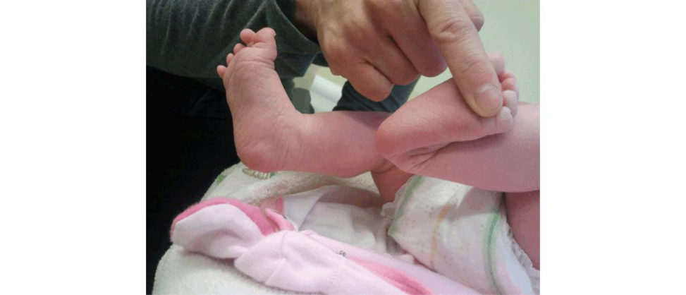
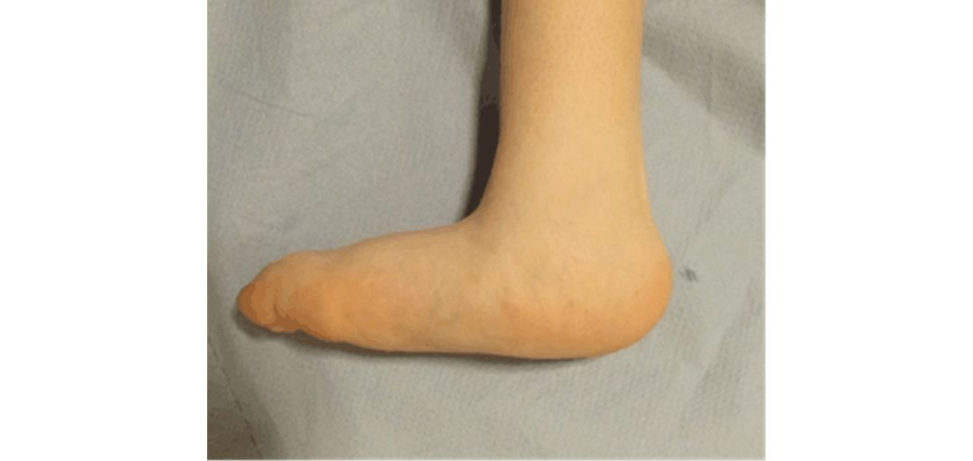
The clinical photograph shows features that are very suggestive of congenital calcaneovalgus foot, which is a benign soft -tissue contracture of the foot secondary to intrauterine foetal packaging. There is an excessive dorsiflexion of the foot to the extent that the dorsum of the foot touches the frontal aspect of the leg. The deformity is usually passively correctable. Although it may look similar to CVT for the inexperienced, the differences are clear: in CVT the hindfoot is a rigid equinus/valgus (not a flexible calcaneovalgus) (see Figure 18.36).
How would you manage it?
It is important to rule out similar deformities (CVT and neurologic foot deformities) and associated conditions posteromedial bowing and dislocated hips). In a classical flexible congenital calcaneovalgus foot the diagnosis is usually obvious and further investigation sare not required. However, radiographs can be useful to exclude CVT and posteromedial bowing, and US to exclude DDH. I usually reassure parents and advise them on simple stretching manoeuvres and it usually resolves over 6 months. In resistant cases serial casting may be used to expedite correction (particularly if the foot cannot be plantar flexed beyond neutral).
This is a radiograph of another child with the same condition ( Figure 18.37). Any comments?
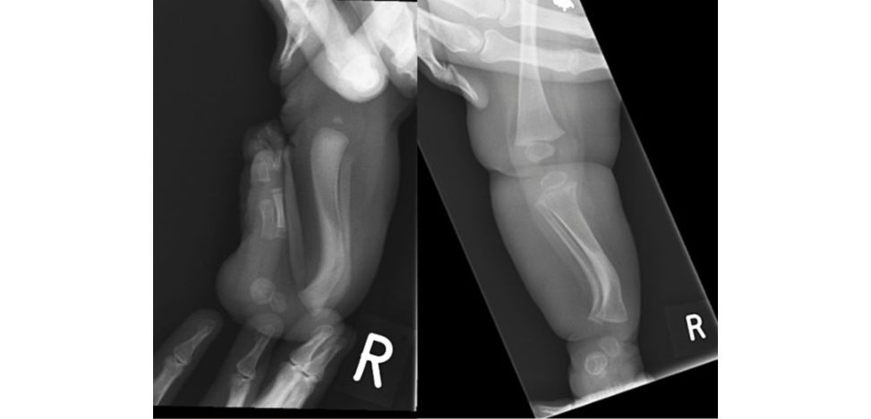
This clinical photograph shows a posteromedial bowing of the tibia (the apex of the deformity is pointing posterior and medial). It is often called physiologic bowing of tibia and it is thought to be a result of intrauterine foetal packaging. It is often associated with congenital calcaneovalgus foot. The two conditions may occur together or independently. In most cases the bowing resolves spontaneously over 3–5 years (Figure 18.38). However, it needs to be followed up, as LL Dis not uncommon.
You followed-up this patient and these are his X-rays over the last 3 years (Figure 18.39– 18.41) (see also Table 18.9).
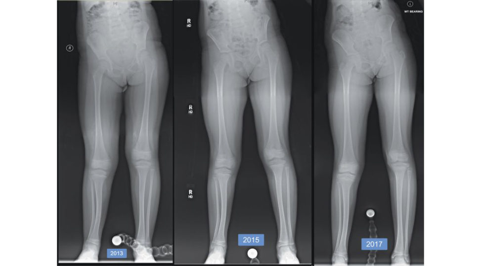
The X-rays show gradual improvement of the deformity (as expected), but there is still residual deformity and LL Das evident by the pelvic tilt. I would like to measure the LL Dand the angular deformity. I would like to have lateral views as well.
Show me how you would do your measurement.
My measurements indicate that there is a LLD of 4 cm (mainly from tibia 3.4 cm) and a coronal angular deformity (valgus) of 15° at the junction between the distal and middle third of the tibia. I do not have lateral views to measure how much deformity there is at the sagift al plane. I would like to take a full history to know the childage, height, any comorbidity, expectations and previous consultations and treatments. I also need to check her skeletal age.
She was 8 years and 10 months when she had the X-ray in 2017, her height was 134 cm, she is fit and healthy. She and her parents will be guided by your advice.
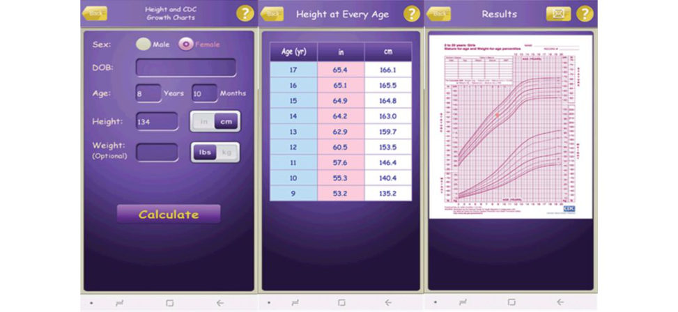
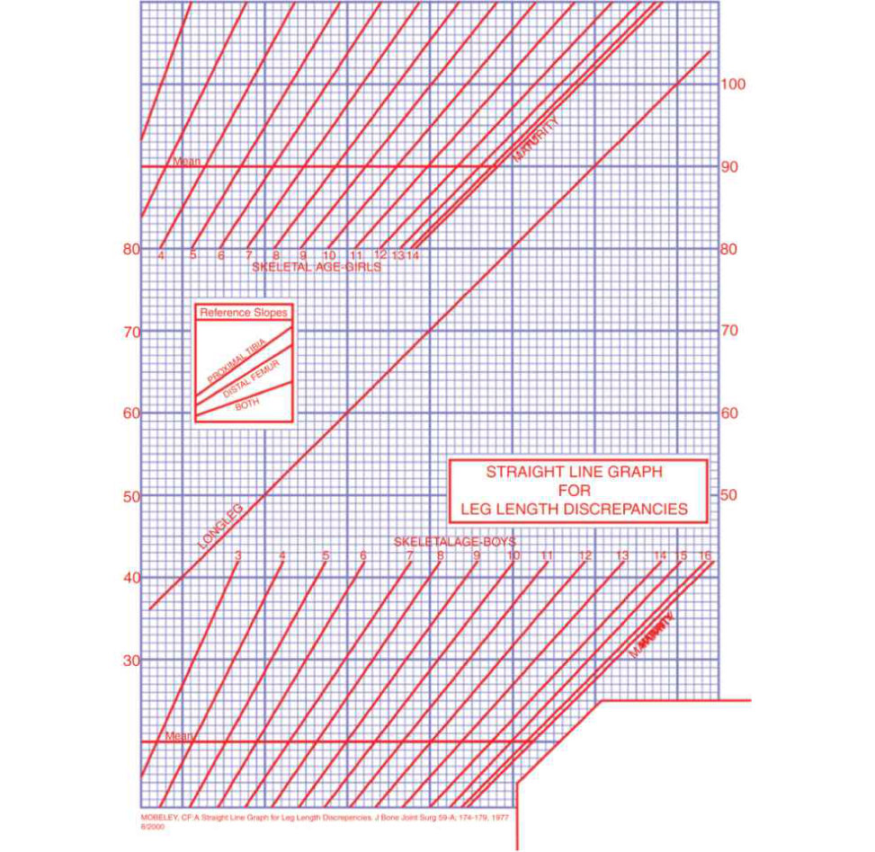
OK, I need to estimate her height and the LL Dat maturity, then plan my management. I use the multiplier app on my phone to do so, but I also double-check it with the charts to ensure they give comparable results (Figure 18.42, 18.43) (see Authors’ note). So, her estimated height at maturity would be 166 cm, which is just above the 50th centile. The estimated full LLD would be 5.1 cm, whereas the tibia LLD would be 4.3 cm. I expect almost equal results using a different method; for example, the rule of thumb method (Menelaus method). There is an increase in the full LLD of 6 mm and tibial LLD of 5 mm e very 2 years from the table above or 3 and 2.5 mm every year. On average, girls finish growth at 14 years of age, so she has 5 years and 2 months of growth. Simple calculation reveals a full LLD of 5.5 cm and tibia LLD of 4.5 cm, which are very close to the multiplier methods.
What will be your management plan?
My objectives are to correct all deformities. In this child, this includes LL Dand the distal tibia deformity. As for the LLD, I want to achieve equal leg lengths at skeletal maturity without excessive risk, morbidity or height reduction. My options are: 1. Shoe raise. 2. Epiphysiodesis of the long side. 3. Limb lengthening of the short side. 4. A combination of all. Given the significant LL Dand the deformity of the distal tibia (15° of valgus), my preferred option will be lengthening of the short leg with gradual correction of the leg deformity using a circular frame. I will use a double-stack frame with two osteotomies; one to correct the deformity and another at the proximal tibial metaphysis.
What is your rationale for this? Why not correct and lengthen at the deformity site?
I could but the bone healing at the site of deformity in this condition is not ideal for lengthening and may end in delayed union and poor regeneration.
The radiograph below shows what has been done (Figure 18.44). Any criticism?
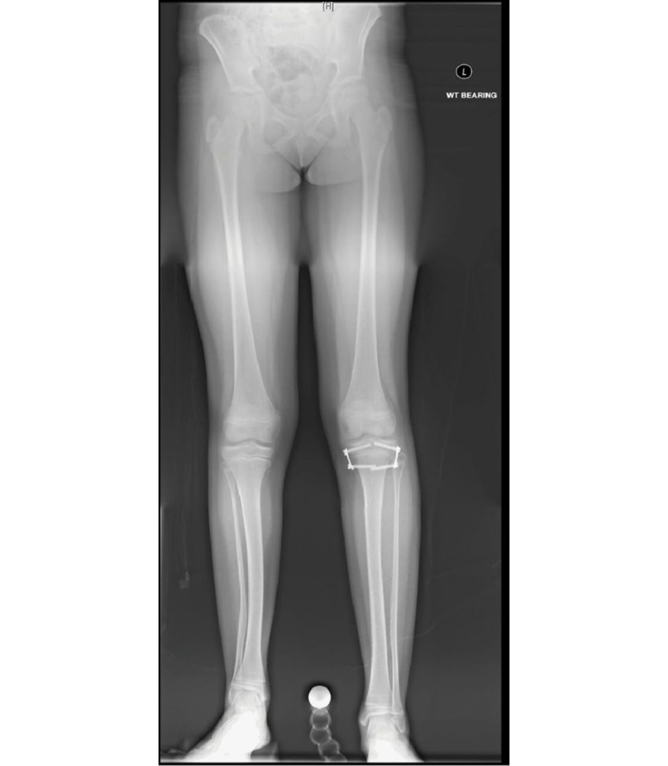
The surgeon chose to slow the growth of the left leg. It is a small operation, revisable and given the remaining few years of growth, it is potentially ablet o equalize the leg. It is a good option; however, it has drawbacks. The child will be shorter and the distal tibial deformity has not been addressed.
Figure 18.35 A newborn with left foot deformity.
Figure 18.36 Rocker boft om foot in congenital vertical talus.
Figure 18.37 Lower limb radiograph.
Figure 18.38 Resolution of posteromedial bowing.
Figure 18.39 Followup X-rays.
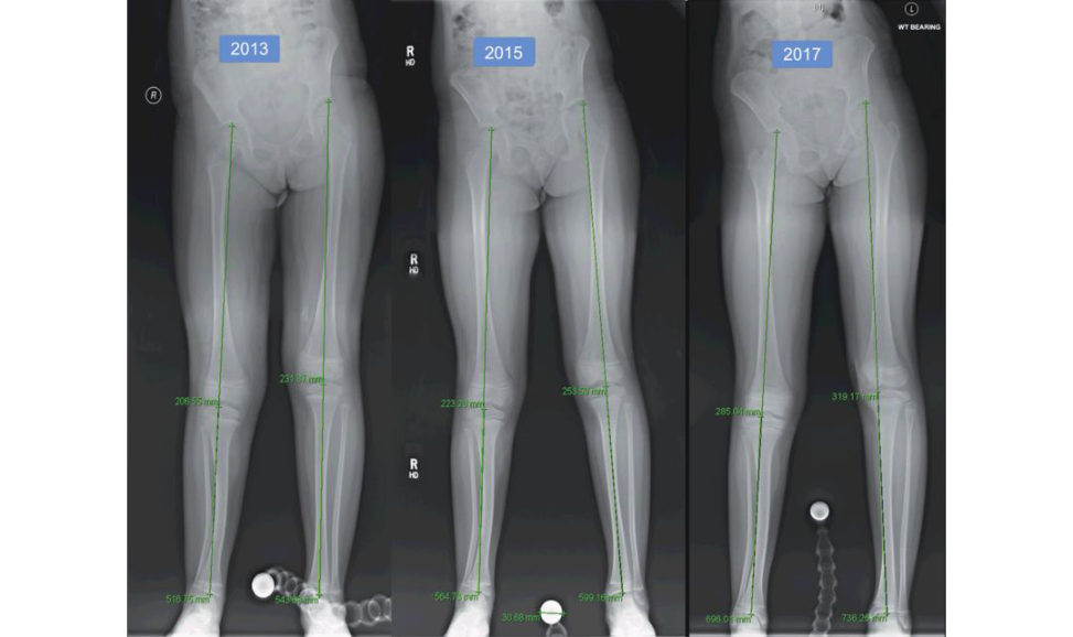
Figure 18.40 LLD following posteromedial bowing.
Table 18.9 Leg length discrepancy table.
Dates Segments Right Left Δ
2013 Total 51.7 54.4 2.7
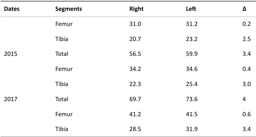
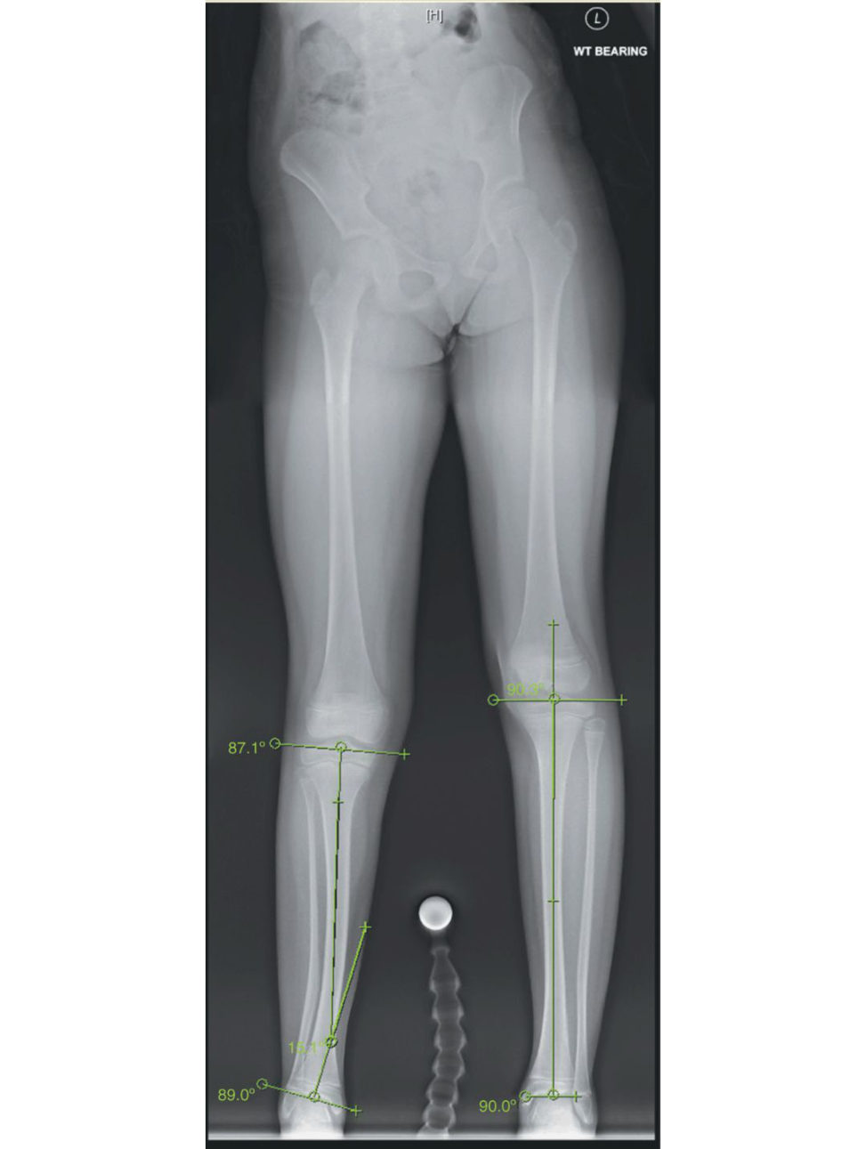
Figure 18.41 Tibial alignment measurements.
Figure 18.42 Estimation of height at maturity using the multiplier app.
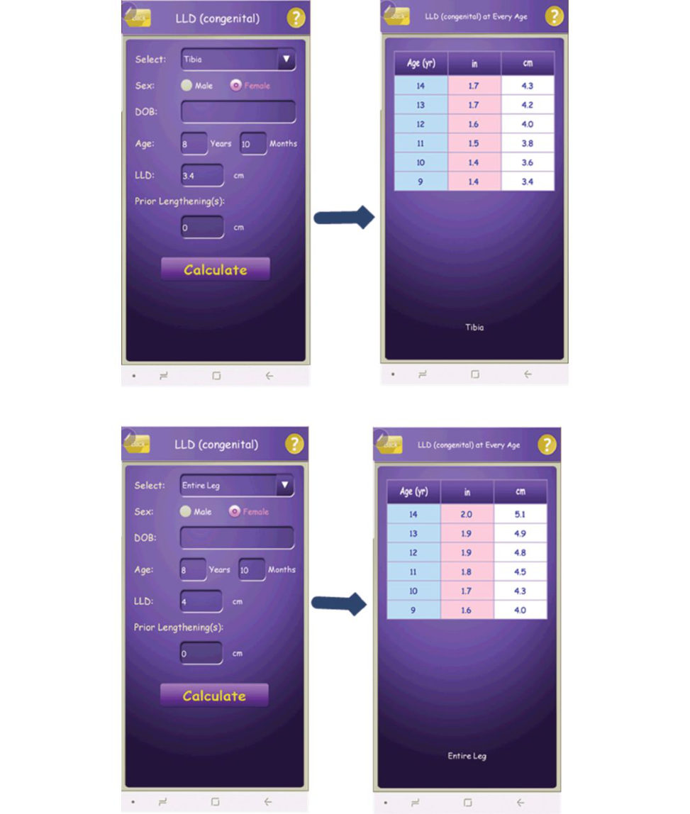
Figure 18.43 Estimation of LL Dat maturity using the multiplier method.
Authors’ note: 7
Limb reconstruction in general and lengthening in particular is an important topic not only in the paediatric section but in the adult section aswell. The viva started with a simple case of congenital calcaneovalgus foot, which all candidates must recognize and come with a sensible plan. The viva progressed into a more complicated area of posteromedial tibial bowing and subsequent deformity and LLD. The candidate was doing very well and he clearly had a good understanding of how to manage LL Dand deformity with a very good pass.
He was not derailed when he realized that the patient did not have what he had suggested.
He pointed out the pros and cons of what had been done and explained why he would have chosen a different plan.
Please be objective and sensitive when you criticize other people’s operation and do not be very negative even if you think the choice of operation or the technique was completely wrong.
We recommend you familiarize yourself with the multiplier app You can download it from:
htip s://itunes.apple.com/us/app/multiplier/id460335161?m t=8
Try to practise using the Mosley chart method (Figure 18.45) to do the calculation for the above patient.
More information is available on the following website:
www.pedipod.com/Chapters/StepByStep.asp#
Figure 18.44 Proximal tibia epiph ysiodesis using eight plates.
Figure 18.45 Blank Mosley chart.
Candidate 6#
These are AP and lateral views of a 13-year-old child (Figure 18.46) who was brought in after a football injury. How would you manage such an injury?
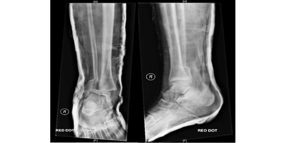
Figure 18.46 Ankle injury.
I always approach these types of injuries according to ATLS protocols. It is very tempting to start with the ankle fracture or the deformed limb and it is important to resist this temptation.
You did your primary and secondary survey, and this is the only injury that this child has.
I ensure adequate analgesia onboard when I assess this child with substantive trauma. I look for obvious swelling, bruises, wounds, colour of the foot and toes. The X-ray indicates the skin on the medial side of the ankle might be overstretched and there may be impending skin breakdown that requires immediate fracture reduction. I f eel for skin temperature, pulses, tenderness. I assess the movement of the joints that are not involved to ensure that there are no unexpected problems. I would not move the ankle joint at this stage as it is expected to be painful. I assess the neurovascular status of the limb using special tests to test for nerve injuries.
Indeed, you have found the skin over the medial malleolus is overstretched, white and about to tear.
Then we need to reduce the fracture immediately to prevent this fracture from becoming an open fracture, which has more comorbidity. If theatre space is immediately available, I will take the patient to the operating room; otherwise, I may need to do it in the emergency room under sedation or Entonox.
Theatre is available, but the patient had food an hour ago.
I think this a limb-threatening condition and justifies a rapid sequence induction anaesthesia (to prevent aspiration). I will discuss this with my anaesthetic colleague and we should proceed to perform the procedure. Of course, other preoperative preparation and consenting should be done appropriately and timely to prevent any delay in performing surgery. These physeal injuries are usually stable after reduction and the y need closed reduction and a well-moulded cast application.
This is what has been done (Figure 18.47), any thoughts?
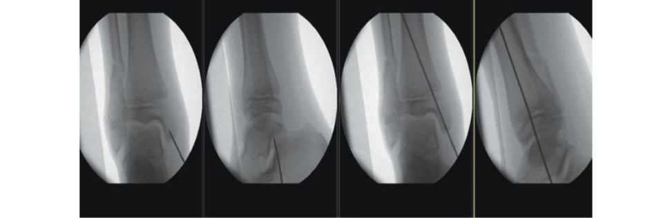
Figure 18.47 Ankle facture fixation.
The surgeon reduced the fracture nicely and stabilized with a single K-wire crossing the physis. As I mentioned, my experience with these fractures is that they are relatively stable, and K- wire is not usually required unless the fracture is not stable. The concern is that the K-wire may cause physeal damage.
What would you do next?
My postoperative instruction ist o keep the foot elevated on aB raun frame, to mobilize non- weight-bearing. I would like to check the skin and the wound in 48 hours or so to ensure the skin is healing well. The patient will be discharged when deemed safe and I would like to see him in a week’s time for wound check and X-ray. If everything is satisfactory, I will plan to remove the K-wire in 6 weeks’ time. As the risk of growth plate disturbance is high in these fractures, I would like to follow this patient until I am sure that there is no growth arrest.
This is the ankle X-ray after 8 months (Figure 18.48).
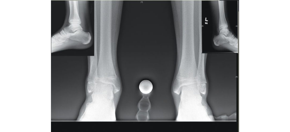
Figure 18.48 Eight months postoperative.
It is difficult to be certain, but I think the patient is developing a growth arrest on the right. I cannot see the growth plate very clearly and there is more valgus around the ankle. I would like to get more imaging (CT scan or MRI scan).
There was no CT scan or MRI scan but there are longlegs alignment views and some measurements (Figure 18.49).
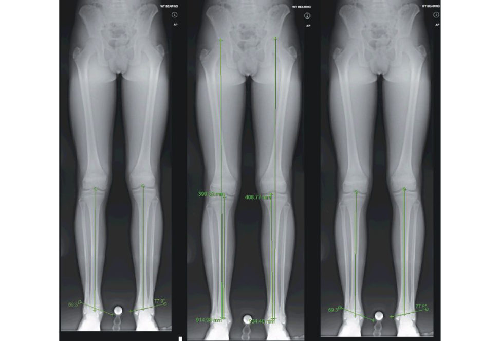
The longleg alignment views confirm my initial suspicions that there is a growth arrest which led to a shortening of almost 1 cm and distal tibial valgus deformity of at least 10°. How old is the patient now?
Figure 18.49 Longleg views measurements.
15 years old.
So, he still has two years or more to go. A hand X-ray to establish his skeletal age will be valuable. My thought is to do bilateral distal tibial and fibular epiph ysiodesis to stop the deformity and the LLD from geting worse.
Would you consider correcting the distal tibial deformity?
If he is asymptomatic and the reis a good subtalar join movement I probably would not; however, if he is symptomatic and the reis a limited subtalar joint movement that does not compensate for the deformity then I would.
The patient underwent left distal tibia and fibular epiph ysiodesis and right distal tibial medial hemiepiphysiodesis. These are his pictures a year after (Figure 18.50), any thoughts?
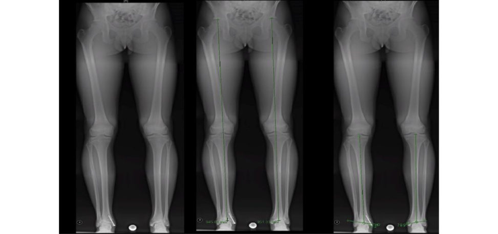
I think this is a very good idea and it seems to have worked. The LLD reduced and the deformity was reduced. I thought about it; however, I was not sure whether the right distal tibia physis is still reliable to correct deformity.
Figure 18.50 Postoperative left distal tibia and fibula hemiepiph ysiodesis and right distal tibia medial hemiepiphysiodesis.
Authors’ note: 8
Physeal injuries are common in children. They are relatively stable after reduction; however, this is not always true. Instability is far more dangerous than a small wire crossing the physis. The above fracture was unstable and using a smooth K-wire to stabilize was the right thing to do. K-wire rarely causes growth arrest (take the supracondylar fracture as an example). Several technical tips can even reduce the risk further, such as using the smallest possible size, smooth wire, burst driver mode rather than continuous cooling the heat generated by friction, etc...
These clinical photographs (Figure 18.51) are for an 18-month-old child who is about to have surgery. Could you tell me what the pictures show and what the surgery involves?
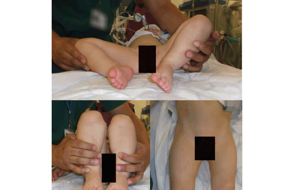
Figure 18.51 Clinical photographs of 18-month-old child.
The clinical photographs show the lower parts of a child’s body. The surgeon tries to demonstrate the limited right hip abduction and the Galeazzi sign (the apparent shortening of the right femur) and the abnormal skin crease on the left proximal femur. All are signs of hip dislocation. In an 18-month-old child, I anticipate the surgery will be hip adductors release, open reduction, pelvic osteotomy (with or without femoral osteotomy) and application of hip spic a.
This is a plain pelvis X-ray of the above baby (Figure 18.52). What can you say about the X- ray?
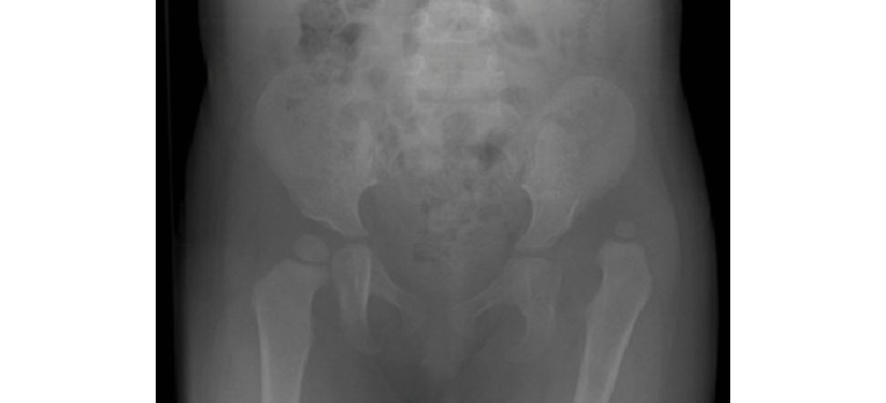
Figure 18.52 Plain pelvic X-ray.
The radiograph confirms my initial thought that the baby has a left hip dislocation. The femoral nucleus is smaller on the left side and has created a false acetabulum. The true acetabulum is shallow and not very well formed when compared to the other side. If I draw Hilgenreiner and Perkin lines, the head will be siting in the upper lateral quadrants. There is a break in the inferior and lateral Shent onlines (Figure 18.53).
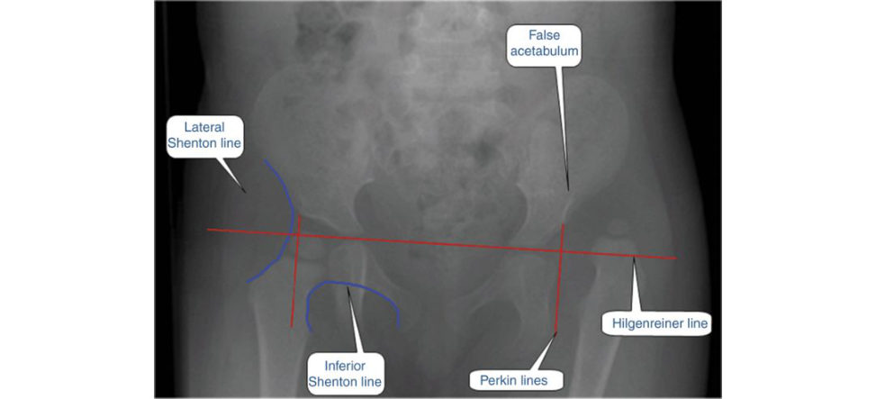
Figure 18.53 Plain pelvis X-ray of the hip of an 18-month-old child.
What would you do if you were the surgeon?
He is 19 months old. I will be surprised if this hip can be reduced closed. However, there is always apa tien t who does not follow the rule. So, I would perform EUA and trial of closed reduction. If it is reduced, stable and has a large zone of safety, I would perform a hip arthrogram to assess how good my reduction is.
Tell me what you mean by zone of safety?
This term was coined by Ramsey and associates [39] and refers to the range of motion in which the hip remains reduced in comparison to the maximum range of motion. The wider the range, the better. Two important facts are to be considered: 1. The average movement of the hip in a hip spica is 15°; so, it is important to keep the hip away from the line of dislocation by about 20° to reduce the risk of dislocation inside the spica. 2. The risk of AVN increases with the hip put at the maximum range of motion so it is safe to be 20° inside the maximum range of motion. So, the safety zone is the range of motion (c one of motion) within 15 –20° of the maximum range of motion and dislocation range. The maximum range of motion can be increased by adductor (to increase abduction) and psoas (to increase extension) tenotomy. These are not needed in all cases (Figures 18.54 and 18.55).
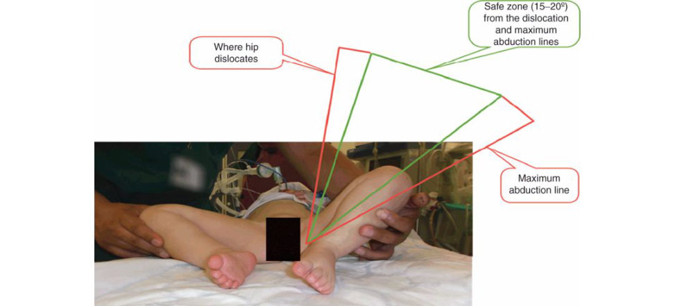
Figure 18.54 Coronal safe zone.
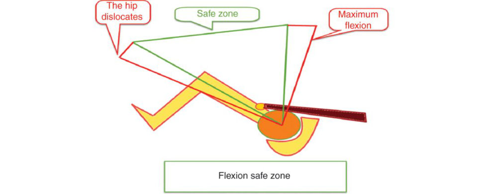
Figure 18.55 Sagift al safe zone.
You tried to reduce it closed and did a hip arthrogram. Figure 18.56 shows the arthrogram. Do you want to comment on the findings?
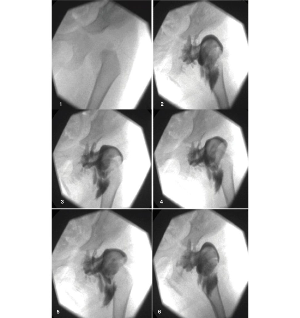
The arthrogram nicely shows the full size of the femoral head, which is much bigger than the ossified nucleus. The femoral head is not reduced in any of the below images and it remained articulating with a false acetabulum on the ileum. The capsule is distended and has the classical hourglass shape. This happens because as the head grows the femoral side becomes bigger while the isthmus of the capsule remains small. The isthmus is further constricted by the Chinese trap effect as well as by pressure from the iliopsoas tendon (visible in pictures 2 and 6). The ligamentum teres is thickened and elongated (pictures 2–6). None of the pictures demonstrate a reduced hip and there is significant medial dye pool. So, I believe this hip is not reducible.
Figure 18.56 Hip arthrogram of a 19-month-old child.
What prevents reduction?
There are several anatomical structures that can prevent reduction. These can be summarized as: A. Extra-articular structures: 1. Iliopsoas tendon. 2. Capsular constriction. B. Intra-articular (2 ligaments + 2 pathological structures):
1. Elongated ligamentum teres.
2. Thickened transverse acetabular ligament giving the acetabular cartilage the classic horse-shoe structure.
3. Pulvinar (fibro-fatiy tissues filled the acetabulum ).
4. Inverted limbus.
What is the limbus?
There is no anatomical structure called limbus (or neolimbus), but it is the name given to the deformed and moulded labrum with the attached cartilaginous r oof of the acetabulum pushed into the acetabulum preventing the femoral head reduction.
So, what would you do in the above situation?
I would proceed to open reduction of the hip through the anterior hip approach (Smith Peterson). This is combined with adductors tenotomy and over the brim psoas muscle release. Pelvic osteotomy is often needed and femoral osteotomy may be needed as well.
Take me through how would you do open reduction throu ghan anterior hip approach? Authors’ note: 9 DD His one of the commonest paediatric topics that have been featured in the exam. It is not the candidates’ favourite, particularly if they have not done a paediatric orthopaedic job. The topic is nicely covered in the postgraduate paediatric orthopaedic book and the relevant section in the third edition. You do not need to know all the ins and outs of the topic as longas you know the principles. Treatment options depend on the age of child at presentation, reducibility of hip, stability after reduction anda mount of acetabular dysplasia. Authors’ note: 10 The FRCS (Trauma and Orthopaedic) exam is easy topass, but ironically it is also easy to fail. Having helped hundreds of candidates topass their exam, we find it is all about preparation: ‘If you fail to prepare, then prepare to fail’. Core knowledge and skills are important, but what is most important is how to present them clearly and confidently. This will come with practice. The Postgraduate Orthopaedic faculty wish you all the best for your exam. Table 18.10 Dislocated hip management. Age groups Treatment
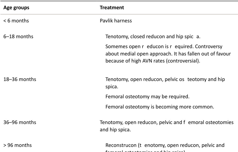
vs.
Leave it unreduced.
Have some answers about controversial points, including:
- Routine screening: currently int heUK all infants have clinical exam and only patient sat risk/with abnormal exam will have an ultrasound scan.
- Traction before closed reduction: different units follow different protocols. Supporters suggested that traction reduces the need for open reduction and decreased AVN rate, other studies show similar results with and without traction mainly duet o better understanding of treatment principles, which encourage gentle reduction, the use of the human position to maintain reduction, and a voiding extreme positions for immobilization.
- Medial vs. anterior approach for open reduction.
- Pelvic osteotomy types and indications.
- Reconstructive must have concentric reduction.
ARe directional: Salter, Pemberton, double (Sutherland), triple (Steel) and periacetabular (Ganz/PAO).
B. Reshaping Dega and Pemberton (Pemberton osteotomy mainly directional but can change the shape as well).
- Salvage: Shelf and Chiari.
References
1. Alshryda S, JonesS, Banaszkiewicz P. Postgraduate Paediatric Orthopaedics: The Candidate’s Guide to the FRCS (Tr and Orth) Examination . Cambridge: Cambridge University Press; 2014.
2. Loder RT Richards BS, Shapiro PS, Reznick LR, Aronson DD. Acute slipped capital femoral epiphysis: the importance of physeal stability. J Bone Joint Surg Am. 1993;75(8):1134–1140.
3. Alshryda ST sang K, Chytas A, et al. Evidence based treatment for unstable slipped upper femoral epiphysis: systematic review and exploratory patient level analysis. The Surgeon. 2018;16(1):46–54.
4. Alshryda ST sang K, Ahmed M, Adepapo A, Montgomery R. Severe slipped upper femoral epiphysis;
fish osteotomy versus pinning-in-situ: an eleven year perspective The Surgeon. 2014;12(5):244–248.
5. Jerre R, Billing L, Hans sonG, Wallin J. The contralateral hip inpatients primarily treated for unilateral slipped upper femoral epiphysis. Long-term follow-up of 61 hips. J Bone Joint Surg Br. 1994;76(4):563–567.
6. Phillips PM, Phadnis J, Willoughby R, Hunt L. Posterior sloping angle as a predictor of contralateral slip in slipped capital femoral epiphysis. J Bone Joint Surg Am. 2013;95(2):146–150.
7. Stasikelis PJ, Sullivan CM Phillips WA, Polard JA. Slipped capital femoral epiphysis. Prediction of contralateral involvement. J Bone Joint Surg Am. 1996; 78(8):1149–1155.
8. Parsch K, WellerS, Parsch D. Open reduction and smooth Kir schner wire fixation for unstable slipped capital femoral epiphysis. J Pediatr Orthop. 2009;29(1):1–8.
9. Ziebarth K, Zilkens C, SpencerS, Leunig M, Ganz R, Kim Y-J. Capital realignment for moderate and severe SCFE using a modified Dunn procedure. Clin Orthop Relat Res. 2009;467(3):704–716.
10. Naseem H, Chatterji ST sang K, Hakimi M, Chytas A, Alshryda S. Treatment of stable slipped capital femoral epiphysis: systematic review and exploratory patient level analysis. J Orthop Traumatol.
2017;18:469.
11. British Orthopaedic Association . BOAST11. Supracondylar fractures of the humerus in children. 2014.
12. Isaacs H, Handelsman JE, Badenhorst M, Pickering A. The muscles in clubfoot – a histological histochemical and electron microscopic study. J Bone Joint Surg Br. 1977;59B(4):465–472.
13. Gray DH, Katz JM. A histochemical study of muscle in clubfoot. J Bone Joint Surg Br. 1981;63B(3):417–423.
14. Wynne-Davies R. Genetic and environmental factors in the etiology of talipes equinovarus. Clin Orthop
Relat Res. 1972;84:9–13.
15. Sharon-Weiner M, Sukenik-Halevy R, Tepper R, Fishman A, Biron-Shental T, Markovitch O. Diagnostic accuracy, work-up, and outcomes of pregnancies with clubfoot detected by prenatal sonography. Prenat
Diagn. 2017;37(8):754–763.
16. Ponseti IV, Morcuende JA. Current management of idiopathic clubfoot questionnaire: a multic enter study. J Pediatr Orthop. 2004;24(4):448.
17. Ponseti IV, Smoley EN. Congenital clubfoot: the result of treatment. J Bone Joint Surg Am.
1963;45(A(2)):261–344.
18. Huntley JS. What is the best treatment for paediatric trigger thumb (acquired thumb flexion contracture)? InS Alshryda, JS Huntley, P Banaszkiewicz (Eds.), Paediatric Orthopaedics: An Evidence-Based Approach to Clinical Questions . Cham: Springer; 2016.
19. Caird MS, Flynn JM, Leung YL, Millman JE, D’Italia JG, Dormans JP. Factors distinguishing septic arthritis from transient synovitis of the hip in children. A prospective study. J Bone Joint Surg Am.
2006;88(6):1251–1257.
20. Maharajan K, Patro DK, Menon J, et al. Serum procalcitonin is a sensitive and specific marker in the diagnosis of septic arthritis anda cute osteomyelitis . J Orthop Surg Res. 2013;8:19.
21. Zhao J, Zhang S, Zhang L, et al. Serum procalcitonin levels as a diagnostic marker for septic arthritis: a meta-analysis. Am JEm erg Med. 2017;35(8):1166–1171.
22. Kocher MS, Zurakowski D, Kasser JR. Differentiating between septic arthritis and transient synovitis of the hip in children: an evidence-based clinical prediction algorithm. J Bone Joint Surg Am.
1999;81(12):1662–1670.
23. Kocher MS, Mandi gaR, Zurakowski D, Barnewolt C, Kasser JR. Validation of a clinical prediction rule for the differentiation between septic arthritis and transient synovitis of the hip in children. J Bone Joint Surg
Am. 2004;86A(8):1629–1635.
24. Herring JA, Kim HT, BrowneR. Legg–Calve–Perthes disease. Part II: Prospective multic enter study of the effect of treatment on outcome. J Bone Joint Surg Am. 2004;86A(10):2121–2134.
25. Wiig O, Terjesen T, Svenningsen S. Prognostic factors and outcome of treatment in Perthes’ disease: a prospective study of 368 patients with five-year follow-up. J Bone Joint Surg Br. 2008;90(10):1364–1371.
26. Wright JG. Evidence-Based Orthopaedics. The Best Answers to Clinical Questions , ed. J. Wright. Philadelphia: Saunders; 2009.
27. Daly K, Bruce C, Catterall A. Lateral shelf acetabuloplasty in Perthes’ disease. A review of the end of growth. J Bone Joint Surg Br. 1999;81(3):380–384.
28. Domzalski MEG luting J, Bowen JR, Litile ton AG. Lateral acetabular growth stimulation following a labral support procedure in Legg–Calve–Perthes disease. J Bone Joint Surg Am. 2006;88(7):1458–1466.
29. Bankes MJ, Catterall A, Hashemi-Nejad A. Valgus extension osteotomy for ‘hinge abduction ’ in
Perthes’ disease. Results at maturity and factors influencing the radiological outcome. J Bone Joint Surg
Br. 2000;82(4):548–554.
30. Seg evE, Ezra E, Wientraub S, Yaniv M, Hayek S, He moY. Treatment of severe late-onset Perthes’
disease with soft tissue release and articulated hip distraction revisited at skeletal maturity. J Child
Orthop. 2007;1(4):229–235.
31. Laklouk MA, Hosny GA. Hinged distraction of the hip joint in the treatment of Perthes disease:
evaluation a t skeletal maturity. J Pediatr Orthop B. 2012;21(5):386–393.
32. Davids JR, Blackhurst DW, Allen Jr BL. Clinical evaluation of bo wed legs in children. J Pediatr Orthop B.
2000;9(4):278–284.
33. Levin eAM, Drennan JC. Physiological bowing and tibia v ara. The metaphyseal–diaphyseal angle in the measurement of bowleg deformities . J Bone Joint Surg Am. 1982;64(8):1158–1163.
34. O’Neill DA, MacEwen GD. Early roentgenographic evaluation of bowlegged children. J Pediatr Orthop.
1982;2(5):547–553.
35. McCarthy JJ, Betz RR, Kim AD avids JR, Davidson RS. Early radiographic differentiation of infantile tibia vara from physiologic bowing using the femoral–tibial ratio . J Pediatr Orthop. 2001;21(4):545–548.
36. Herring JA (Ed.). Tachdjians Pediatric Orthopaedics, Vol. 1. Philadelphia: Saunders Elsevier; 2008.
37. Raney EM, Topoleski TA, Yaghoubian R, Guidera KJ, Marshall JG. Orthotic treatment of infantile tibia vara. J Pediatr Orthop. 1998;18(5):670–674.
38. Richards BS, Katz DE, Sims JB. Effectiveness of brace treatment in early infantile Bloun t’s disease. J Ped
Orthopaed. 1998;18(3):374–380.
39. Ramsey PL, Lass erS, MacEwen GD. Congenital dislocation of the hip: use of the P avlik harness in the child during the first six months of life. 1976. J Bone Joint Surg Am. 2002;84A(8):1478; discussion 1478.
Section 5